Recombinant Mouse CCL9 protein (rFc Tag)
Species
Mouse
Purity
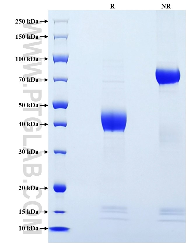
>90 %, SDS-PAGE
Tag
rFc Tag
Activity
not tested
Cat no : Eg3336
Validation Data Gallery
Product Information
| Purity | >90 %, SDS-PAGE |
| Endotoxin | <0.1 EU/μg protein, LAL method |
| Activity |
Not tested |
| Expression | HEK293-derived Mouse CCL9 protein Gln22-Gln122 (Accession# Q3U9T8) with a rabbit IgG Fc tag at the C-terminus. |
| GeneID | 20308 |
| Accession | Q3U9T8 |
| PredictedSize | 37.8 kDa |
| SDS-PAGE | 38-48 kDa, reducing (R) conditions |
| Formulation | Lyophilized from 0.22 μm filtered solution in PBS, pH 7.4. Normally 5% trehalose and 5% mannitol are added as protectants before lyophilization. |
| Reconstitution | Briefly centrifuge the tube before opening. Reconstitute at 0.1-0.5 mg/mL in sterile water. |
| Storage Conditions |
It is recommended that the protein be aliquoted for optimal storage. Avoid repeated freeze-thaw cycles.
|
| Shipping | The product is shipped at ambient temperature. Upon receipt, store it immediately at the recommended temperature. |
Background
Mouse C-C motif ligand 9 (CCL9), alternatively named macrophage inflammatory protein 1γ (MIP-1γ), was identified in 1995 and is homologous to the mouse CCL6, as well as human CCL23 and CCL15. It is noted that CCL9 is also known by various names such as macrophage inflammatory protein-1 gamma (MIP-1ɣ), macrophage inflammatory protein-related protein-2 (MRP-2) and CCF18 in rodents. Monocytes and myeloid cell lines produce large quantities of CCL9 , as do dendritic cells and T cells, in particular Th1 type T cells. Despite high baseline levels in circulation, it has become apparent that concentrations of CCL9 vary greatly in specific tissues with profound effects on health. For example, in the bone, CCL9 is produced at even higher levels, and is critical to osteoclast versus osteoblast differentiation of macrophages. There are also indications of a timed specific induction of CCL9 in skin wound healing and follicle-associated epithelium of the gut.
References:
1. Łazarczyk, Marzena et al. (2023) Curr Issues Mol Biol. 45(4):3446-3461.
2. Niu, Boning et al. (2024) Acta Pharm Sin B.14(8):3711-3729.
3. Schneberger, D et al. (2020) Environ Dis.5(4):93-99.
