Tested Applications
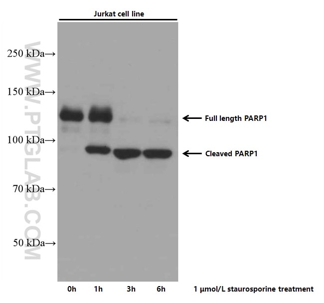
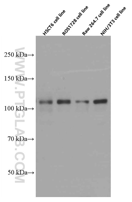
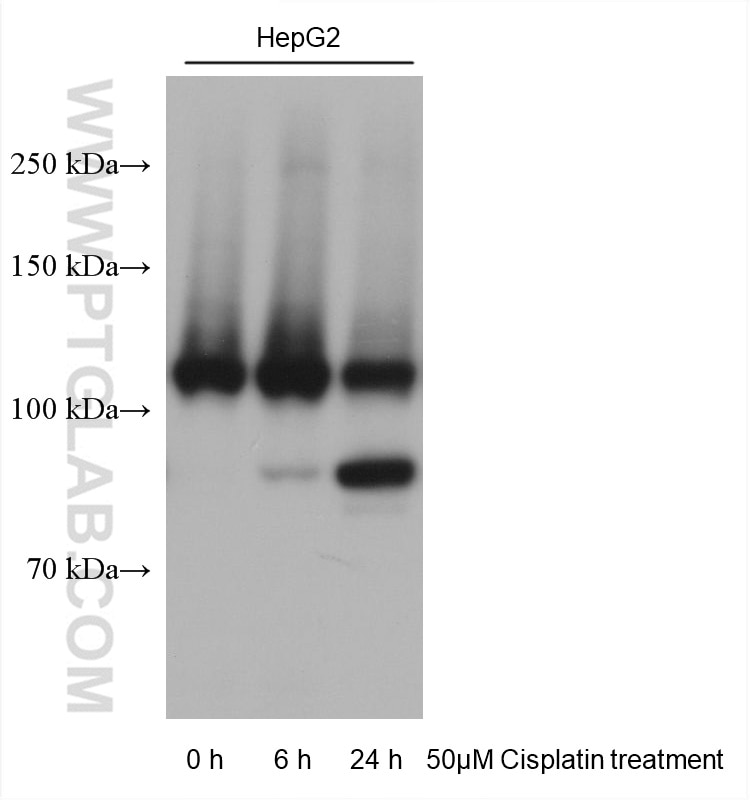
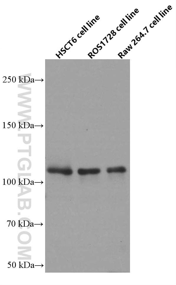
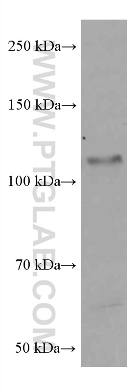
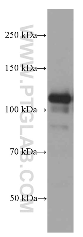
| Positive WB detected in | Jurkat cells, RAW 264.7 cells, HeLa cells, HSC-T6 cells, HepG2 cells, ROS1728 cells, NIH/3T3 cells |
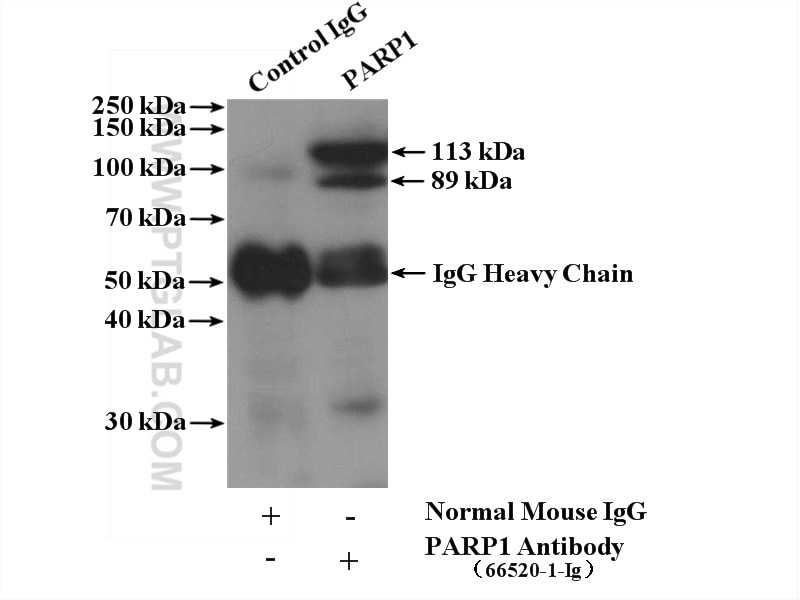
| Positive IP detected in | K-562 cells |
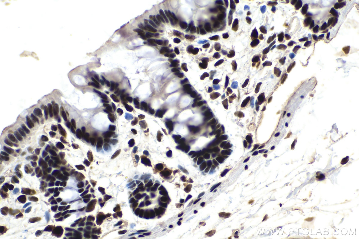
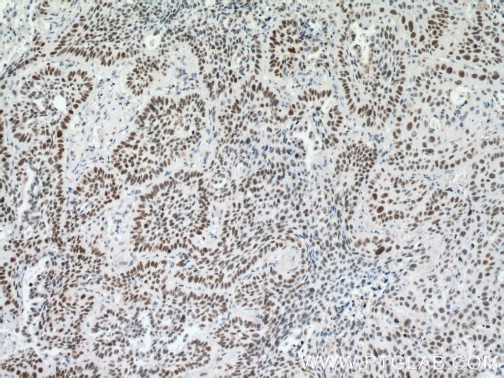
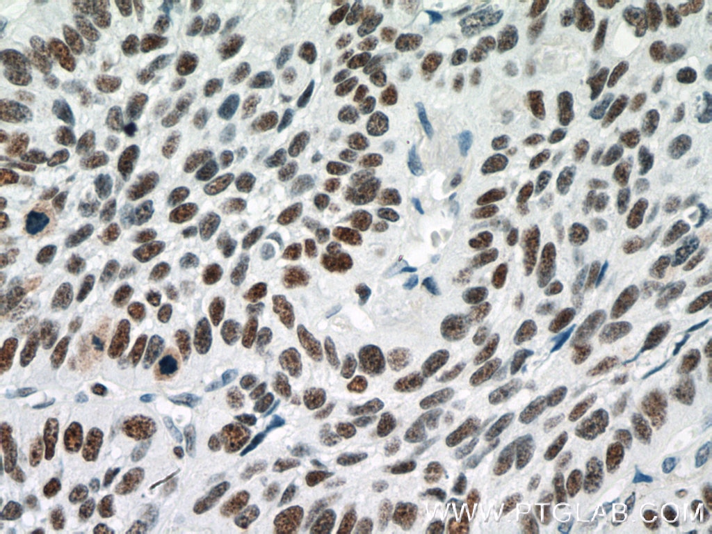
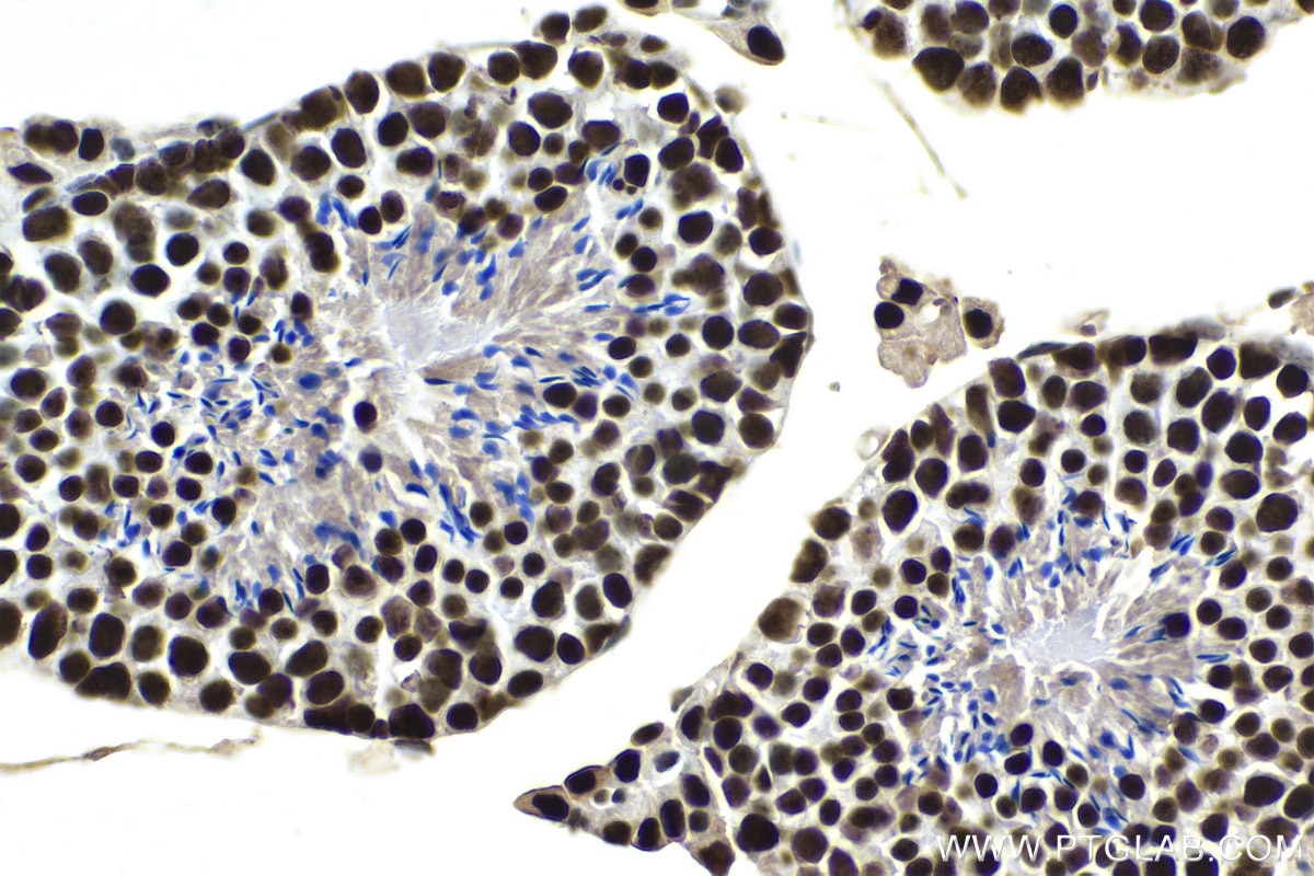
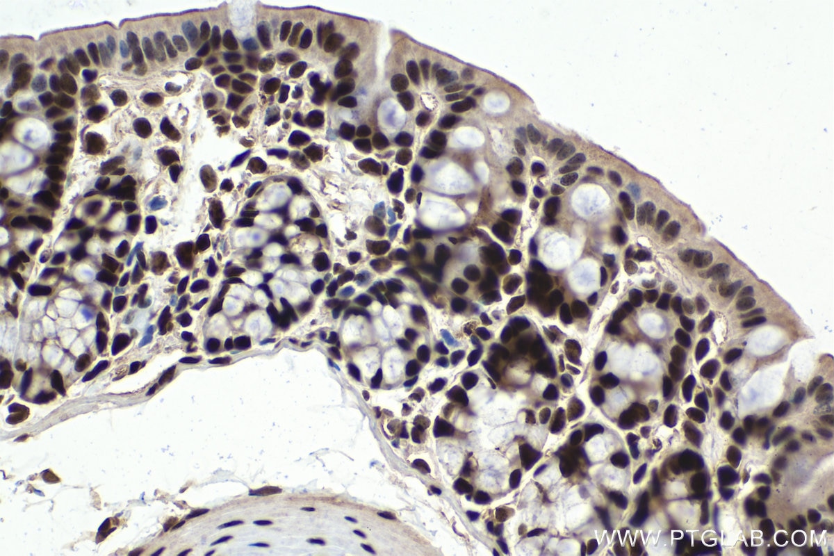
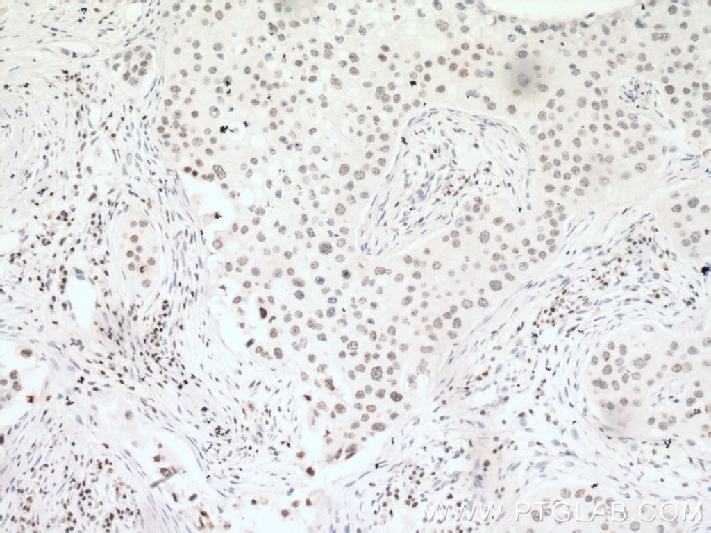
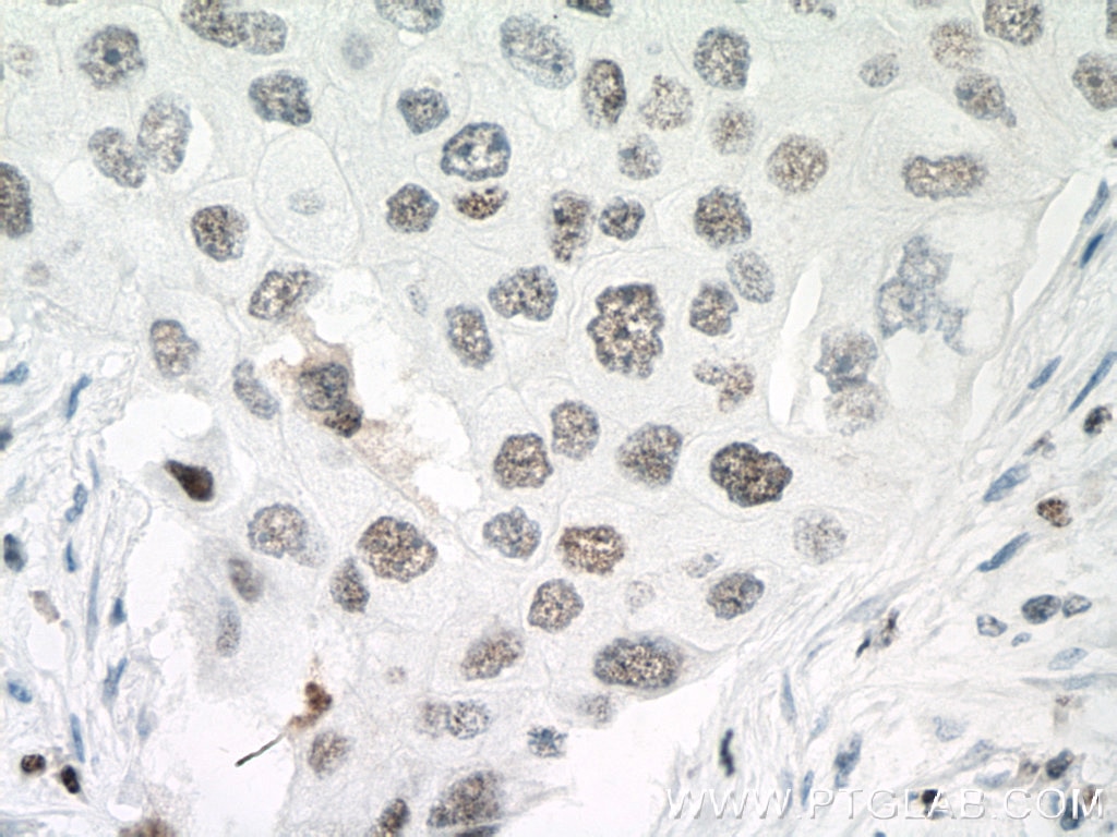
| Positive IHC detected in | human lung cancer tissue, mouse colon tissue, mouse testis tissue, human breast cancer tissue, rat colon tissue Note: suggested antigen retrieval with TE buffer pH 9.0; (*) Alternatively, antigen retrieval may be performed with citrate buffer pH 6.0 |
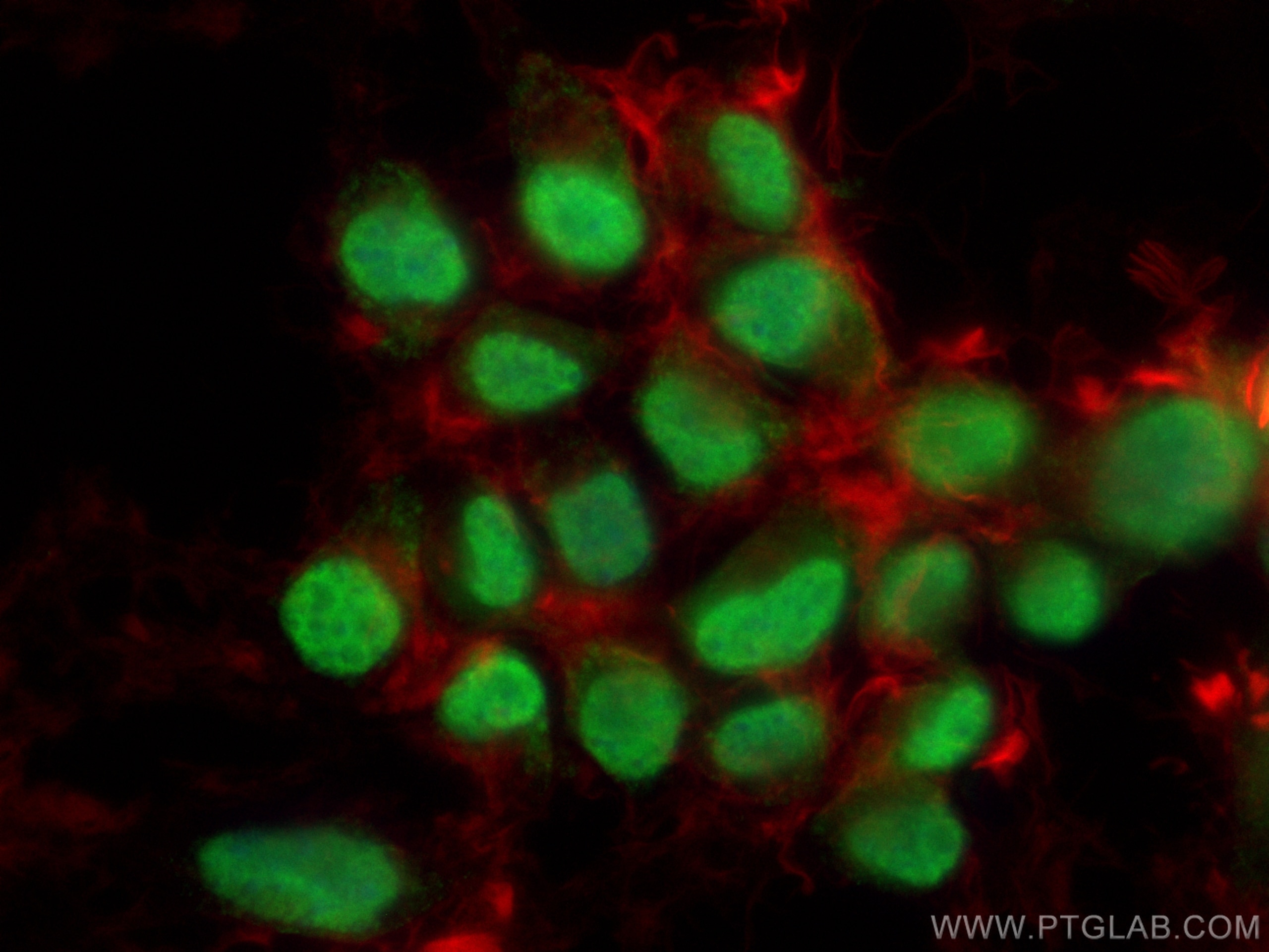
| Positive IF/ICC detected in | Neuro-2a cells |
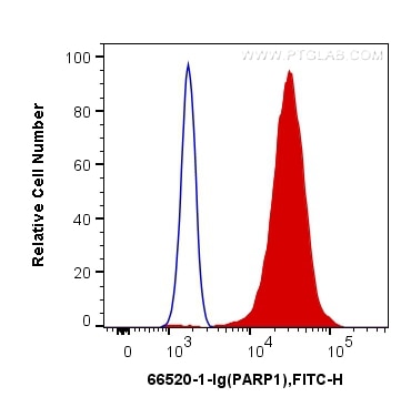
| Positive FC (Intra) detected in | HeLa cells, Jurkat cells |
Recommended dilution
| Application | Dilution |
|---|---|
| Western Blot (WB) | WB : 1:5000-1:50000 |
| Immunoprecipitation (IP) | IP : 0.5-4.0 ug for 1.0-3.0 mg of total protein lysate |
| Immunohistochemistry (IHC) | IHC : 1:100-1:1200 |
| Immunofluorescence (IF)/ICC | IF/ICC : 1:200-1:800 |
| Flow Cytometry (FC) (INTRA) | FC (INTRA) : 0.20 ug per 10^6 cells in a 100 µl suspension |
| It is recommended that this reagent should be titrated in each testing system to obtain optimal results. | |
| Sample-dependent, Check data in validation data gallery. | |
Published Applications
| KD/KO | See 6 publications below |
| WB | See 141 publications below |
| IHC | See 10 publications below |
| IF | See 10 publications below |
| IP | See 7 publications below |
| CoIP | See 3 publications below |
Product Information
66520-1-Ig targets PARP1 in WB, IHC, IF/ICC, FC (Intra), IP, CoIP, ELISA applications and shows reactivity with human, mouse, rat samples.
| Tested Reactivity | human, mouse, rat |
| Cited Reactivity | human, mouse, rat, canine, chicken, zebrafish |
| Host / Isotype | Mouse / IgG1 |
| Class | Monoclonal |
| Type | Antibody |
| Immunogen |
CatNo: Ag19173 Product name: Recombinant human PARP1 protein Source: e coli.-derived, PET28a Tag: 6*His Domain: 1-327 aa of BC037545 Sequence: MAESSDKLYRVEYAKSGRASCKKCSESIPKDSLRMAIMVQSPMFDGKVPHWYHFSCFWKVGHSIRHPDVEVDGFSELRWDDQQKVKKTAEAGGVTGKGQDGIGSKAEKTLGDFAAEYAKSNRSTCKGCMEKIEKGQVRLSKKMVDPEKPQLGMIDRWYHPGCFVKNREELGFRPEYSASQLKGFSLLATEDKEALKKQLPGVKSEGKRKGDEVDGVDEVAKKKSKKEKDKDSKLEKALKAQNDLIWNIKDELKKVCSTNDLKELLIFNKQQVPSGESAILDRVADGMVFGALLPCEECSGQLVFKSDAYYCTGDVTAWTKCMVKTQT Predict reactive species |
| Full Name | poly (ADP-ribose) polymerase 1 |
| Calculated Molecular Weight | 1014 aa, 113 kDa |
| Observed Molecular Weight | 113-116 kDa, 85-89 kDa |
| GenBank Accession Number | BC037545 |
| Gene Symbol | PARP1 |
| Gene ID (NCBI) | 142 |
| RRID | AB_2881883 |
| Conjugate | Unconjugated |
| Form | Liquid |
| Purification Method | Protein G purification |
| UNIPROT ID | P09874 |
| Storage Buffer | PBS with 0.02% sodium azide and 50% glycerol, pH 7.3. |
| Storage Conditions | Store at -20°C. Stable for one year after shipment. Aliquoting is unnecessary for -20oC storage. 20ul sizes contain 0.1% BSA. |
Background Information
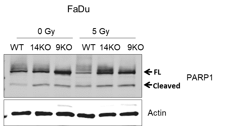
PARP1 (poly(ADP-ribose) polymerase 1) is a nuclear enzyme catalyzing the poly(ADP-ribosyl)ation of many key proteins in vivo. The normal function of PARP1 is the routine repair of DNA damage. Activated by DNA strand breaks, the PARP1 is cleaved into an 85 to 89-kDa COOH-terminal fragment and a 24-kDa NH2-terminal peptide by caspases during the apoptotic process. The appearance of PARP fragments is commonly considered an important biomarker of apoptosis. In addition to caspases, other proteases like calpains, cathepsins, granzymes, and matrix metalloproteinases (MMPs) have also been reported to cleave PARP1 and give rise to fragments ranging from 42-89-kDa. This antibody was generated against the N-terminal region of human PARP1 and it recognizes the full-length as well as the cleavage of the PARP1.
Protocols
| Product Specific Protocols | |
|---|---|
| FC protocol for PARP1 antibody 66520-1-Ig | Download protocol |
| IF protocol for PARP1 antibody 66520-1-Ig | Download protocol |
| IHC protocol for PARP1 antibody 66520-1-Ig | Download protocol |
| IP protocol for PARP1 antibody 66520-1-Ig | Download protocol |
| WB protocol for PARP1 antibody 66520-1-Ig | Download protocol |
| Standard Protocols | |
|---|---|
| Click here to view our Standard Protocols |
Publications
| Species | Application | Title |
|---|---|---|
Nat Commun Cis- and trans-resveratrol have opposite effects on histone serine-ADP-ribosylation and tyrosine induced neurodegeneration. | ||
J Exp Clin Cancer Res PARP1-targeted fluorescence molecular endoscopy as novel tool for early detection of esophageal dysplasia and adenocarcinoma | ||
Biomaterials Urinary exosomes-based Engineered Nanovectors for Homologously Targeted Chemo-Chemodynamic Prostate Cancer Therapy via abrogating IGFR/AKT/NF-kB/IkB signaling. | ||
Arthritis Rheumatol Association of the Polymorphism rs13259960 in SLEAR With Predisposition to Systemic Lupus Erythematosus. |
Reviews
The reviews below have been submitted by verified Proteintech customers who received an incentive for providing their feedback.
FH Rashmi (Verified Customer) (09-25-2024) | Excellent Product
|
FH Vikas (Verified Customer) (07-11-2024) | Used for Immunoblot. Highly recommended product.
|
FH Hadil (Verified Customer) (05-23-2023) | This antibody is a great choice, delivering strong signals and enhanced specificity. It has low background noise, ensuring accurate results. Its efficiency saves valuable time, making it a reliable and versatile tool for various applications. A must-have for researchers seeking reliable and efficient antibody performance.
 |
FH Carly (Verified Customer) (11-17-2020) | Tested using EDTA plasma on an antibody microarray
|
FH MANOHAR (Verified Customer) (12-11-2019) | No nonspecific binding
|