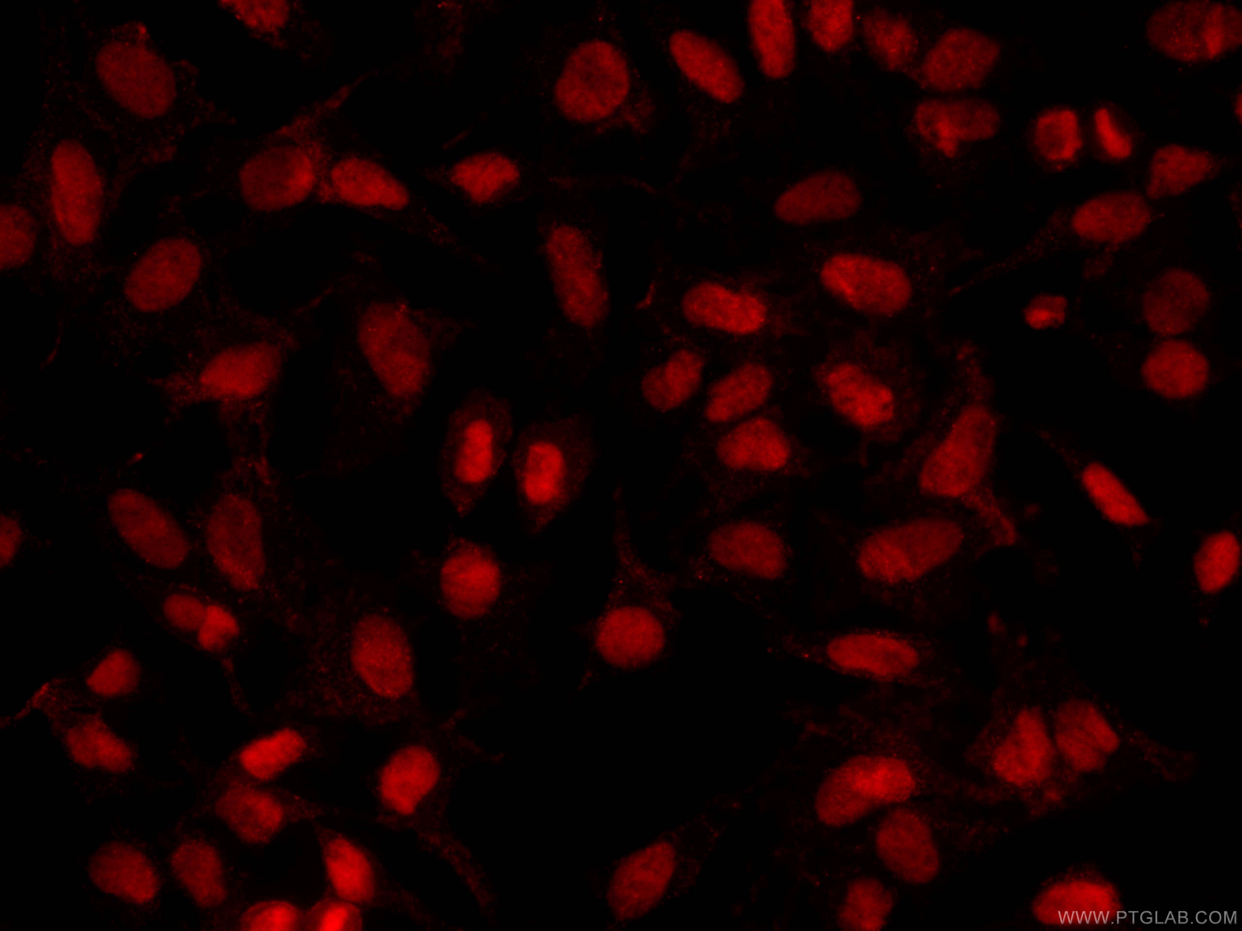
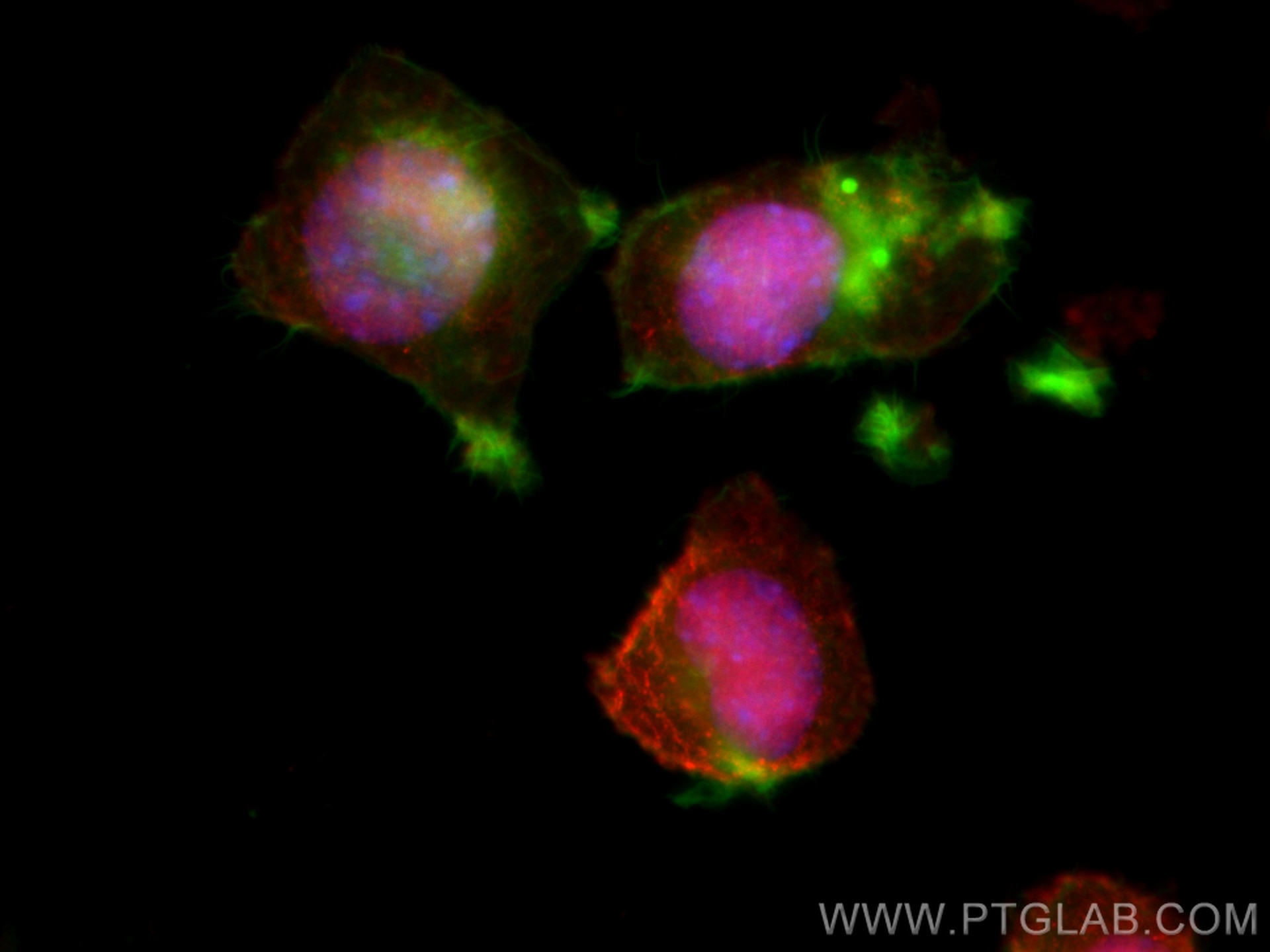
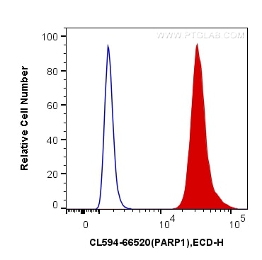
Tested Applications
| Positive IF/ICC detected in | HeLa cells, Neuro-2a cells |
| Positive FC (Intra) detected in | HeLa cells |
Recommended dilution
| Application | Dilution |
|---|---|
| Immunofluorescence (IF)/ICC | IF/ICC : 1:50-1:500 |
| Flow Cytometry (FC) (INTRA) | FC (INTRA) : 0.40 ug per 10^6 cells in a 100 µl suspension |
| It is recommended that this reagent should be titrated in each testing system to obtain optimal results. | |
| Sample-dependent, Check data in validation data gallery. | |
Product Information
CL594-66520 targets PARP1 in IF/ICC, FC (Intra) applications and shows reactivity with human, mouse, rat samples.
| Tested Reactivity | human, mouse, rat |
| Host / Isotype | Mouse / IgG1 |
| Class | Monoclonal |
| Type | Antibody |
| Immunogen |
CatNo: Ag19173 Product name: Recombinant human PARP1 protein Source: e coli.-derived, PET28a Tag: 6*His Domain: 1-327 aa of BC037545 Sequence: MAESSDKLYRVEYAKSGRASCKKCSESIPKDSLRMAIMVQSPMFDGKVPHWYHFSCFWKVGHSIRHPDVEVDGFSELRWDDQQKVKKTAEAGGVTGKGQDGIGSKAEKTLGDFAAEYAKSNRSTCKGCMEKIEKGQVRLSKKMVDPEKPQLGMIDRWYHPGCFVKNREELGFRPEYSASQLKGFSLLATEDKEALKKQLPGVKSEGKRKGDEVDGVDEVAKKKSKKEKDKDSKLEKALKAQNDLIWNIKDELKKVCSTNDLKELLIFNKQQVPSGESAILDRVADGMVFGALLPCEECSGQLVFKSDAYYCTGDVTAWTKCMVKTQT Predict reactive species |
| Full Name | poly (ADP-ribose) polymerase 1 |
| Calculated Molecular Weight | 1014 aa, 113 kDa |
| Observed Molecular Weight | 113-116 kDa, 85-89 kDa |
| GenBank Accession Number | BC037545 |
| Gene Symbol | PARP1 |
| Gene ID (NCBI) | 142 |
| RRID | AB_2883598 |
| Conjugate | CoraLite®594 Fluorescent Dye |
| Excitation/Emission Maxima Wavelengths | 588 nm / 604 nm |
| Excitation Laser | Yellow-Green Laser (561 nm) |
| Form | Liquid |
| Purification Method | Protein G purification |
| UNIPROT ID | P09874 |
| Storage Buffer | PBS with 50% glycerol, 0.05% Proclin300, 0.5% BSA, pH 7.3. |
| Storage Conditions | Store at -20°C. Avoid exposure to light. Stable for one year after shipment. Aliquoting is unnecessary for -20oC storage. |
Background Information
PARP1 (poly(ADP-ribose) polymerase 1) is a nuclear enzyme catalyzing the poly(ADP-ribosyl)ation of many key proteins in vivo. The normal function of PARP1 is the routine repair of DNA damage. Activated by DNA strand breaks, the PARP1 is cleaved into an 85 to 89-kDa COOH-terminal fragment and a 24-kDa NH2-terminal peptide by caspases during the apoptotic process. The appearance of PARP fragments is commonly considered as an important biomarker of apoptosis. In addition to caspases, other proteases like calpains, cathepsins, granzymes and matrix metalloproteinases (MMPs) have also been reported to cleave PARP1 and gave rise to fragments ranging from 42-89-kD. This antibody was generated against the N-terminal region of human PARP1 and it recognizes the full-length as well as the cleavage of the PARP1.
Protocols
| Product Specific Protocols | |
|---|---|
| FC protocol for CL594 PARP1 antibody CL594-66520 | Download protocol |
| IF protocol for CL594 PARP1 antibody CL594-66520 | Download protocol |
| Standard Protocols | |
|---|---|
| Click here to view our Standard Protocols |