

Tested Applications
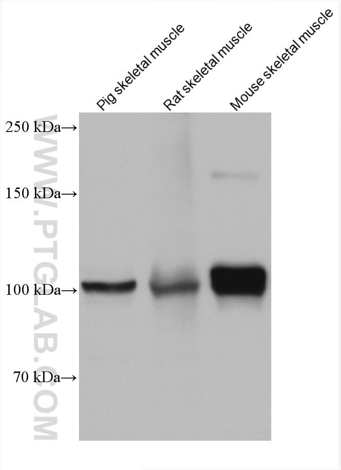
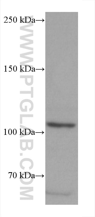
| Positive WB detected in | pig skeletal muscle tissue, HSC-T6 cells, rat skeletal muscle tissue, mouse skeletal muscle tissue |
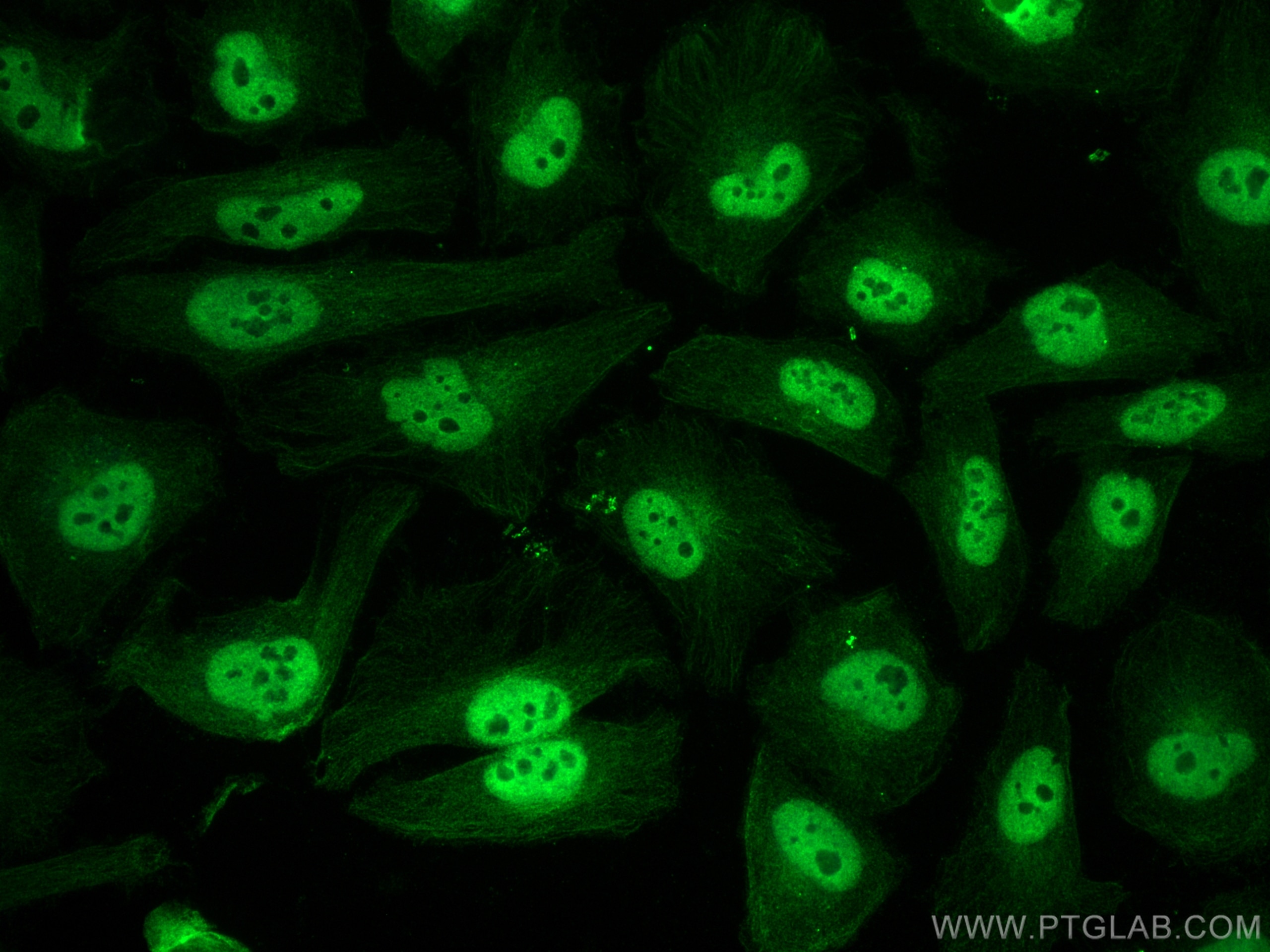
| Positive IF/ICC detected in | HeLa cells |
Recommended dilution
| Application | Dilution |
|---|---|
| Western Blot (WB) | WB : 1:5000-1:50000 |
| Immunofluorescence (IF)/ICC | IF/ICC : 1:1000-1:4000 |
| It is recommended that this reagent should be titrated in each testing system to obtain optimal results. | |
| Sample-dependent, Check data in validation data gallery. | |
Product Information
66369-1-Ig targets PGC1a in WB, IHC, IF/ICC, IP, CoIP, ChIP, ELISA applications and shows reactivity with human, mouse, rat, pig samples.
| Tested Reactivity | human, mouse, rat, pig |
| Cited Reactivity | human, mouse, rat, monkey, chicken, zebrafish, hamster, goat, ducks |
| Host / Isotype | Mouse / IgG1 |
| Class | Monoclonal |
| Type | Antibody |
| Immunogen |
Peptide Predict reactive species |
| Full Name | peroxisome proliferator-activated receptor gamma, coactivator 1 alpha |
| Calculated Molecular Weight | 91 kDa |
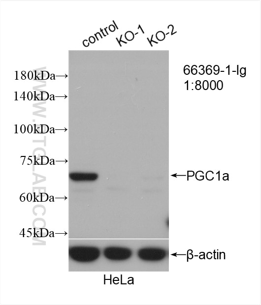
| Observed Molecular Weight | 70 kDa, 100 kDa |
| GenBank Accession Number | NM_013261 |
| Gene Symbol | PGC1a |
| Gene ID (NCBI) | 10891 |
| RRID | AB_2828002 |
| Conjugate | Unconjugated |
| Form | Liquid |
| Purification Method | Protein G purification |
| UNIPROT ID | Q9UBK2 |
| Storage Buffer | PBS with 0.02% sodium azide and 50% glycerol, pH 7.3. |
| Storage Conditions | Store at -20°C. Stable for one year after shipment. Aliquoting is unnecessary for -20oC storage. 20ul sizes contain 0.1% BSA. |
Background Information
PPARGC1A, also named as Peroxisome proliferator-activated receptor gamma coactivator 1-alpha, is a 798 amino acid protein, which Contains 1 RRM (RNA recognition motif) domain and localizes in the nucleus. PPARGC1A is transcriptional coactivator for steroid receptors and nuclear receptors. PPARGC1A can regulate key mitochondrial genes that contribute to the program of adaptive thermogenesis and plays an essential role in metabolic reprogramming in response to dietary availability through coordination of the expression of a wide array of genes involved in glucose and fatty acid metabolism. PPARGC1A exists an isoform in the liver and the molecular weight of it is 77 kDa. Except for a deletion of 127 amino acids at the N terminus, the protein, termed L-PGC-1 α, is identical to PGC-1.
Protocols
| Product Specific Protocols | |
|---|---|
| IF protocol for PGC1a antibody 66369-1-Ig | Download protocol |
| WB protocol for PGC1a antibody 66369-1-Ig | Download protocol |
| Standard Protocols | |
|---|---|
| Click here to view our Standard Protocols |
Publications
What published studies show
Anti-PGC-1α (Catalog No. 66369-1-Ig) has a total of 809 citations. These appear in high-impact journals including Nature, Cell Metabolism, Nature Communications, Nature Metabolism, Science Advances, EMBO Journal, Autophagy, Redox Biology, Theranostics, Bioactive Materials, and Advanced Science, among over 50 others. Associated research fields encompass mitochondrial biology and energy metabolism, cancer, neurodegeneration, cardiovascular disease, diabetes and obesity, inflammation, aging, oxidative stress, ferroptosis, and mitophagy.
| Species | Application | Title |
|---|---|---|
Signal Transduct Target Ther GRP75 triggers white adipose tissue browning to promote cancer-associated cachexia | ||
Signal Transduct Target Ther Short-chain acyl-CoA dehydrogenase initiates mtDNA demethylation and leakage to fuel antitumor immunity in colorectal cancer. | ||
Cell Metab Macrophage PD-1 regulates energy expenditure and metabolic dysfunction under immune checkpoint blockade. | ||
Cell Metab m6A mRNA methylation in brown fat regulates systemic insulin sensitivity via an inter-organ prostaglandin signaling axis independent of UCP1 |
Reviews
What customers say
Users report this antibody performs well for Western Blot on mouse brain tissue, showing a clear band between 60–100 kDa, and for immunofluorescence on fibroblasts. However, several reviewers noted non-specific bands when used on adipocyte cells (both human and 3T3-L1), with unexpected molecular weights. One user also reported the antibody degraded after 9 months stored at -20°C. Overall, it is considered reliable for mouse tissue applications but may require optimization or KO validation for adipocyte studies.
The reviews below have been submitted by verified Proteintech customers who received an incentive for providing their feedback.
Manon (Verified Customer) (01-23-2026) | Good staining of PGC1 on fibroblast
|
Michaela (Verified Customer) (11-24-2023) | Worked well in 15μg mouse brain tissue lysate in Western Blot. Band appears between 60-100 kDa
|
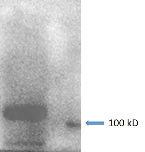
No (Verified Customer) (10-24-2023) | A good antibody to detect endogenous PGC1a.
 |
Mi (Verified Customer) (03-07-2023) | It doesn´t work well in human white and brown adipocyte cells, we got bands at unexpected size. Further KO validation may be needed.
|
Huai-Chin (Verified Customer) (07-11-2022) | There are several non-specific bands. At least there's a dominant band after adipogenesis and we can use this antibody at a fairly high dilution.
|
Mohammad (Verified Customer) (01-21-2020) | It was working with some non-specific bands in lower weight. But after 9 months antibody was degraded in -20.
|





