Tested Applications
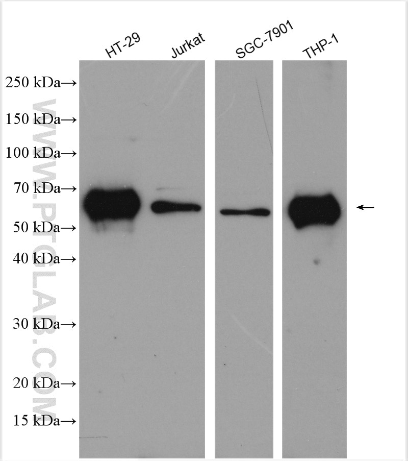
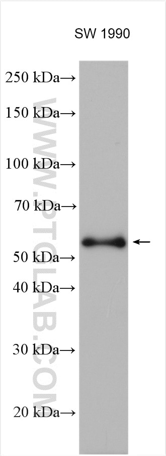
| Positive WB detected in | HT-29 cells, Jurkat cells, SW 1990 cells, THP-1 cells, SGC-7901 cells |
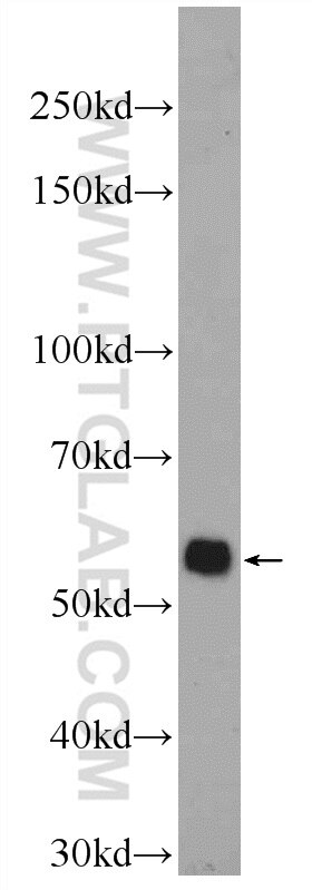
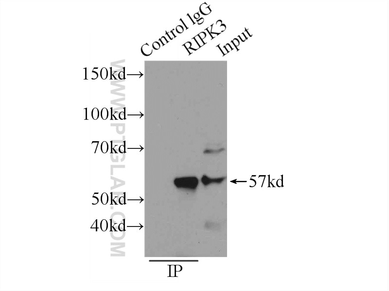
| Positive IP detected in | SW 1990 cells |
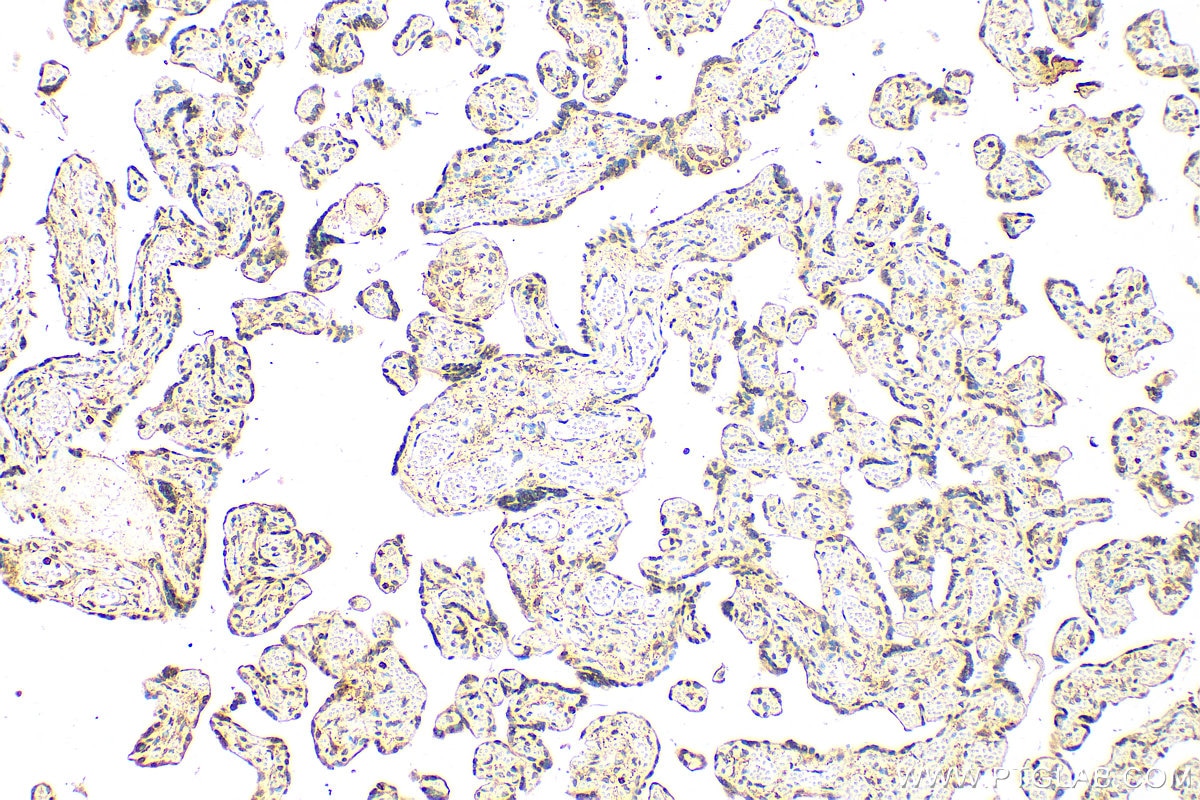
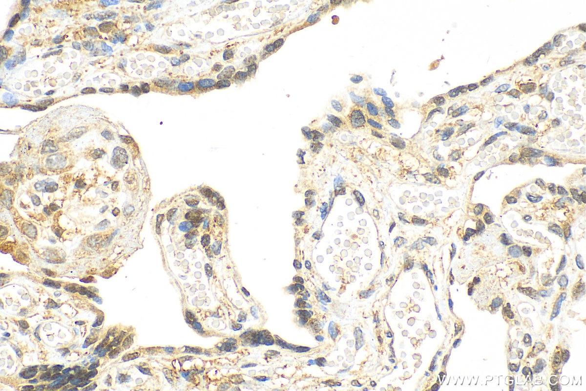
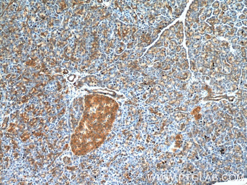
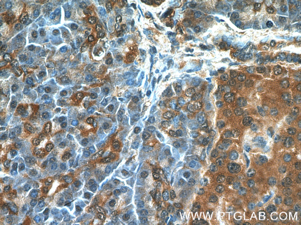
| Positive IHC detected in | human pancreas tissue, human placenta tissue Note: suggested antigen retrieval with TE buffer pH 9.0; (*) Alternatively, antigen retrieval may be performed with citrate buffer pH 6.0 |
| Positive IF/ICC detected in | HT-29 cells |
Recommended dilution
| Application | Dilution |
|---|---|
| Western Blot (WB) | WB : 1:1000-1:4000 |
| Immunoprecipitation (IP) | IP : 0.5-4.0 ug for 1.0-3.0 mg of total protein lysate |
| Immunohistochemistry (IHC) | IHC : 1:100-1:400 |
| Immunofluorescence (IF)/ICC | IF/ICC : 1:50-1:500 |
| It is recommended that this reagent should be titrated in each testing system to obtain optimal results. | |
| Sample-dependent, Check data in validation data gallery. | |
Published Applications
| KD/KO | See 3 publications below |
| WB | See 115 publications below |
| IHC | See 9 publications below |
| IF | See 17 publications below |
| IP | See 5 publications below |
| CoIP | See 4 publications below |
Product Information
17563-1-AP targets RIPK3 in WB, IHC, IF/ICC, IP, CoIP, ELISA applications and shows reactivity with human samples.
| Tested Reactivity | human |
| Cited Reactivity | human, yeast |
| Host / Isotype | Rabbit / IgG |
| Class | Polyclonal |
| Type | Antibody |
| Immunogen |
CatNo: Ag11759 Product name: Recombinant human RIPK3 protein Source: e coli.-derived, PGEX-4T Tag: GST Domain: 1-289 aa of BC062584 Sequence: MSCVKLWPSGAPAPLVSIEELENQELVGKGGFGTVFRAQHRKWGYDVAVKIVNSKAISREVKAMASLDNEFVLRLEGVIEKVNWDQDPKPALVTKFMENGSLSGLLQSQCPRPWPLLCRLLKEVVLGMFYLHDQNPVLLHRDLKPSNVLLDPELHVKLADFGLSTFQGGSQSGTGSGEPGGTLGYLAPELFVNVNRKASTASDVYSFGILMWAVLAGREVELPTEPSLVYEAVCNRQNRPSLAELPQAGPETPGLEGLKELMQLCWSSEPKDRPSFQECLPKTDEVFQM Predict reactive species |
| Full Name | receptor-interacting serine-threonine kinase 3 |
| Calculated Molecular Weight | 518 aa, 57 kDa |
| Observed Molecular Weight | 57-70 kDa |
| GenBank Accession Number | BC062584 |
| Gene Symbol | RIPK3 |
| Gene ID (NCBI) | 11035 |
| RRID | AB_2178659 |
| Conjugate | Unconjugated |
| Form | Liquid |
| Purification Method | Antigen affinity purification |
| UNIPROT ID | Q9Y572 |
| Storage Buffer | PBS with 0.02% sodium azide and 50% glycerol, pH 7.3. |
| Storage Conditions | Store at -20°C. Stable for one year after shipment. Aliquoting is unnecessary for -20oC storage. 20ul sizes contain 0.1% BSA. |
Background Information
Receptor-interacting protein 3 (RIP3, also known as RIPK3) is a serine-threonine protein involved in the regulation of inflammatory signaling and cell death. RIPK3, also named as RIP3, a Ser/Thr kinase of RIP (Receptor Interacting Protein) family, is a nucleocytoplasmic shuttling protein and its unconventional nuclear localization signal (NLS, 442-472 aa) is sufficient to trigger apoptosis in the nucleus (PMID: 18533105). It has 3 isoforms produced by alternative splicing.
What is the molecular weight of RIP3? Is RIP3 post-translationally modified?
The molecular weight of RIP3 is 57 kDa. During the induction of necroptosis, RIP3 migrates slower in SDS-PAGE due to its phosphorylation (PMID: 19524512). Additionally, RIP3 can be a subject of poly-ubiquitination, when targeted for degradation.
Are there any splice isoforms of RIP3?
Apart from full-length RIP3, there are two reported splice isoforms of RIP3: RIP3β and RIP3γ, and 28 and 25 kDa, respectively (PMID: 15896315).
What is the subcellular localization of RIP3?
RIP3 can shuttle between the cytoplasm and the nucleus. Although RIP3 forms necrosomes in the cytoplasm, a recent study suggests that the phosphorylation of RIP3, required for necroptosis, may also occur in the nucleus (PMID: 30271893). Additionally, the induction of necrosis by reactive oxygen species can cause transient translocation of RIP3 to mitochondria (PMID: 25206339).
What is the role of RIP3 in cell death (necroptosis)?
Activation of RIP3 kinase is required for the induction of necroptosis (PMID: 19524512, 19524513 and 19498109). Activation of RIP3 can be induced by interferons, death ligands, or by Toll-like receptors in response to pathogens. That leads to the phosphorylation of RIP3 and the formation of a β-amyloid-like protein complex. Phosphorylated RIP3 acts downstream by phosphorylation of MLKL (PMID: 30131615).
How to study necroptosis in a cell-based system?
The choice of the cell line is important. Many commonly used immortalized cell types are derived from cancers and may have very low RIL3 expression level (PMID: 25952668). Those cell lines are not going to be responsive to necrotic stimuli. A few of the examined cell types have high RIP3 levels: Jurkat, CCRF-CEM, U937, L929 cells, and mouse embryonic fibroblasts (PMID: 19524512). Good necroptosis readouts reflect an increased level of RIP3 protein and its phosphorylation.
Protocols
| Product Specific Protocols | |
|---|---|
| IF protocol for RIPK3 antibody 17563-1-AP | Download protocol |
| IHC protocol for RIPK3 antibody 17563-1-AP | Download protocol |
| IP protocol for RIPK3 antibody 17563-1-AP | Download protocol |
| WB protocol for RIPK3 antibody 17563-1-AP | Download protocol |
| Standard Protocols | |
|---|---|
| Click here to view our Standard Protocols |
Publications
| Species | Application | Title |
|---|---|---|
Acta Pharm Sin B Ligand-based substituent-anchoring design of selective receptor-interacting protein kinase 1 necroptosis inhibitors for ulcerative colitis therapy | ||
Cell Mol Biol Lett Integrated transcriptomics and metabolomics confirms the oxidative stress mechanism of hypothermia-induced neuronal necroptosis | ||
Cancer Lett Lovastatin/SN38 co-loaded liposomes amplified ICB therapeutic effect via remodeling the immunologically-cold colon tumor and synergized stimulation of cGAS-STING pathway | ||
Cancer Lett NR4A1 depletion inhibits colorectal cancer progression by promoting necroptosis via the RIG-I-like receptor pathway | ||
Int J Biol Macromol Antibody-DNA nanostructure conjugates exhibit inhibited effects on CT26 cells and prevent upregulation of PD-1 in the tumor microenvironment. | ||
Microbiol Res Caspase-8 drove apoptosis of BMECs to promote neutrophil infiltration and DE205B clearance in meningitis |