Recombinant Rat C-Reactive Protein/CRP protein (His Tag)
Species
Rat
Purity
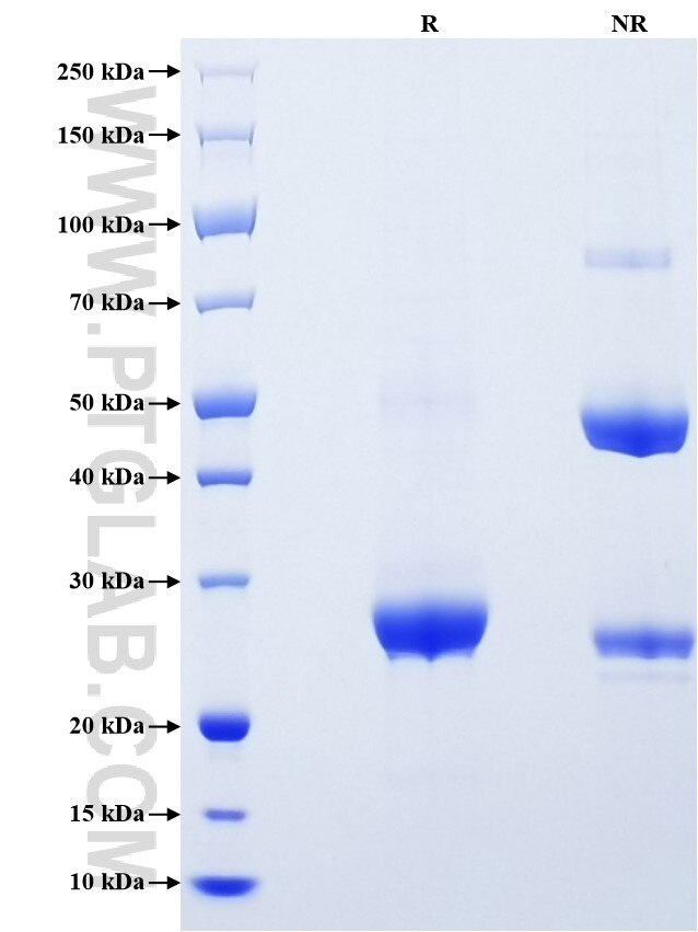
>90 %, SDS-PAGE
Tag
His Tag
Activity
not tested
Cat no : Eg0942
Validation Data Gallery
Product Information
| Purity | >90 %, SDS-PAGE |
| Endotoxin | <0.1 EU/μg protein, LAL method |
| Activity |
Not tested |
| Expression | HEK293-derived Rat C-Reactive Protein protein His20-Ser230 (Accession# P48199) with a His tag at the C-terminus. |
| GeneID | 25419 |
| Accession | P48199 |
| PredictedSize | 24.4 kDa |
| SDS-PAGE | 25-28 kDa, reducing (R) conditions |
| Formulation | Lyophilized from 0.22 μm filtered solution in PBS, pH 7.4. Normally 5% trehalose and 5% mannitol are added as protectants before lyophilization. |
| Reconstitution | Briefly centrifuge the tube before opening. Reconstitute at 0.1-0.5 mg/mL in sterile water. |
| Storage Conditions |
It is recommended that the protein be aliquoted for optimal storage. Avoid repeated freeze-thaw cycles.
|
| Shipping | The product is shipped at ambient temperature. Upon receipt, store it immediately at the recommended temperature. |
Background
C-reactive protein (CRP) is an evolutionary highly conserved member of the pentraxin superfamily of proteins. CRP consists of five identical subunits with molecular weight 20-30 kDa arranged as a cyclic pentamer. CRP exists in at least two conformationally distinct forms, native pentameric CRP (pCRP) and modified/monomeric CRP (mCRP). CRP plays important roles in inflammatory processes and host responses to infection including promoting agglutination, bacterial capsular swelling, phagocytosis, and complement fixation through its Ca2+-dependent binding to phosphorylcholine (PCh). CRP is mainly expressed by hepatocytes and secreted into plasma. The concentration of CRP will increase significantly in plasma during acute phase response to tissue injury, infection, or other inflammatory stimuli.
References:
1. McFadyen JD. et al. (2020).  Subcell Biochem. 94:499-520.
2. Pathak A. et al. (2019). Front Immunol. 10:943.
3. Singh SK. et al. (2019). Front Immunol. 10:1655.
4. Yao Z. et al. (2019). Inflamm Res. 68(10):815-823.
