Validation Data Gallery
![Western blot analysis of N- and C-terminal Spot-tagged GFP added to HEK- 293T cell lysate. Detection with Spot-tag® antibody [28A5] (28a5, ChromoTek) 1:5,000 and Nano-Secondary® alpaca anti-mouse IgG1, recombinant VHH, Alexa Fluor® 488 [CTK0103, CTK0104] (sms1AF488-1, ChromoTek) 1:5,000. Western blot membrane was incubated simultaneously with the primary antibody and Nano-Secondary® (one-step staining).](https://www.ptglab.com/products/pictures/28A5-1.jpg "Western blot analysis of N- and C-terminal Spot-tagged GFP added to HEK- 293T cell lysate. Detection with Spot-tag® antibody [28A5] (28a5, ChromoTek) 1:5,000 and Nano-Secondary® alpaca anti-mouse IgG1, recombinant VHH, Alexa Fluor® 488 [CTK0103, CTK0104] (sms1AF488-1, ChromoTek) 1:5,000. Western blot membrane was incubated simultaneously with the primary antibody and Nano-Secondary® (one-step staining).")
![Western blot analysis of C-terminal Spot-tagged GFP added to HEK-293T cell lysate. Detection with Spot-tag® antibody [28A5] (28a5, ChromoTek) 1:5,000 and anti-mouse secondary antibody HRP 1:1,000.](https://www.ptglab.com/products/pictures/28A5-2.png "Western blot analysis of C-terminal Spot-tagged GFP added to HEK-293T cell lysate. Detection with Spot-tag® antibody [28A5] (28a5, ChromoTek) 1:5,000 and anti-mouse secondary antibody HRP 1:1,000.")
![Western blot analysis of different cell lysates +/- 50 ng C-terminal Spot-tagged GFP: human (HEK-293T), yeast (S.cerevisiae), insect (High Five™), bacteria (E.coli) and plant (A.thaliana). Detection with Spot-tag® antibody [28A5] (28a5, ChromoTek) 1:5,000 and Nano-Secondary® alpaca anti-mouse IgG1, recombinant VHH, Alexa Fluor® 488 [CTK0103, CTK0104] (sms1AF488-1, ChromoTek) 1:5,000. Western blot membrane was incubated simultaneously with the primary antibody and Nano-Secondary® (one-step staining).](https://www.ptglab.com/products/pictures/Result-28a5_20210803100622724.png "Western blot analysis of different cell lysates +/- 50 ng C-terminal Spot-tagged GFP: human (HEK-293T), yeast (S.cerevisiae), insect (High Five™), bacteria (E.coli) and plant (A.thaliana). Detection with Spot-tag® antibody [28A5] (28a5, ChromoTek) 1:5,000 and Nano-Secondary® alpaca anti-mouse IgG1, recombinant VHH, Alexa Fluor® 488 [CTK0103, CTK0104] (sms1AF488-1, ChromoTek) 1:5,000. Western blot membrane was incubated simultaneously with the primary antibody and Nano-Secondary® (one-step staining).")
![ELISA detection of N- and C-terminal Spot-tagged GFP with Spot-tag® antibody [28A5] (28a5, ChromoTek). N- and C-terminal Spot-tagged GFP was coated on a microtiter plate, blocked and detected with 1 nM Spot-tag® antibody [28A5] (28a5, ChromoTek) and anti-mouse IgG antibody AP.](https://www.ptglab.com/products/pictures/Result-28a5_20210803100844572.png "ELISA detection of N- and C-terminal Spot-tagged GFP with Spot-tag® antibody [28A5] (28a5, ChromoTek). N- and C-terminal Spot-tagged GFP was coated on a microtiter plate, blocked and detected with 1 nM Spot-tag® antibody [28A5] (28a5, ChromoTek) and anti-mouse IgG antibody AP.")
Product Information
Mouse monoclonal antibody to Spot-tag®
| Description | Spot-tag® mouse monoclonal [28A5] for WB, ELISA |
| Applications | WB, ELISA |
| Host/IsoType | Mouse/IgG1 |
| Specificity/Target | Spot-Tag® (PDRVRAVSHWSS) at the N-terminus, C-terminus, or internal site of the fusion protein |
| Conjugate | Unconjugated |
| Recommended Dilution | WB: 1:1,000 - 1:5,000 ELISA: 1:5,000 - 1:10,000 |
| Purification Method | Protein A purification |
| Type | Primary Antibody |
| Class | Monoclonal |
| Format | Monoclonal mouse antibody |
| Immunogen | Spot-tagged fusion protein |
| Clone No. | 28A5 |
| Storage Buffer | PBS, 50% glycerol |
| Storage Condition | Shipped on ice. Aliquot upon receipt and store at -20°C/-4°F. Avoid freeze-thaw cycles. |
| Form | Liquid |
| Size | 20 μL; 100 μL |
| RRID | AB_2892245 |
Documentation
| SDS |
|---|
| 28a5_SDS_mouse anti Spot-tag® antibody [28A5] (EN) |
| Datasheet |
|---|
| mouse anti Spot-tag® antibody (28A5) Datasheet |
Publications
| Application | Title |
|---|---|
iScience Monitoring extracellular ion and metabolite dynamics with recombinant nanobody-fused biosensors | |
Methods Mol Biol Measurement of Molecular Height Using Cell Surface Optical Profilometry (CSOP) | |
Mol Cell Anti-apoptotic MCL-1 promotes long-chain fatty acid oxidation through interaction with ACSL1 | |
Open Biol The cytosolic form of dual localized BolA family protein Bol3 is important for adaptation to iron starvation in Aspergillus fumigatus |
Reviews
The reviews below have been submitted by verified Proteintech customers who received an incentive for providing their feedback.
FH Alex (Verified Customer) (09-23-2024) | Used for Western blotting of primary rat cortical neurons expressig spot-tagged protein. Diluted in 6% milk, 4C, ON. Very good detection, but some nonspecific bands appear when pushed to max so not suitable for detection of extremely low expressing spot-tagged protein.
|
FH Janine (Verified Customer) (06-13-2024) | Excellent detection (tested dilution 1:1000). I used 5%BSA instead of 5% milk but the performance was still very good.
|
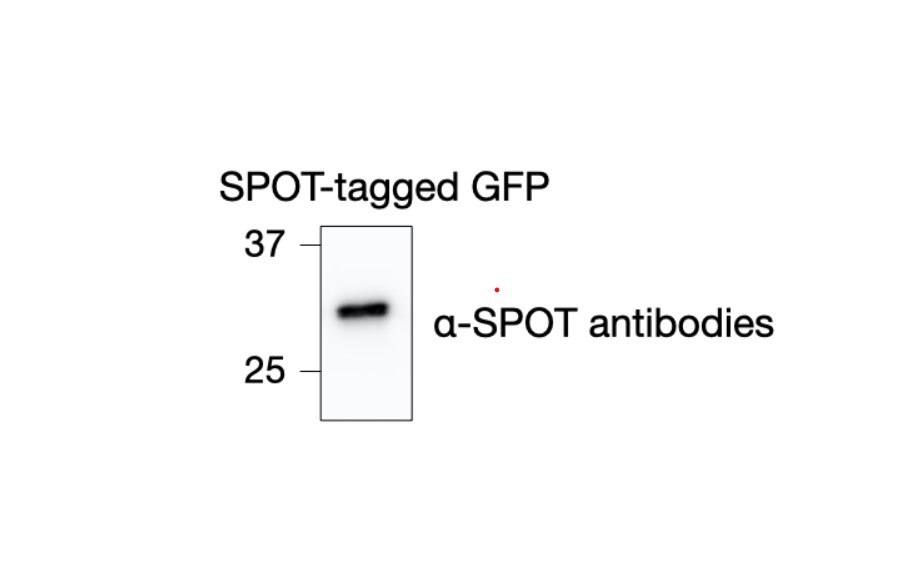
FH Tsimafei (Verified Customer) (06-01-2022) | mESCs express HA-SPOT-GFP protein. 20µg of total protein extract were loaded on the gel. Membrane was probed with anti-SPOT antibodies (1:1000 dilution in 3% milk) at 4C, ON.
 |
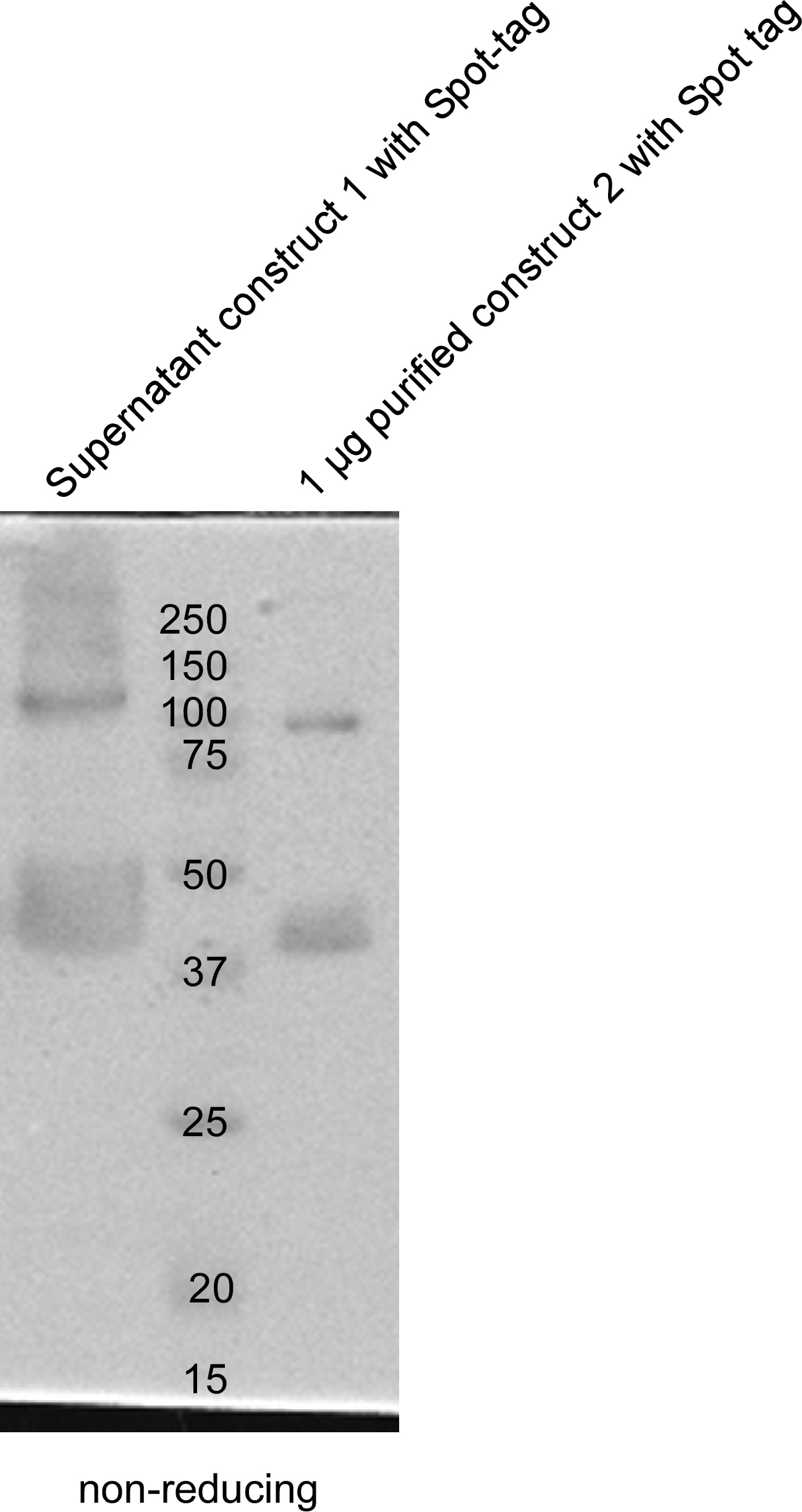
FH Daniela (Verified Customer) (04-11-2022) | The 28A5 Spot-antibody was tested in Western blots of Expi-293F supernatants and purified proteins in a 1:5000 dilution using 5% non-fat milk (Biozym)/TBST (or 1x Rotiblock solution (Roth). Incubation was either 2 h at RT or ON at 4°C. Secondary HRP-antibody was incubated for 1 h at RT. The antibody is working fine, monomers, dimers, oligomers of the construct can be distinguished.
 |
