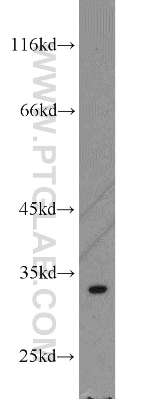
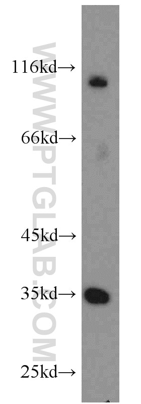
WB analysis of K-562 using 15486-1-AP (same clone as 15486-1-PBS)
K-562 cells were subjected to SDS PAGE followed by western blot with 15486-1-AP (THTPA antibody) at dilution of 1:800 incubated at room temperature for 1.5 hours. This data was developed using the same antibody clone with 15486-1-PBS in a different storage buffer formulation.
K-562 cells were subjected to SDS PAGE followed by western blot with 15486-1-AP (THTPA antibody) at dilution of 1:800 incubated at room temperature for 1.5 hours. This data was developed using the same antibody clone with 15486-1-PBS in a different storage buffer formulation.
WB analysis of HeLa using 15486-1-AP (same clone as 15486-1-PBS)
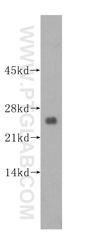
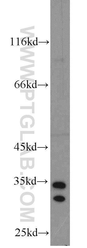
WB result of THTPA antibody (15486-1-AP; 1:1000; incubated at room temperature for 1.5 hours) with sh-Control and sh-THTPA transfected HeLa cells. This data was developed using the same antibody clone with 15486-1-PBS in a different storage buffer formulation.
WB result of THTPA antibody (15486-1-AP; 1:1000; incubated at room temperature for 1.5 hours) with sh-Control and sh-THTPA transfected HeLa cells. This data was developed using the same antibody clone with 15486-1-PBS in a different storage buffer formulation.
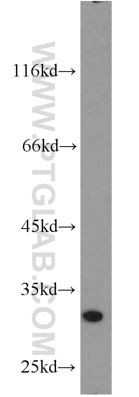
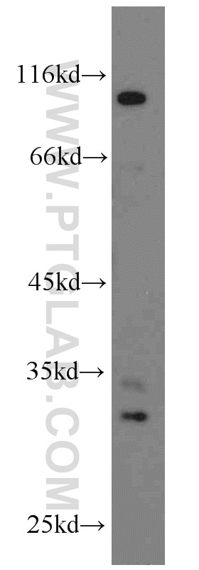
WB analysis of human brain using 15486-1-AP (same clone as 15486-1-PBS)
human brain tissue were subjected to SDS PAGE followed by western blot with 15486-1-AP (THTPA antibody) at dilution of 1:500 incubated at room temperature for 1.5 hours. This data was developed using the same antibody clone with 15486-1-PBS in a different storage buffer formulation.
human brain tissue were subjected to SDS PAGE followed by western blot with 15486-1-AP (THTPA antibody) at dilution of 1:500 incubated at room temperature for 1.5 hours. This data was developed using the same antibody clone with 15486-1-PBS in a different storage buffer formulation.
WB analysis of A431 using 15486-1-AP (same clone as 15486-1-PBS)
A431 cells were subjected to SDS PAGE followed by western blot with 15486-1-AP (THTPA antibody) at dilution of 1:800 incubated at room temperature for 1.5 hours. This data was developed using the same antibody clone with 15486-1-PBS in a different storage buffer formulation.
A431 cells were subjected to SDS PAGE followed by western blot with 15486-1-AP (THTPA antibody) at dilution of 1:800 incubated at room temperature for 1.5 hours. This data was developed using the same antibody clone with 15486-1-PBS in a different storage buffer formulation.
WB analysis of mouse uterus using 15486-1-AP (same clone as 15486-1-PBS)
mouse uterus tissue were subjected to SDS PAGE followed by western blot with 15486-1-AP (THTPA antibody) at dilution of 1:800 incubated at room temperature for 1.5 hours. This data was developed using the same antibody clone with 15486-1-PBS in a different storage buffer formulation.
mouse uterus tissue were subjected to SDS PAGE followed by western blot with 15486-1-AP (THTPA antibody) at dilution of 1:800 incubated at room temperature for 1.5 hours. This data was developed using the same antibody clone with 15486-1-PBS in a different storage buffer formulation.
WB analysis of mouse testis using 15486-1-AP (same clone as 15486-1-PBS)
mouse testis tissue were subjected to SDS PAGE followed by western blot with 15486-1-AP (THTPA antibody) at dilution of 1:800 incubated at room temperature for 1.5 hours. This data was developed using the same antibody clone with 15486-1-PBS in a different storage buffer formulation.
mouse testis tissue were subjected to SDS PAGE followed by western blot with 15486-1-AP (THTPA antibody) at dilution of 1:800 incubated at room temperature for 1.5 hours. This data was developed using the same antibody clone with 15486-1-PBS in a different storage buffer formulation.
WB analysis of PC-3 using 15486-1-AP (same clone as 15486-1-PBS)
PC-3 cells were subjected to SDS PAGE followed by western blot with 15486-1-AP (THTPA antibody) at dilution of 1:800 incubated at room temperature for 1.5 hours. This data was developed using the same antibody clone with 15486-1-PBS in a different storage buffer formulation.
PC-3 cells were subjected to SDS PAGE followed by western blot with 15486-1-AP (THTPA antibody) at dilution of 1:800 incubated at room temperature for 1.5 hours. This data was developed using the same antibody clone with 15486-1-PBS in a different storage buffer formulation.
WB analysis of HeLa using 15486-1-AP (same clone as 15486-1-PBS)
HeLa cells were subjected to SDS PAGE followed by western blot with 15486-1-AP (THTPA antibody) at dilution of 1:800 incubated at room temperature for 1.5 hours. This data was developed using the same antibody clone with 15486-1-PBS in a different storage buffer formulation.
HeLa cells were subjected to SDS PAGE followed by western blot with 15486-1-AP (THTPA antibody) at dilution of 1:800 incubated at room temperature for 1.5 hours. This data was developed using the same antibody clone with 15486-1-PBS in a different storage buffer formulation.
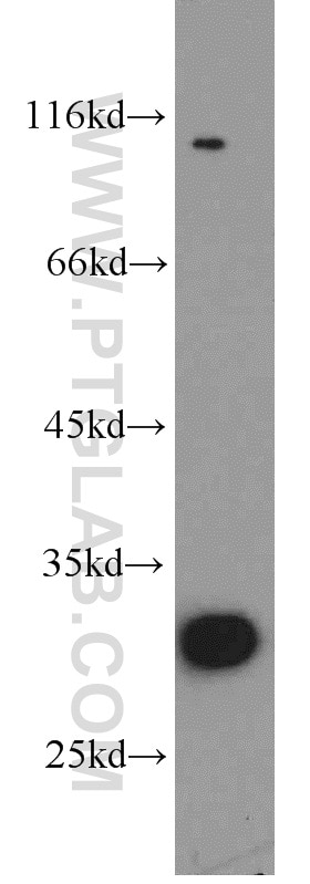
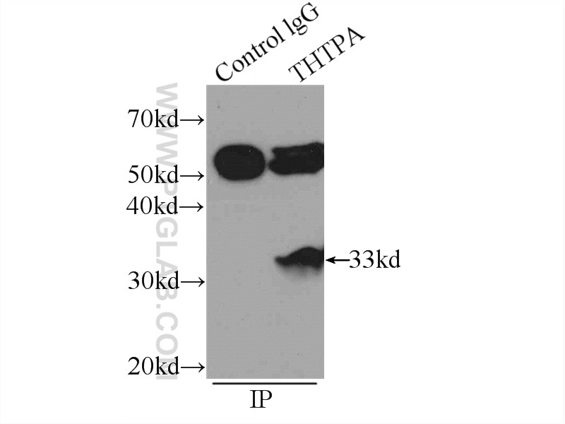
IP experiment of K-562 using 15486-1-AP (same clone as 15486-1-PBS)
IP result of anti-THTPA (IP:15486-1-AP, 3ug; Detection:15486-1-AP 1:800) with K-562 cells lysate 1720ug. This data was developed using the same antibody clone with 15486-1-PBS in a different storage buffer formulation.
IP result of anti-THTPA (IP:15486-1-AP, 3ug; Detection:15486-1-AP 1:800) with K-562 cells lysate 1720ug. This data was developed using the same antibody clone with 15486-1-PBS in a different storage buffer formulation.
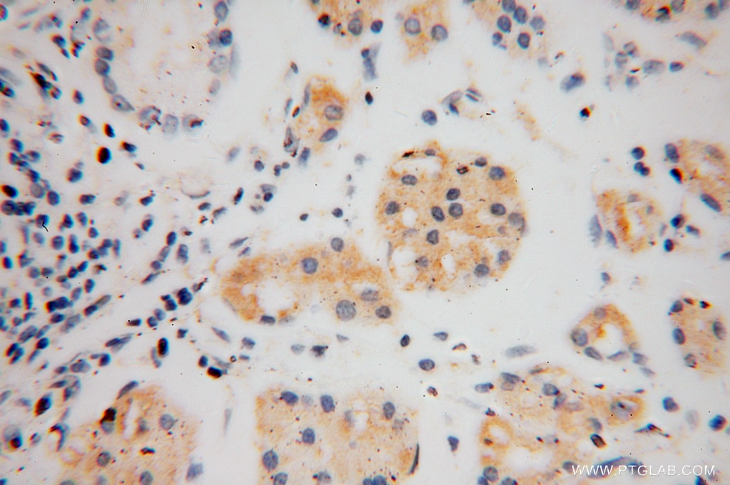
IHC staining of human stomach cancer using 15486-1-AP (same clone as 15486-1-PBS)
Immunohistochemical analysis of paraffin-embedded human stomach cancer using 15486-1-AP (THTPA antibody) at dilution of 1:50 (under 40x lens). This data was developed using the same antibody clone with 15486-1-PBS in a different storage buffer formulation.
Immunohistochemical analysis of paraffin-embedded human stomach cancer using 15486-1-AP (THTPA antibody) at dilution of 1:50 (under 40x lens). This data was developed using the same antibody clone with 15486-1-PBS in a different storage buffer formulation.
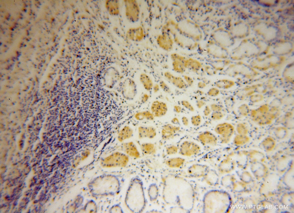
IHC staining of human stomach cancer using 15486-1-AP (same clone as 15486-1-PBS)
Immunohistochemical analysis of paraffin-embedded human stomach cancer using 15486-1-AP (THTPA antibody) at dilution of 1:50 (under 10x lens). This data was developed using the same antibody clone with 15486-1-PBS in a different storage buffer formulation.
Immunohistochemical analysis of paraffin-embedded human stomach cancer using 15486-1-AP (THTPA antibody) at dilution of 1:50 (under 10x lens). This data was developed using the same antibody clone with 15486-1-PBS in a different storage buffer formulation.
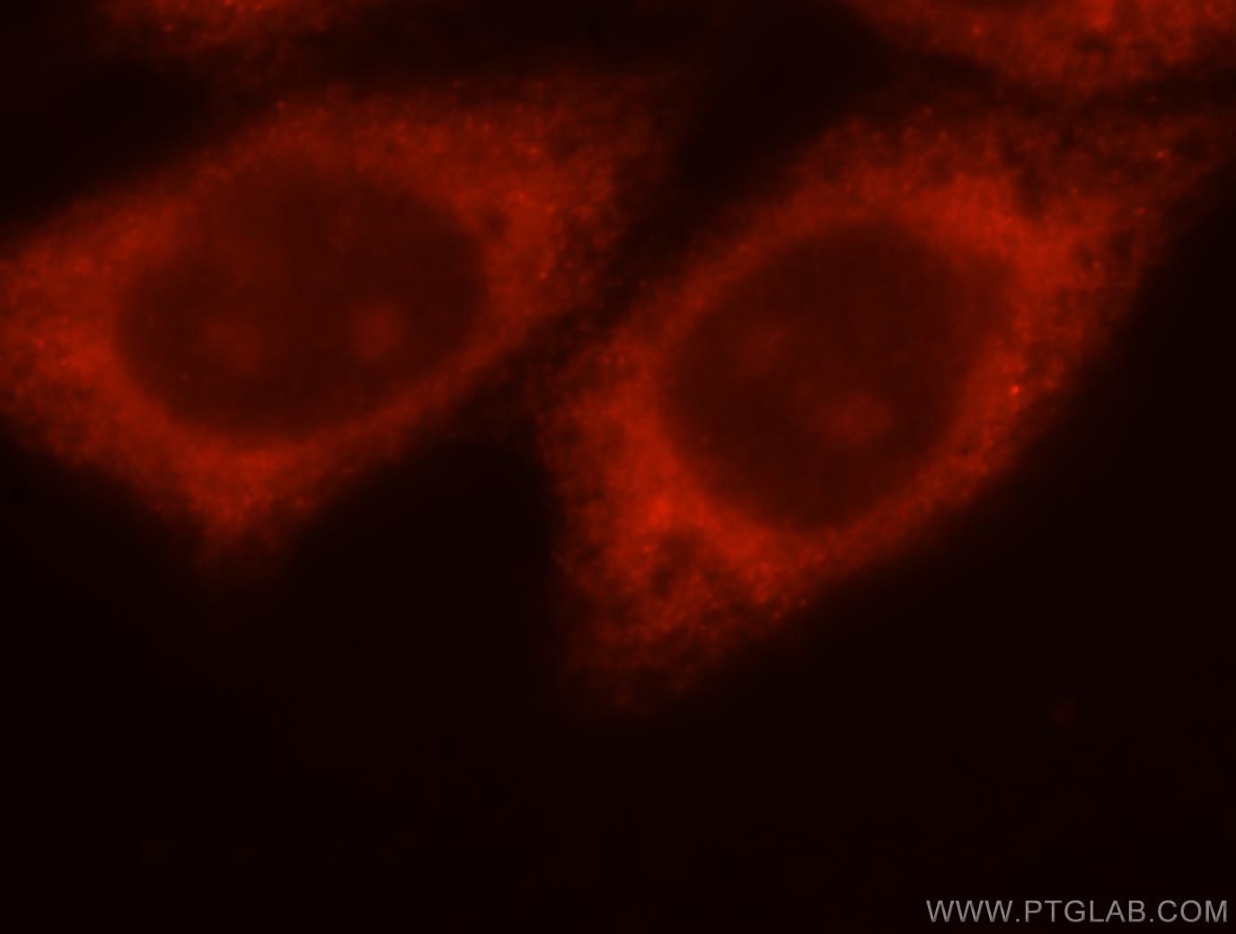
IF Staining of HepG2 using 15486-1-AP (same clone as 15486-1-PBS)
Immunofluorescent analysis of HepG2 cells, using THTPA antibody 15486-1-AP at 1:25 dilution and Rhodamine-labeled goat anti-rabbit IgG (red). This data was developed using the same antibody clone with 15486-1-PBS in a different storage buffer formulation.
Immunofluorescent analysis of HepG2 cells, using THTPA antibody 15486-1-AP at 1:25 dilution and Rhodamine-labeled goat anti-rabbit IgG (red). This data was developed using the same antibody clone with 15486-1-PBS in a different storage buffer formulation.
The Proteintech guarantee covers Proteintech antibodies in any species and any application, including those not listed on the datasheet. If the antibody doesn’t perform, you can receive a hassle-free refund or credit note.
WB analysis of K-562 using 15486-1-AP (same clone as 15486-1-PBS)
K-562 cells were subjected to SDS PAGE followed by western blot with 15486-1-AP (THTPA antibody) at dilution of 1:800 incubated at room temperature for 1.5 hours. This data was developed using the same antibody clone with 15486-1-PBS in a different storage buffer formulation.
WB analysis of HeLa using 15486-1-AP (same clone as 15486-1-PBS)
WB result of THTPA antibody (15486-1-AP; 1:1000; incubated at room temperature for 1.5 hours) with sh-Control and sh-THTPA transfected HeLa cells. This data was developed using the same antibody clone with 15486-1-PBS in a different storage buffer formulation.
WB analysis of human brain using 15486-1-AP (same clone as 15486-1-PBS)
human brain tissue were subjected to SDS PAGE followed by western blot with 15486-1-AP (THTPA antibody) at dilution of 1:500 incubated at room temperature for 1.5 hours. This data was developed using the same antibody clone with 15486-1-PBS in a different storage buffer formulation.
WB analysis of A431 using 15486-1-AP (same clone as 15486-1-PBS)
A431 cells were subjected to SDS PAGE followed by western blot with 15486-1-AP (THTPA antibody) at dilution of 1:800 incubated at room temperature for 1.5 hours. This data was developed using the same antibody clone with 15486-1-PBS in a different storage buffer formulation.
WB analysis of mouse uterus using 15486-1-AP (same clone as 15486-1-PBS)
mouse uterus tissue were subjected to SDS PAGE followed by western blot with 15486-1-AP (THTPA antibody) at dilution of 1:800 incubated at room temperature for 1.5 hours. This data was developed using the same antibody clone with 15486-1-PBS in a different storage buffer formulation.
WB analysis of mouse testis using 15486-1-AP (same clone as 15486-1-PBS)
mouse testis tissue were subjected to SDS PAGE followed by western blot with 15486-1-AP (THTPA antibody) at dilution of 1:800 incubated at room temperature for 1.5 hours. This data was developed using the same antibody clone with 15486-1-PBS in a different storage buffer formulation.
WB analysis of PC-3 using 15486-1-AP (same clone as 15486-1-PBS)
PC-3 cells were subjected to SDS PAGE followed by western blot with 15486-1-AP (THTPA antibody) at dilution of 1:800 incubated at room temperature for 1.5 hours. This data was developed using the same antibody clone with 15486-1-PBS in a different storage buffer formulation.
WB analysis of HeLa using 15486-1-AP (same clone as 15486-1-PBS)
HeLa cells were subjected to SDS PAGE followed by western blot with 15486-1-AP (THTPA antibody) at dilution of 1:800 incubated at room temperature for 1.5 hours. This data was developed using the same antibody clone with 15486-1-PBS in a different storage buffer formulation.
IHC Figures
IHC staining of human stomach cancer using 15486-1-AP (same clone as 15486-1-PBS)
Immunohistochemical analysis of paraffin-embedded human stomach cancer using 15486-1-AP (THTPA antibody) at dilution of 1:50 (under 40x lens). This data was developed using the same antibody clone with 15486-1-PBS in a different storage buffer formulation.
IHC staining of human stomach cancer using 15486-1-AP (same clone as 15486-1-PBS)
Immunohistochemical analysis of paraffin-embedded human stomach cancer using 15486-1-AP (THTPA antibody) at dilution of 1:50 (under 10x lens). This data was developed using the same antibody clone with 15486-1-PBS in a different storage buffer formulation.
IP Figures
IP experiment of K-562 using 15486-1-AP (same clone as 15486-1-PBS)
IP result of anti-THTPA (IP:15486-1-AP, 3ug; Detection:15486-1-AP 1:800) with K-562 cells lysate 1720ug. This data was developed using the same antibody clone with 15486-1-PBS in a different storage buffer formulation.
IF/ICC Figures
IF Staining of HepG2 using 15486-1-AP (same clone as 15486-1-PBS)
Immunofluorescent analysis of HepG2 cells, using THTPA antibody 15486-1-AP at 1:25 dilution and Rhodamine-labeled goat anti-rabbit IgG (red). This data was developed using the same antibody clone with 15486-1-PBS in a different storage buffer formulation.
The species listed in Tested Reactivity are in-house verified and applicable species. For unlisted species, please refer to the homology analysis of the immunogen sequence and related species. For rabbit polyclonal antibodies, homology >70% is recommended. For mouse monoclonal antibodies and rabbit recombinant antibodies, homology >90% is recommended. Generally, the higher the homology, the greater the applicability. However, there will be certain differences in protein expression in different species, tissues or cells. Therefore, the homology analysis results are for reference only and do not serve as a guarantee.
At Proteintech, we pride ourselves on our antibody quality, customer service and transparency. As such, we are comparing our antibodies with other vendors, enabling easy identification and comparisons of key data to help you choose the suitable antibody for your needs.
We have selected the top cited antibodies from these vendors for you to compare.
Proteintech
KD/KO VALIDATED
THTPA Polyclonal antibody
Catalog Number
15486-1-PBS
Citations
-
Dilutions
Applications
WB, IHC, IF/ICC, IP, Indirect ELISA
Reactivity
human, mouse, rat
Product Guarantee
Covers any species including not listed on datasheet
Covers any applications including not listed on datasheet
 at dilution of 1:800 incubated at room temperature for 1.5 hours. This data was developed using the same antibody clone with 15486-1-PBS in a different storage buffer formulation.")
 with sh-Control and sh-THTPA transfected HeLa cells. This data was developed using the same antibody clone with 15486-1-PBS in a different storage buffer formulation.")
 at dilution of 1:500 incubated at room temperature for 1.5 hours. This data was developed using the same antibody clone with 15486-1-PBS in a different storage buffer formulation.")
 at dilution of 1:800 incubated at room temperature for 1.5 hours. This data was developed using the same antibody clone with 15486-1-PBS in a different storage buffer formulation.")
 at dilution of 1:800 incubated at room temperature for 1.5 hours. This data was developed using the same antibody clone with 15486-1-PBS in a different storage buffer formulation.")
 at dilution of 1:800 incubated at room temperature for 1.5 hours. This data was developed using the same antibody clone with 15486-1-PBS in a different storage buffer formulation.")
 at dilution of 1:800 incubated at room temperature for 1.5 hours. This data was developed using the same antibody clone with 15486-1-PBS in a different storage buffer formulation.")
 at dilution of 1:800 incubated at room temperature for 1.5 hours. This data was developed using the same antibody clone with 15486-1-PBS in a different storage buffer formulation.")
 with K-562 cells lysate 1720ug. This data was developed using the same antibody clone with 15486-1-PBS in a different storage buffer formulation.")
 at dilution of 1:50 (under 40x lens). This data was developed using the same antibody clone with 15486-1-PBS in a different storage buffer formulation.")
 at dilution of 1:50 (under 10x lens). This data was developed using the same antibody clone with 15486-1-PBS in a different storage buffer formulation.")
. This data was developed using the same antibody clone with 15486-1-PBS in a different storage buffer formulation.")