Filter:


Tested Applications
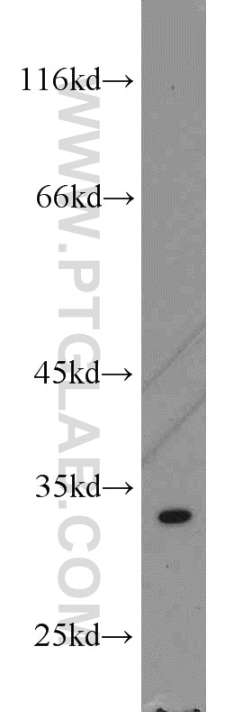
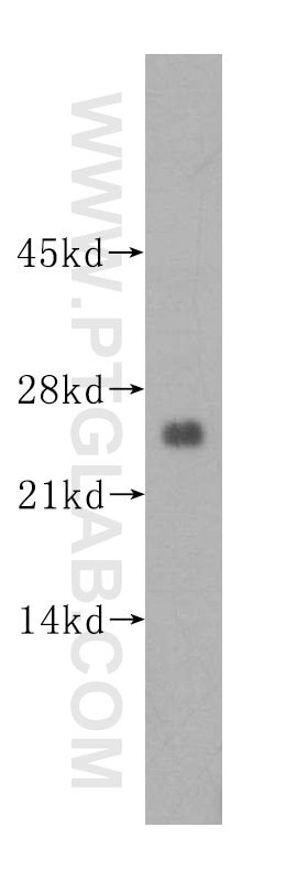
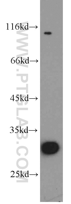
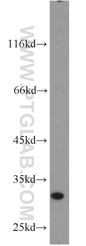
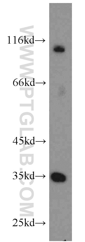
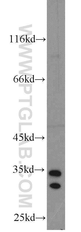
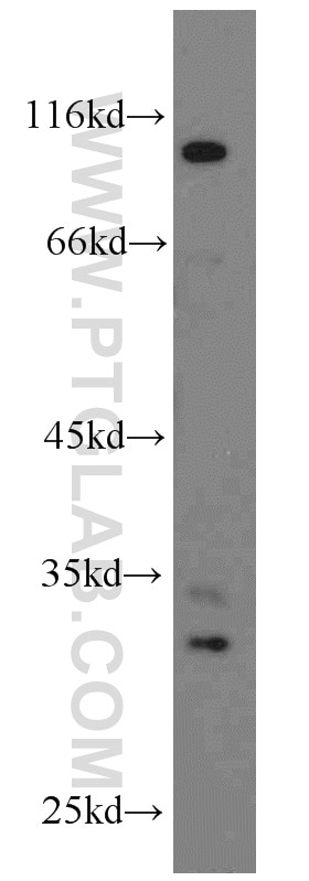
| Positive WB detected in | K-562 cells, A431 cells, HeLa cells, human brain tissue, mouse testis tissue, mouse uterus tissue, PC-3 cells |
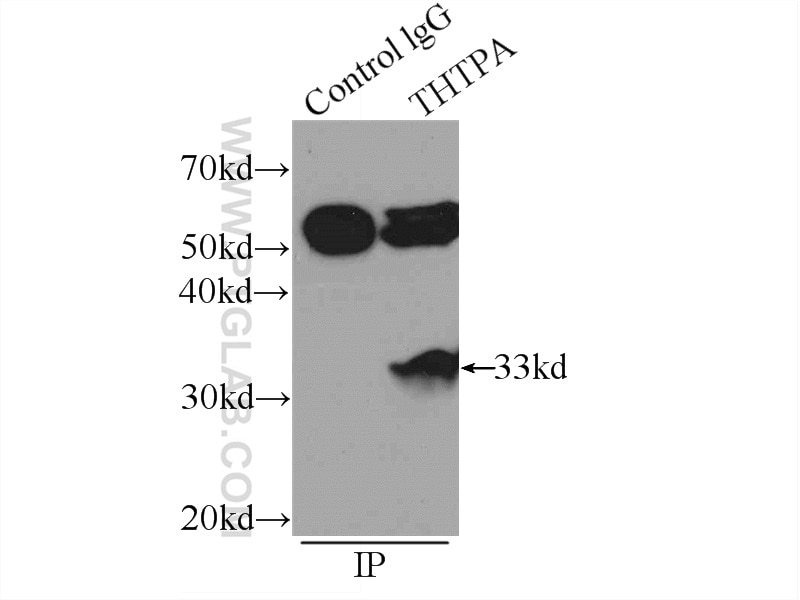
| Positive IP detected in | K-562 cells |
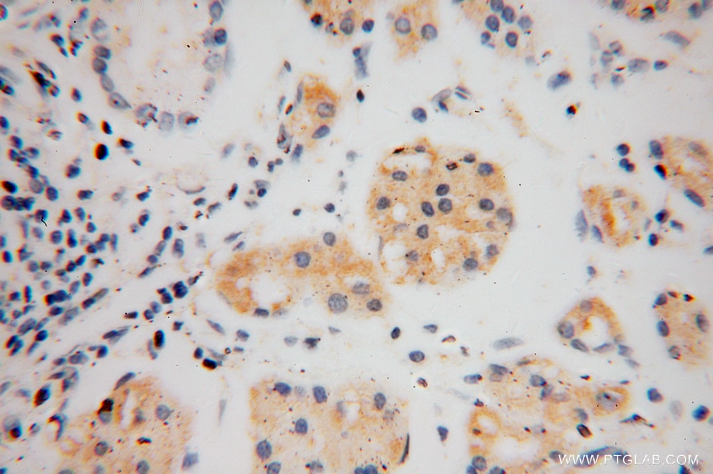
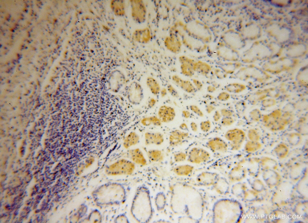
| Positive IHC detected in | human stomach cancer tissue Note: suggested antigen retrieval with TE buffer pH 9.0; (*) Alternatively, antigen retrieval may be performed with citrate buffer pH 6.0 |
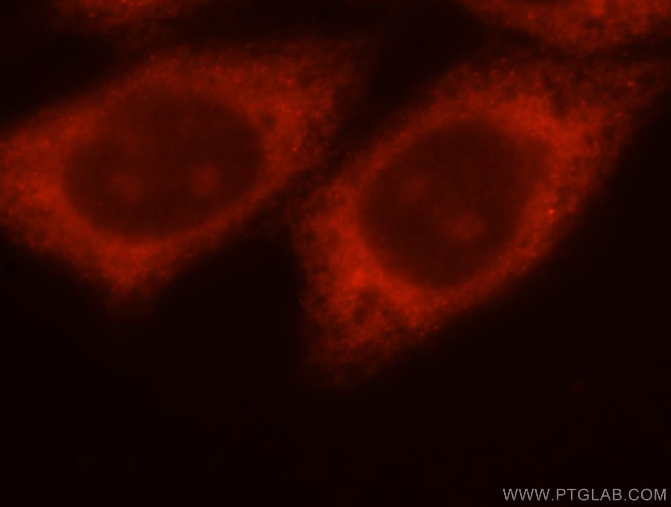
| Positive IF/ICC detected in | HepG2 cells |
Recommended dilution
| Application | Dilution |
|---|---|
| Western Blot (WB) | WB : 1:500-1:1000 |
| Immunoprecipitation (IP) | IP : 0.5-4.0 ug for 1.0-3.0 mg of total protein lysate |
| Immunohistochemistry (IHC) | IHC : 1:20-1:200 |
| Immunofluorescence (IF)/ICC | IF/ICC : 1:10-1:100 |
| It is recommended that this reagent should be titrated in each testing system to obtain optimal results. | |
| Sample-dependent, Check data in validation data gallery. | |
Published Applications
| WB | See 1 publications below |
Product Information
15486-1-AP targets THTPA in WB, IHC, IF/ICC, IP, ELISA applications and shows reactivity with human, mouse, rat samples.
| Tested Reactivity | human, mouse, rat |
| Cited Reactivity | human |
| Host / Isotype | Rabbit / IgG |
| Class | Polyclonal |
| Type | Antibody |
| Immunogen |
CatNo: Ag7834 Product name: Recombinant human THTPA protein Source: e coli.-derived, PGEX-4T Tag: GST Domain: 1-230 aa of BC002984 Sequence: MAQGLIEVERKFLPGPGTEERLQELGGTLEYRVTFRDTYYDTPELSLMQADHWLRRREDSGWELKCPGAAGVLGPHTEYKELTAEPTIVAQLCKVLRADGLGAGDVAAVLGPLGLQEVASFVTKRSAWKLVLLGADEEEPQLRVDLDTADFGYAVGEVEALVHEEAEVPTALEKIHRLSSMLGVPAQETAPAKLIVYLQRFRPQDYQRLLEVNSSRERPQETEDPDHCLG Predict reactive species |
| Full Name | thiamine triphosphatase |
| Calculated Molecular Weight | 26 kDa |
| Observed Molecular Weight | 26-35 kDa |
| GenBank Accession Number | BC002984 |
| Gene Symbol | THTPA |
| Gene ID (NCBI) | 79178 |
| RRID | AB_2203183 |
| Conjugate | Unconjugated |
| Form | Liquid |
| Purification Method | Antigen affinity purification |
| UNIPROT ID | Q9BU02 |
| Storage Buffer | PBS with 0.02% sodium azide and 50% glycerol, pH 7.3. |
| Storage Conditions | Store at -20°C. Stable for one year after shipment. Aliquoting is unnecessary for -20oC storage. 20ul sizes contain 0.1% BSA. |
Protocols
| Product Specific Protocols | |
|---|---|
| IF protocol for THTPA antibody 15486-1-AP | Download protocol |
| IHC protocol for THTPA antibody 15486-1-AP | Download protocol |
| IP protocol for THTPA antibody 15486-1-AP | Download protocol |
| WB protocol for THTPA antibody 15486-1-AP | Download protocol |
| Standard Protocols | |
|---|---|
| Click here to view our Standard Protocols |