
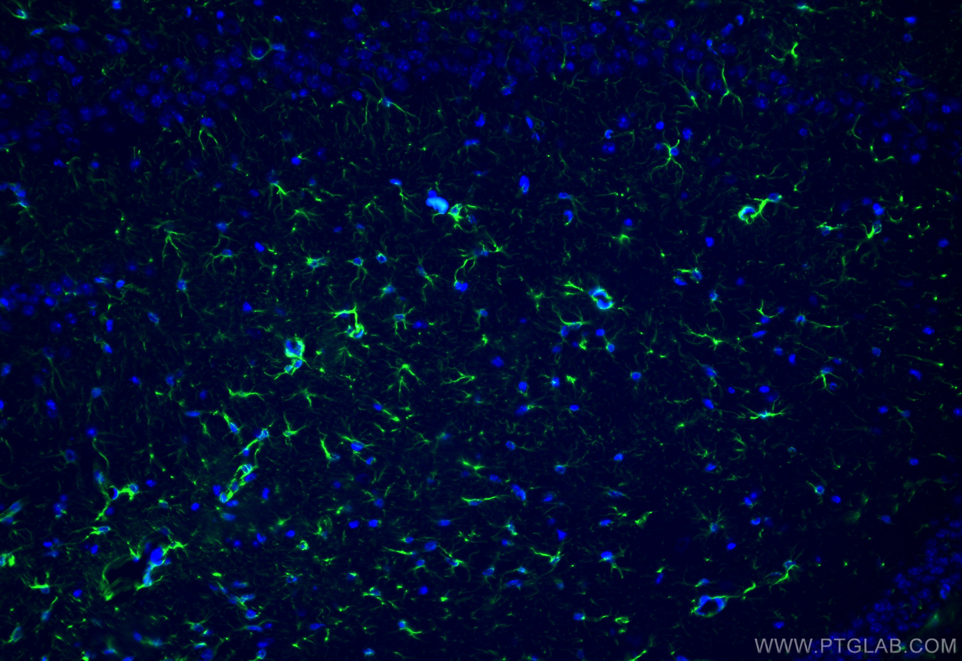

Tested Applications
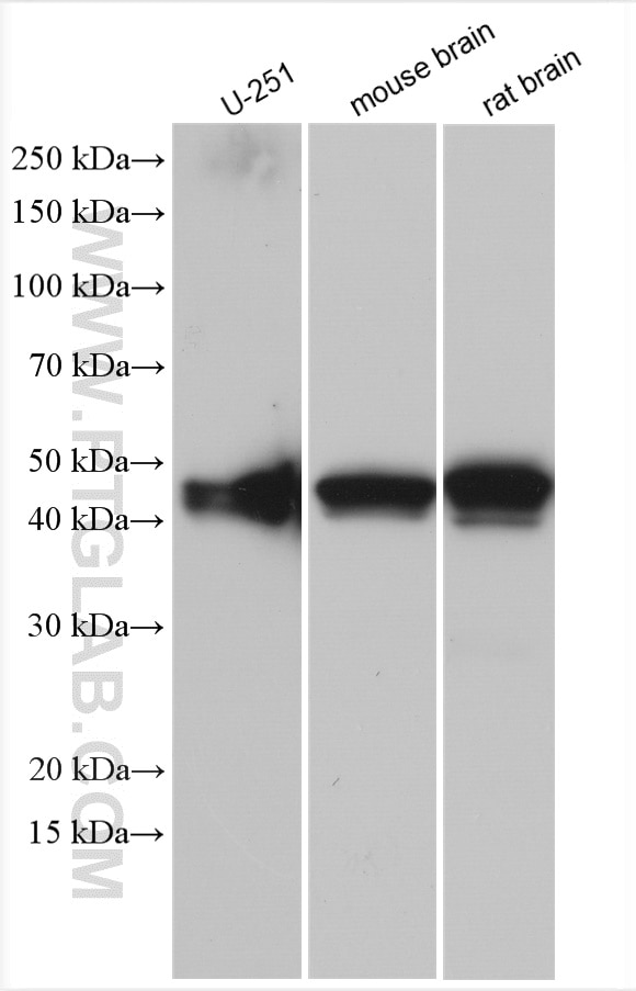
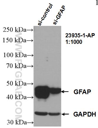
| Positive WB detected in | U-251 cells, mouse brain tissue, rat brain tissue |
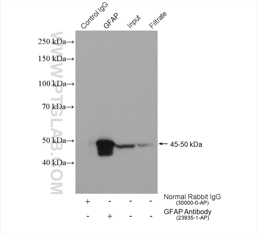
| Positive IP detected in | mouse brain tissue |
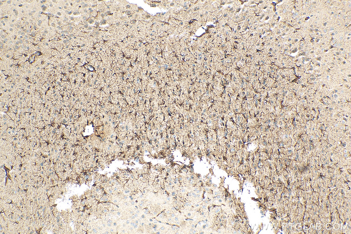
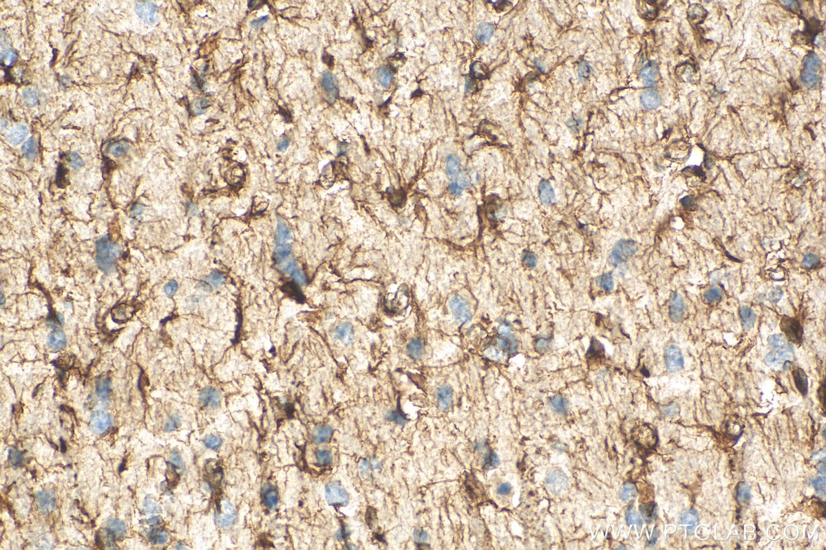
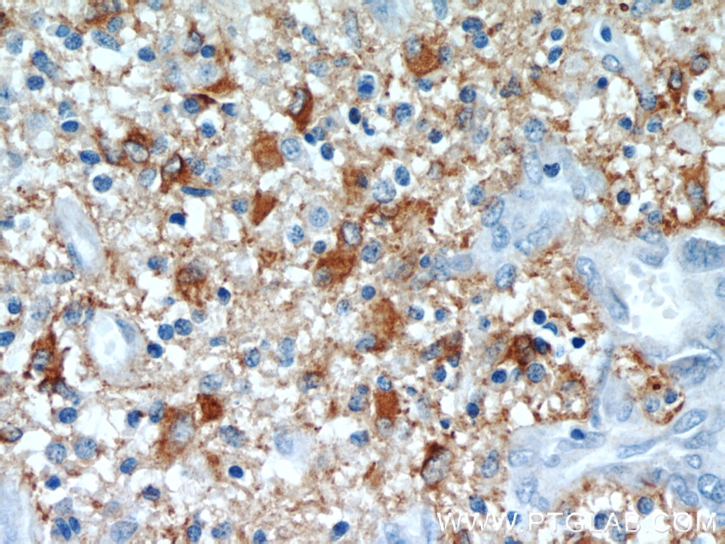
| Positive IHC detected in | mouse brain tissue, human gliomas tissue Note: suggested antigen retrieval with TE buffer pH 9.0; (*) Alternatively, antigen retrieval may be performed with citrate buffer pH 6.0 |
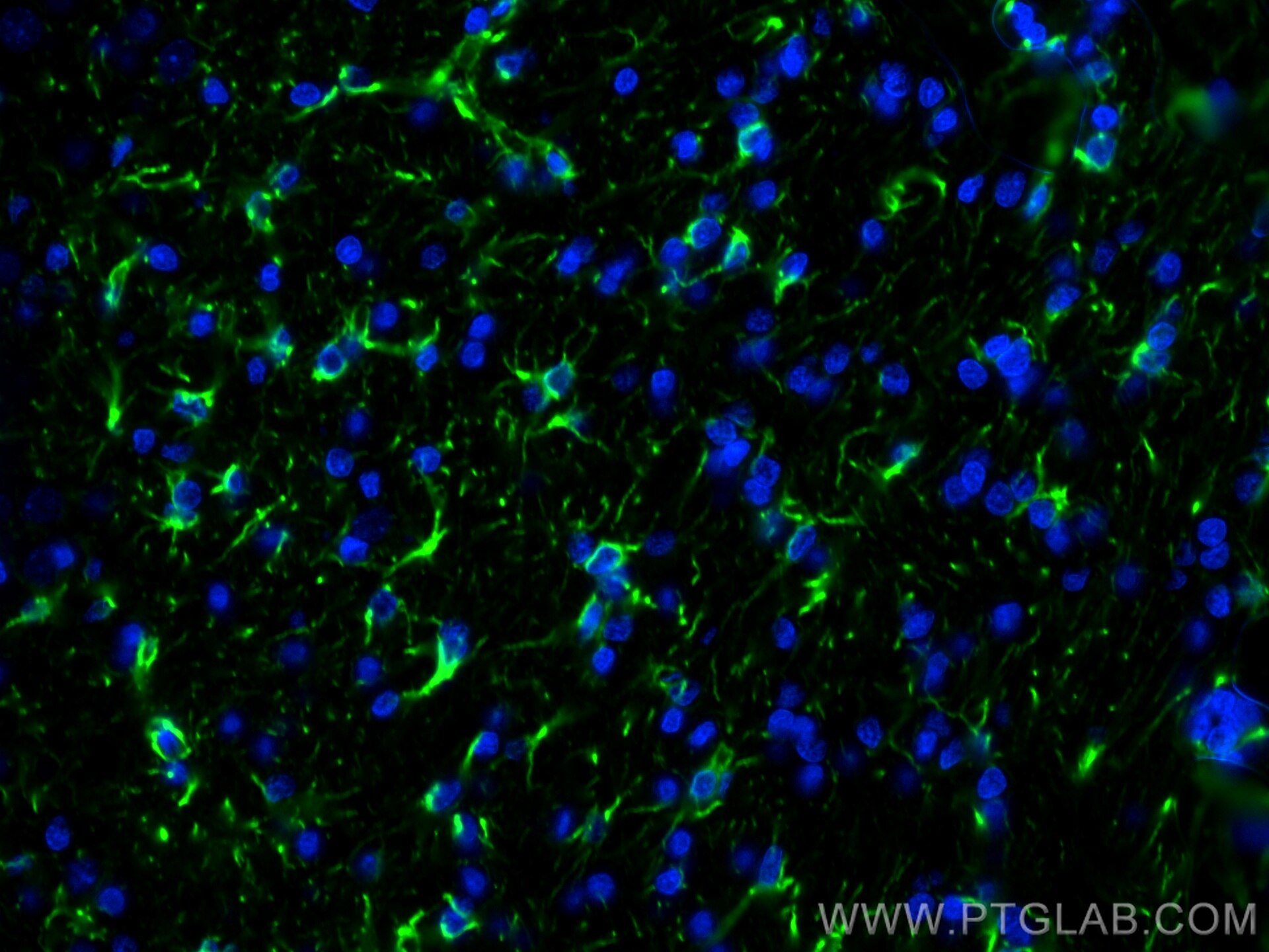
| Positive IF-P detected in | rat brain tissue |
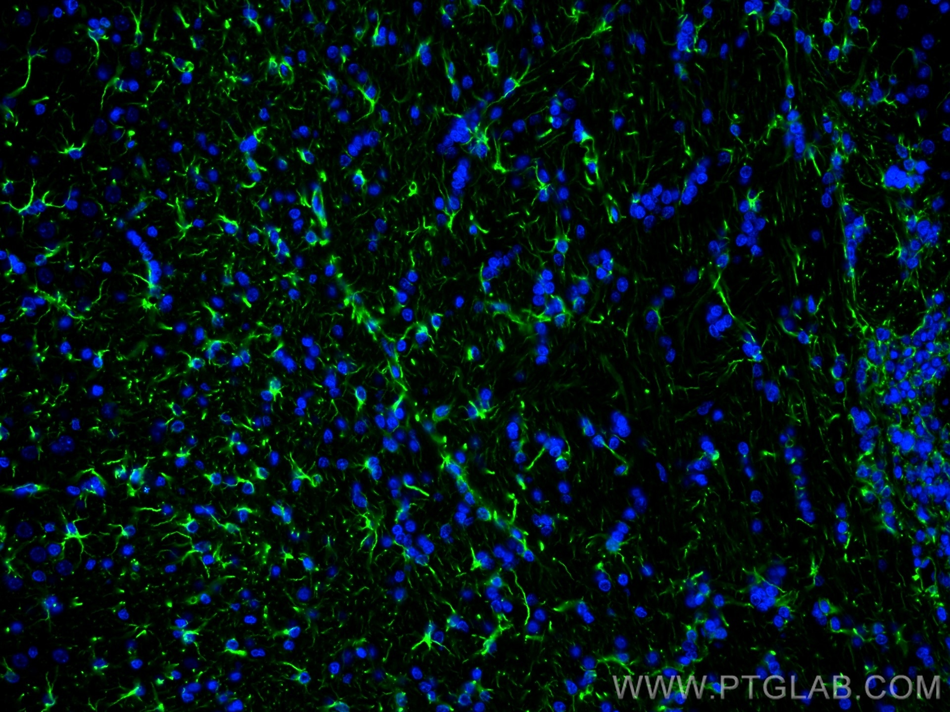
| Positive IF-Fro detected in | mouse brain tissue |
Recommended dilution
| Application | Dilution |
|---|---|
| Western Blot (WB) | WB : 1:5000-1:50000 |
| Immunoprecipitation (IP) | IP : 0.5-4.0 ug for 1.0-3.0 mg of total protein lysate |
| Immunohistochemistry (IHC) | IHC : 1:200-1:800 |
| Immunofluorescence (IF)-P | IF-P : 1:500-1:2000 |
| Immunofluorescence (IF)-FRO | IF-FRO : 1:50-1:500 |
| It is recommended that this reagent should be titrated in each testing system to obtain optimal results. | |
| Sample-dependent, Check data in validation data gallery. | |
Product Information
23935-1-AP targets GFAP in WB, IHC, IF-P, IF-Fro, IP, ELISA applications and shows reactivity with human, mouse, rat samples.
| Tested Reactivity | human, mouse, rat |
| Cited Reactivity | human, mouse, rat, macaque |
| Host / Isotype | Rabbit / IgG |
| Class | Polyclonal |
| Type | Antibody |
| Immunogen |
CatNo: Ag20853 Product name: Recombinant human GFAP protein Source: e coli.-derived, PET30a Tag: 6*His Domain: 1-432 aa of BC013596 Sequence: MERRRITSAARRSYVSSGEMMVGGLAPGRRLGPGTRLSLARMPPPLPTRVDFSLAGALNAGFKETRASERAEMMELNDRFASYIEKVRFLEQQNKALAAELNQLRAKEPTKLADVYQAELRELRLRLDQLTANSARLEVERDNLAQDLATVRQKLQDETNLRLEAENNLAAYRQEADEATLARLDLERKIESLEEEIRFLRKIHEEEVRELQEQLARQQVHVELDVAKPDLTAALKEIRTQYEAMASSNMHEAEEWYRSKFADLTDAAARNAELLRQAKHEANDYRRQLQSLTCDLESLRGTNESLERQMREQEERHVREAASYQEALARLEEEGQSLKDEMARHLQEYQDLLNVKLALDIEIATYRKLLEGEENRITIPVQTFSNLQIRETSLDTKSVSEGHLKRNIVVKTVEMRDGEVIKESKQEHKDVM Predict reactive species |
| Full Name | glial fibrillary acidic protein |
| Calculated Molecular Weight | 432 aa, 50 kDa |
| Observed Molecular Weight | 45-50 kDa |
| GenBank Accession Number | BC013596 |
| Gene Symbol | GFAP |
| Gene ID (NCBI) | 2670 |
| RRID | AB_2879367 |
| Conjugate | Unconjugated |
| Form | Liquid |
| Purification Method | Antigen affinity purification |
| UNIPROT ID | P14136 |
| Storage Buffer | PBS with 0.02% sodium azide and 50% glycerol, pH 7.3. |
| Storage Conditions | Store at -20°C. Stable for one year after shipment. Aliquoting is unnecessary for -20oC storage. 20ul sizes contain 0.1% BSA. |
Background Information
Function
GFAP (Glial fibrillary acidic protein) is a type III intermediate filament (IF) protein specific to the central nervous system (CNS). GFAP is one of the main components of the intermediate filament network in astrocytes and has been proposed as playing a role in cell migration, cell motility, maintaining mechanical strength, and in mitosis.
Tissue specificity
GFAP is expressed in central nervous system cells, predominantly in astrocytes. GFAP is commonly used as an astrocyte marker. However, GFAP is also present in peripheral glia and in non-CNS cells, including fibroblasts, chondrocytes, lymphocytes, and liver stellate cells (PMID: 21219963).
Involvement in disease
Mutations in GFAP lead to Alexander disease (OMIM: 203450), an autosomal dominant CNS disorder. The mutations present in affected individuals are thought to be gain-of-function.
Upregulation of GFAP is a hallmark of reactive astrocytes, in which GFAP is present in hypertrophic cellular processes. Reactive astrogliosis is present in many neurological disorders, such as stroke, various neurodegenerative diseases (including Alzheimer's and Parkinson's disease), and neurotrauma.
Isoforms
Astrocytes express 10 different isoforms of GFAP that differ in the rod and tail domains (PMID: 25726916), which means that they differ in molecular size. Isoform expression varies during the development and across different subtypes of astrocytes. Not all isoforms are upregulated in reactive astrocytes.
Post-translational modifications
Intermediate filament proteins are regulated by phosphorylation. Six phosphorylation sites have been identified in GFAP protein, at least some of which are reported to control filament assembly (PMID: 21219963).
Cellular localization
GFAP localizes to intermediate filaments and stains well in astrocyte cellular processes.
Protocols
| Product Specific Protocols | |
|---|---|
| IF protocol for GFAP antibody 23935-1-AP | Download protocol |
| IHC protocol for GFAP antibody 23935-1-AP | Download protocol |
| IP protocol for GFAP antibody 23935-1-AP | Download protocol |
| WB protocol for GFAP antibody 23935-1-AP | Download protocol |
| Standard Protocols | |
|---|---|
| Click here to view our Standard Protocols |
Publications
What published studies show
The GFAP Polyclonal antibody (Cat# 23935-1-AP) has 26 citations spanning journals including Nature, Cell, Cell Metabolism, Theranostics, Science Advances, and Scientific Reports. Research fields covered include glioblastoma and brain metastasis, neurodegeneration (Parkinson's, Alzheimer's), cerebral ischemia, retinal degeneration, gut-brain axis, neuroinflammation, myelin biology, and astrocyte function. Applications cited include WB, IF, and IHC across human, mouse, rat, and macaque species.
| Species | Application | Title |
|---|---|---|
Cell Metab Metabolic regulation of the glioblastoma stem cell epitranscriptome by malate dehydrogenase 2 | ||
Theranostics CD146(+) pericyte-like lung cancer brain metastatic stem cells promote tumor angiogenesis through dual regulatory effects on the VEGF/VEGFR axis. | ||
J Exp Clin Cancer Res RBM10 deficiency promotes brain metastasis by modulating sphingolipid metabolism in a BBB model of EGFR mutant lung adenocarcinoma | ||
Adv Sci (Weinh) Understanding Glioblastoma Dynamics Using 3D Organoids and Engineered Extracellular Matrix. |
Reviews
What customers say
The single review for this product is highly positive. A researcher rated it 5 stars and reported excellent performance in immunofluorescence using a 1:100 dilution on patient-derived glioblastoma stem-like cells, simply stating "Works wonderfully!" Overall, the feedback suggests the antibody delivers reliable results in IF applications with straightforward optimization.
The reviews below have been submitted by verified Proteintech customers who received an incentive for providing their feedback.
Autumn (Verified Customer) (02-08-2024) | Works wonderfully!
|










