

Tested Applications
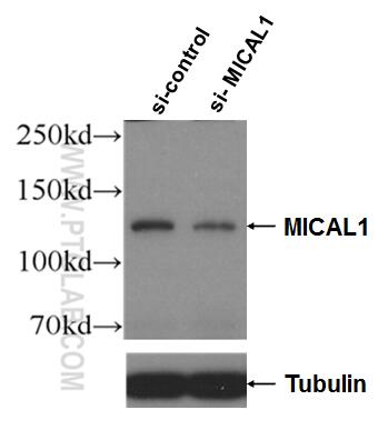
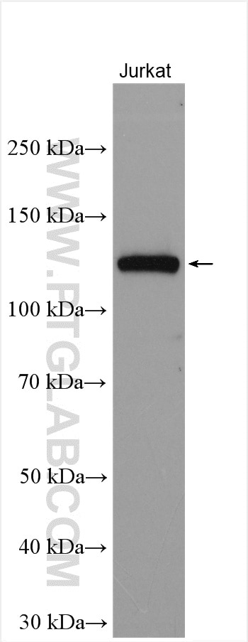
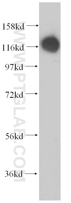
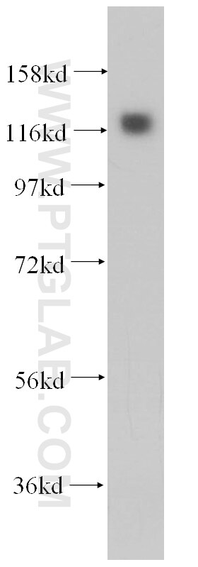
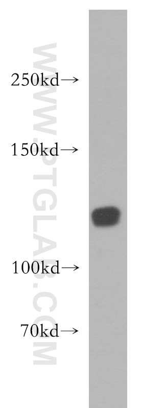
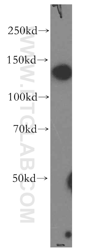
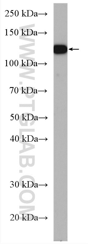
| Positive WB detected in | Jurkat cells, HEK-293 cells, HeLa cells, human brain tissue, T-47D cells |
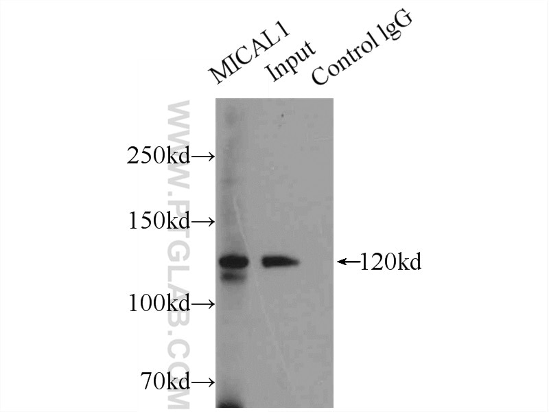
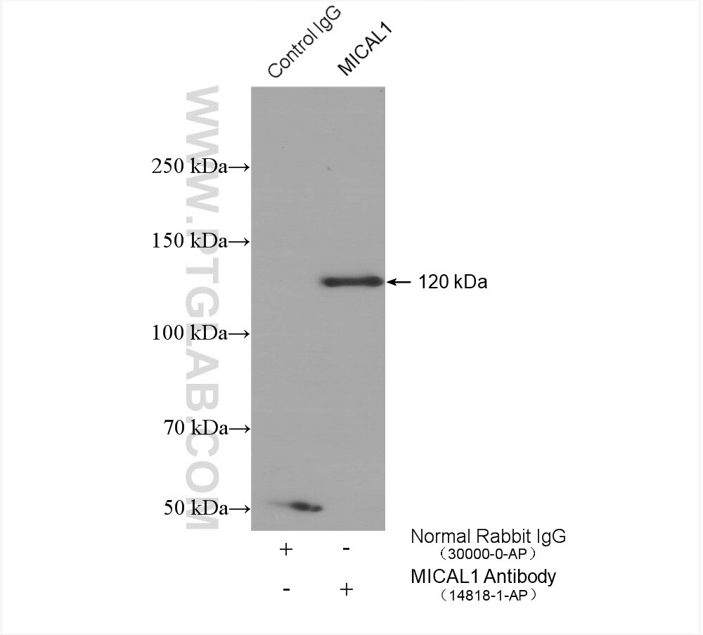
| Positive IP detected in | HeLa cells, T-47D cells |
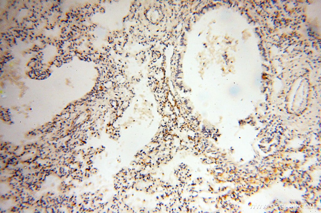
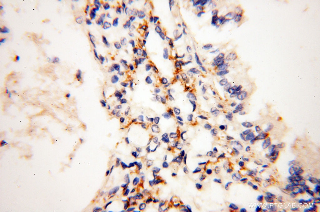
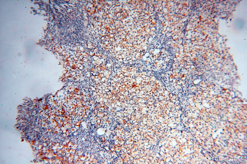
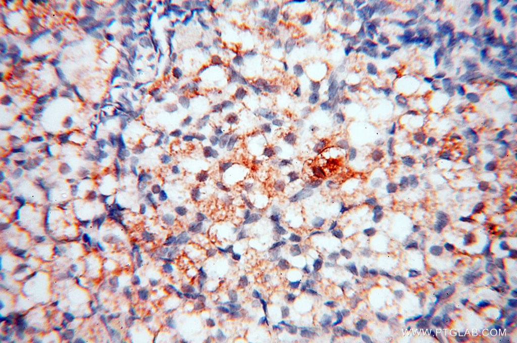
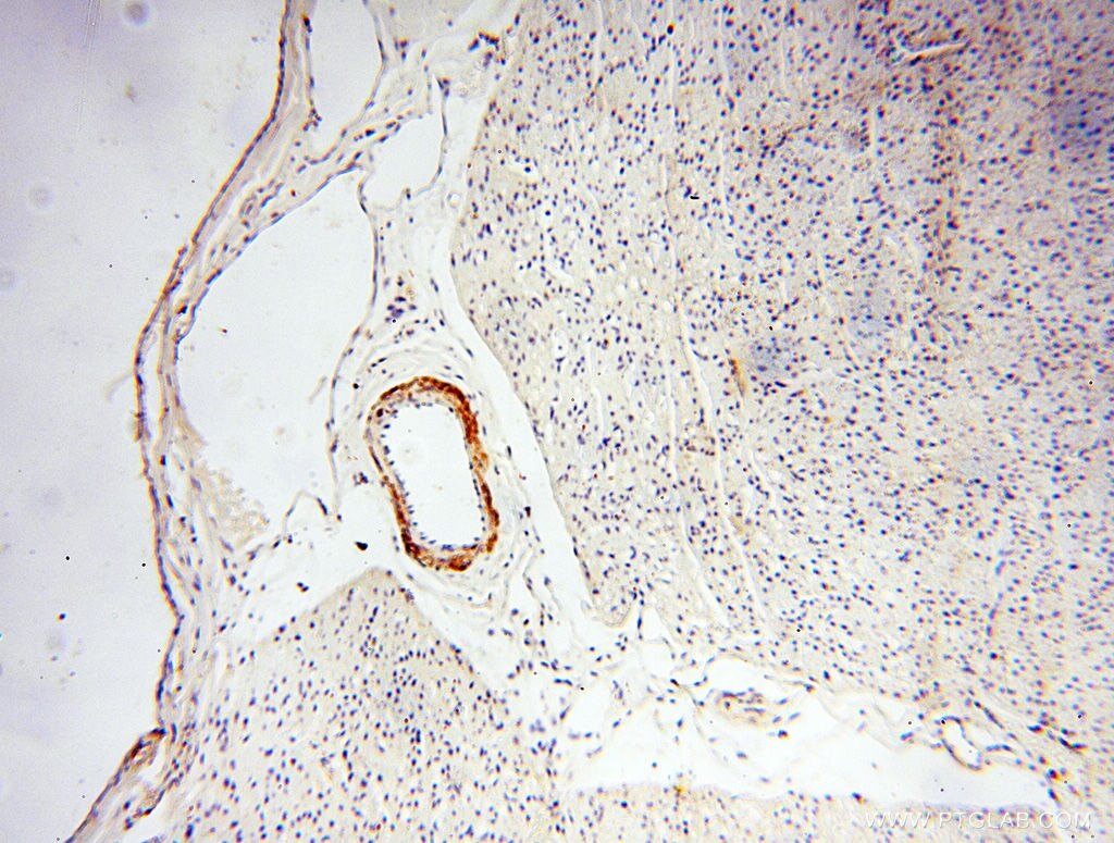
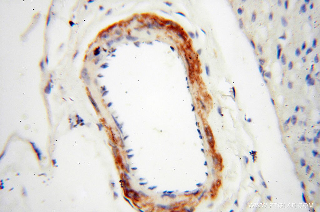
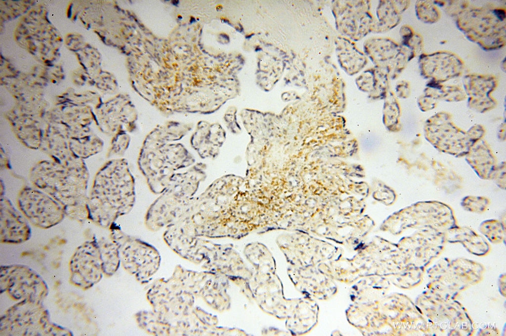
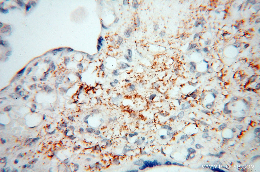
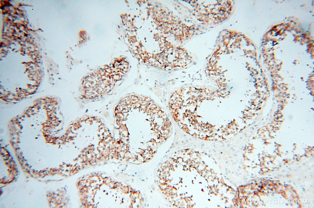
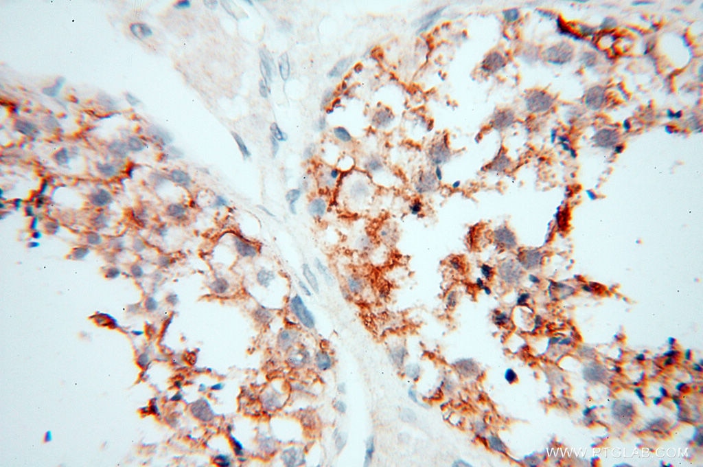
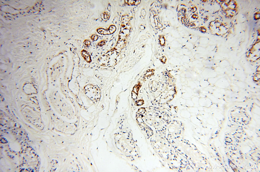
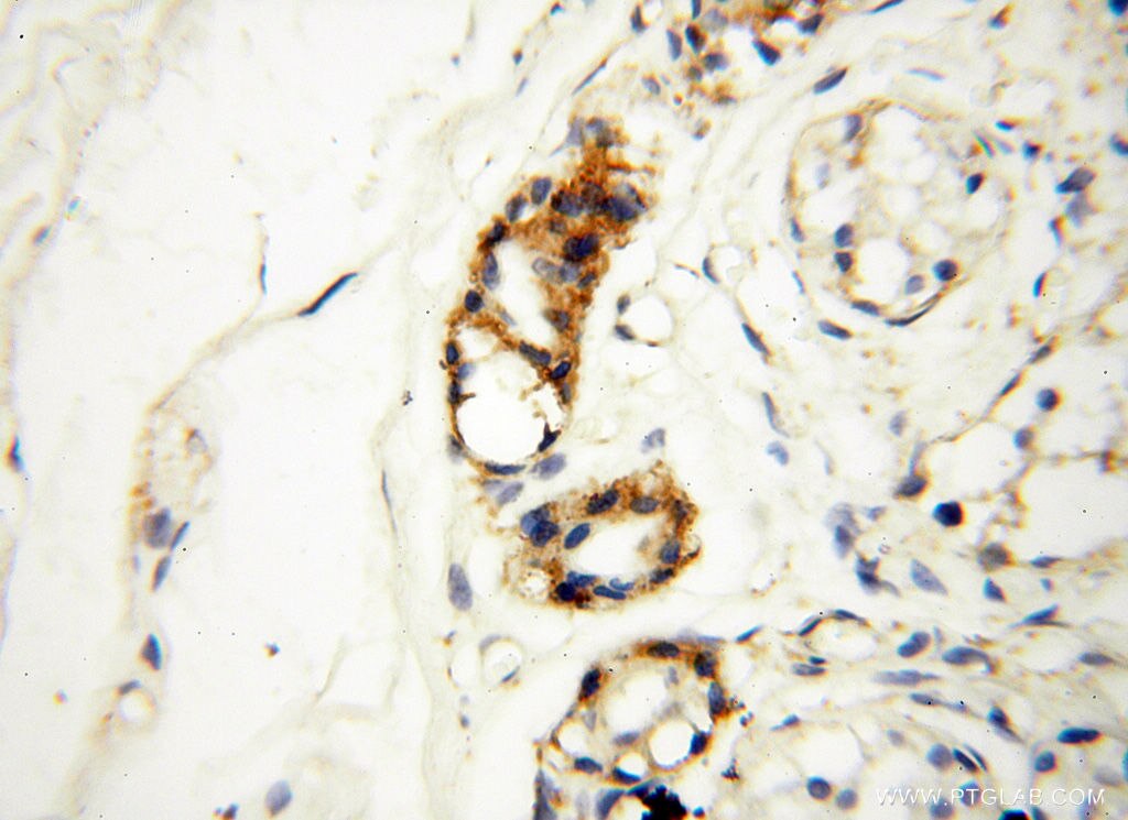
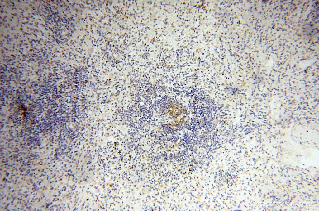
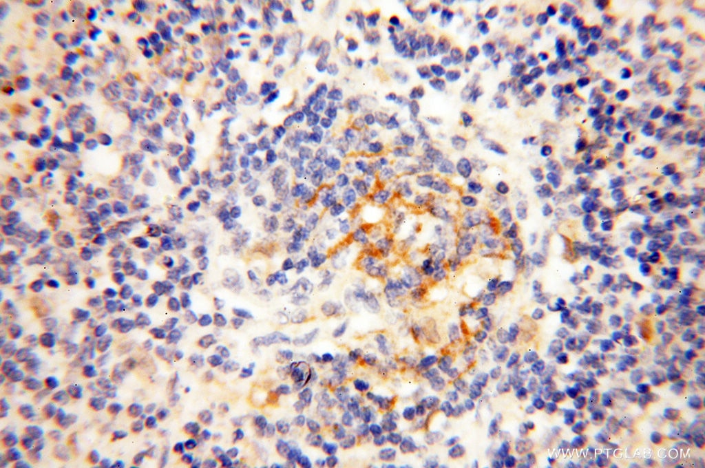
| Positive IHC detected in | human lung tissue, human heart tissue, human ovary tissue, human placenta tissue, human skin tissue, human spleen tissue, human testis tissue Note: suggested antigen retrieval with TE buffer pH 9.0; (*) Alternatively, antigen retrieval may be performed with citrate buffer pH 6.0 |
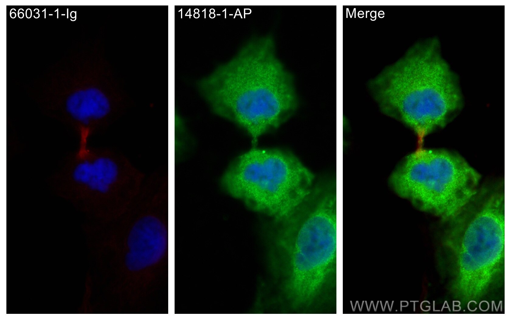
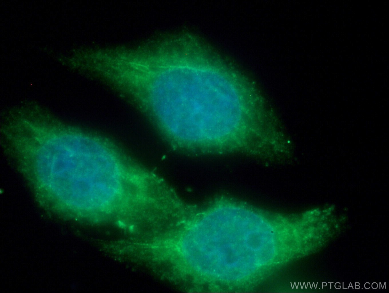
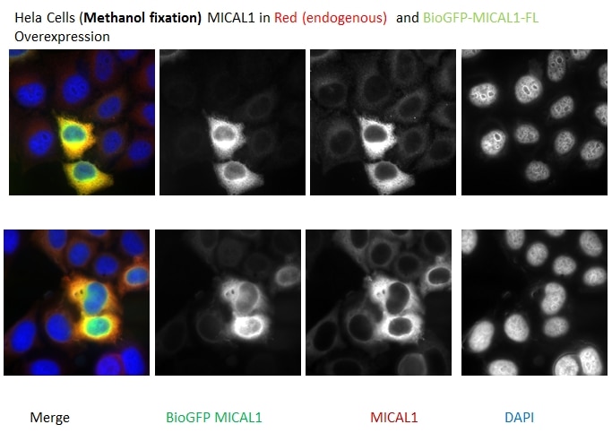
| Positive IF/ICC detected in | A549 cells, HeLa cells |
Recommended dilution
| Application | Dilution |
|---|---|
| Western Blot (WB) | WB : 1:2000-1:16000 |
| Immunoprecipitation (IP) | IP : 0.5-4.0 ug for 1.0-3.0 mg of total protein lysate |
| Immunohistochemistry (IHC) | IHC : 1:20-1:200 |
| Immunofluorescence (IF)/ICC | IF/ICC : 1:200-1:800 |
| It is recommended that this reagent should be titrated in each testing system to obtain optimal results. | |
| Sample-dependent, Check data in validation data gallery. | |
Product Information
14818-1-AP targets MICAL1 in WB, IHC, IF/ICC, IP, CoIP, ELISA applications and shows reactivity with human samples.
| Tested Reactivity | human |
| Cited Reactivity | human, mouse |
| Host / Isotype | Rabbit / IgG |
| Class | Polyclonal |
| Type | Antibody |
| Immunogen |
CatNo: Ag6578 Product name: Recombinant human MICAL1 protein Source: e coli.-derived, PET28a Tag: 6*His Domain: 718-1067 aa of BC052983 Sequence: HRSCFRCHTCEATLWPGGYEQHPGDGHFYCLQHLPQTDHKAEGSDRGPESPELPTPSENSMPPGLSTPTASQEGAGPVPDPSQPTRRQIRLSSPERQRLSSLNLTPDPEMEPPPKPPRSCSALARHALESSFVGWGLPVQSPQALVAMEKEEKESPFSSEEEEEDVPLDSDVEQALQTFAKTSGTMNNYPTWRRTLLRRAKEEEMKRFCKAQTIQRRLNEIEAALRELEAEGVKLELALRRQSSSPEQQKKLWVGQLLQLVDKKNSLVAEEAELMITVQELNLEEKQWQLDQELRGYMNREENLKTAADRQAEDQVLRKLVDLVNQRDALIRFQEERRLSELALGTGAQG Predict reactive species |
| Full Name | microtubule associated monoxygenase, calponin and LIM domain containing 1 |
| Calculated Molecular Weight | 118 kDa |
| Observed Molecular Weight | 120 kDa |
| GenBank Accession Number | BC052983 |
| Gene Symbol | MICAL1 |
| Gene ID (NCBI) | 64780 |
| RRID | AB_2143754 |
| Conjugate | Unconjugated |
| Form | Liquid |
| Purification Method | Antigen affinity purification |
| UNIPROT ID | Q8TDZ2 |
| Storage Buffer | PBS with 0.02% sodium azide and 50% glycerol, pH 7.3. |
| Storage Conditions | Store at -20°C. Stable for one year after shipment. Aliquoting is unnecessary for -20oC storage. 20ul sizes contain 0.1% BSA. |
Background Information
MICALs (Molecules Interacting with CasL) are atypical multidomain flavoenzymes with diverse cellular functions.There are three known isoforms, MICAL1, MICAL2 and MICAL3, as well as the MICAL-like proteins MICAL-L1 and MICAL-L2. MICAL1 has four conserved domains: an N-terminal flavin adenine dinucleotide (FAD) binding domain, a calponin homology (CH) domain, a Lin11, Isl-1 and Mec-3 (LIM) domain and a C-terminal coiled-coil (CC) domain. MICAL1 is reported to regulate actin stress fibers and be required for normal actin organization. It may also be involved in apoptosis through binding with NDR (nuclear Dbf2-related) kinases. This antibody specially recognizes MICAL1.
Protocols
| Product Specific Protocols | |
|---|---|
| IF protocol for MICAL1 antibody 14818-1-AP | Download protocol |
| IHC protocol for MICAL1 antibody 14818-1-AP | Download protocol |
| IP protocol for MICAL1 antibody 14818-1-AP | Download protocol |
| WB protocol for MICAL1 antibody 14818-1-AP | Download protocol |
| Standard Protocols | |
|---|---|
| Click here to view our Standard Protocols |
Publications
What published studies show
The MICAL1 polyclonal antibody (Cat# 14818-1-AP) has 29 citations published in journals including Nature Communications, Science Advances, Cell Reports, Developmental Cell, Oncogene, Journal of Cell Biology, Journal of Cell Science, Cancer Letters, BMC Cancer, Redox Biology, and others. Research fields span actin cytoskeleton regulation, cytokinesis, cancer biology (breast, gastric, colorectal, renal, pancreatic), oxidative stress, diabetic nephropathy, neuroscience, and immune response.
| Species | Application | Title |
|---|---|---|
Redox Biol Methionine oxidation of actin cytoskeleton attenuates traumatic memory retention via reactivating dendritic spine morphogenesis | ||
Sci Adv F-actin disassembly factor MICAL1 binding to Myosin Va mediates cargo unloading during cytokinesis.
| ||
Cell Death Dis STK38-mediated feedback loop regulation of the hedgehog pathway governing tumor heterogeneity in renal papillary carcinoma. | ||
Cancer Lett Unraveling pancreatic ductal adenocarcinoma immune prognostic signature through a naive B cell gene set. | ||
Cancer Lett MICAL1 regulates actin cytoskeleton organization, directional cell migration and the growth of human breast cancer cells as orthotopic xenograft tumours.
|
Reviews
What customers say
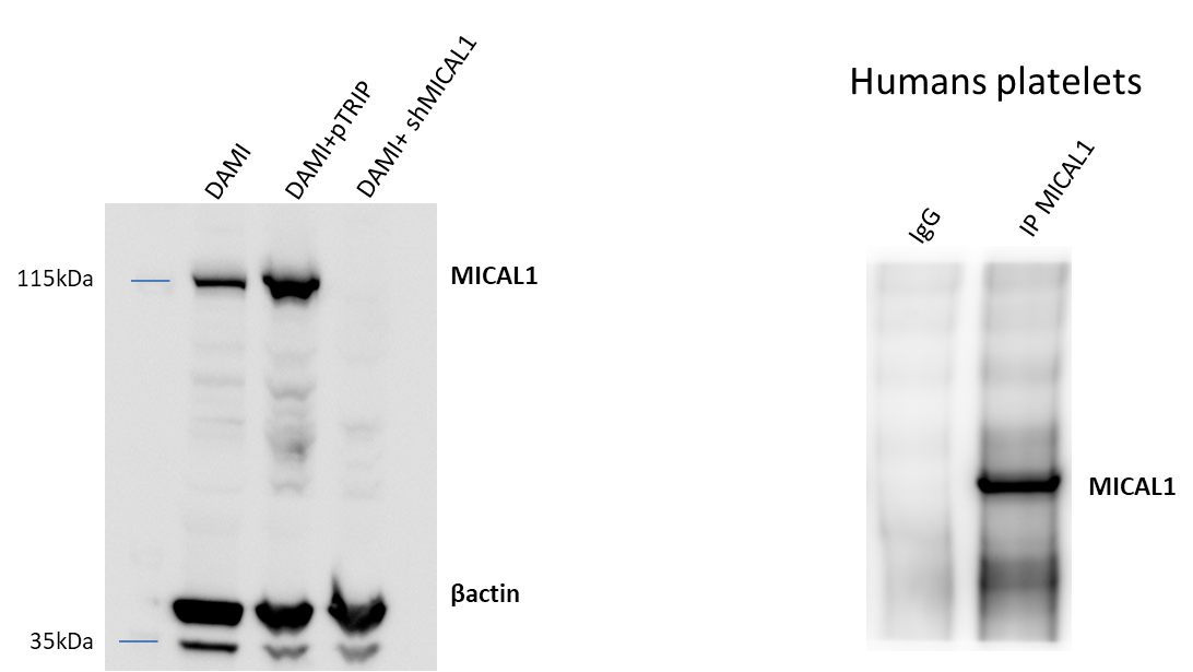
Users report mixed results with this antibody. One reviewer achieved good Western Blot results in H4 cells at 1:500 dilution (4/5). Another rated it 5/5, using 1:1000 for WB and 2 µg for IP on human platelets/megacaryocytic cells. A third reviewer (3/5) noted a faint band at the expected MICAL1 molecular weight (~118–120 kDa) in MDCK GII cells, even with extended exposure. Overall, the antibody works well for WB and IP in some cell types, but signal intensity may vary depending on the sample.
The reviews below have been submitted by verified Proteintech customers who received an incentive for providing their feedback.
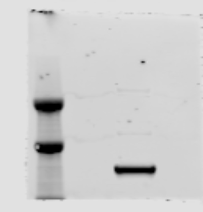
Kincaid (Verified Customer) (06-02-2024) | Tested antibody in H4 cells. Diluted aMICAL1 1:500 in 3% BSA, diluted in TBS-tween (1%).
 |
Christelle (Verified Customer) (11-21-2023) | For wb: used dilution 1/1000 For IP: used 2µg of antibody
 |
Joleen (Verified Customer) (06-03-2019) | Shows band at where MICAL1 is expected but is faint. This image is a merge image of the membrane exposed for 120s and a stain free image to visualize the molecular weight. Even with long exposure, the band is faint.
 |



























