


Tested Applications
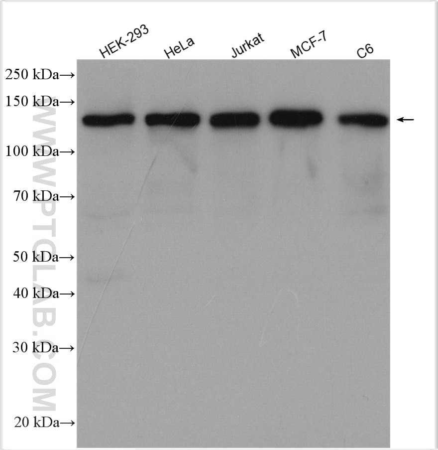
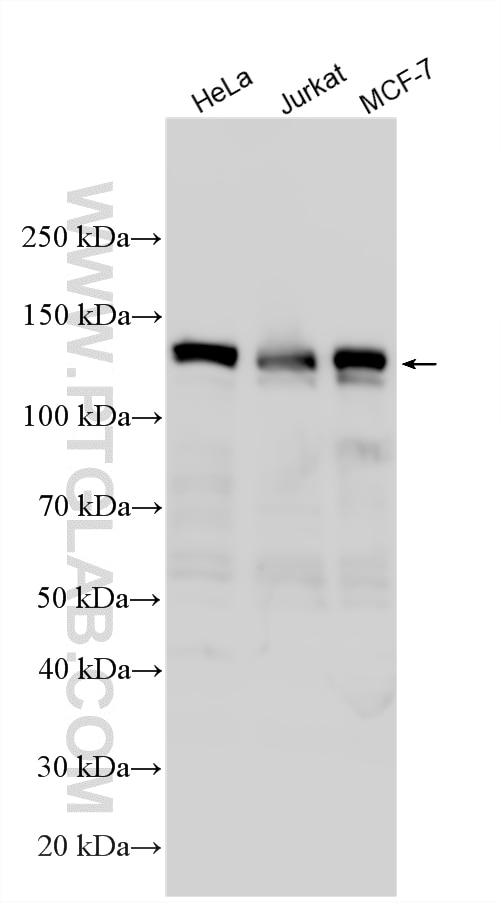
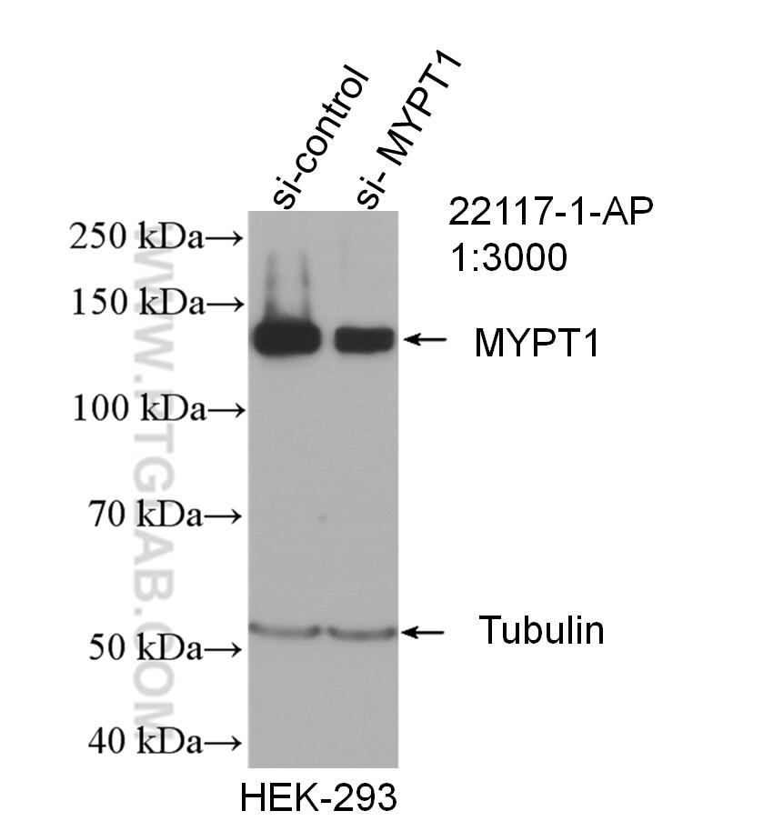
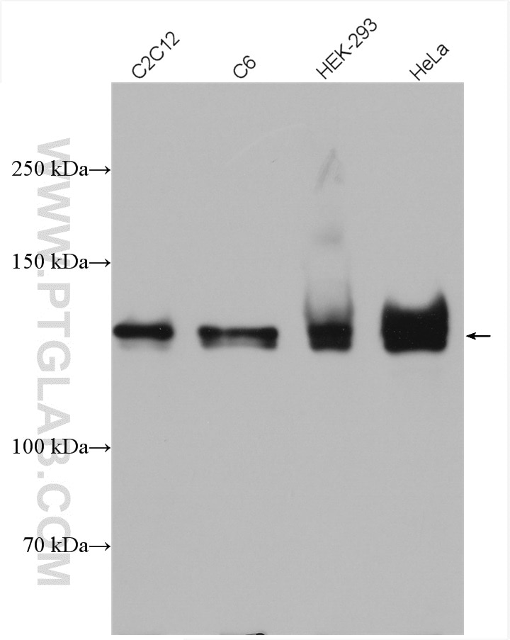
| Positive WB detected in | HEK-293 cells, C2C12 cells, HeLa cells, C6 cells, Jurkat cells, MCF-7 cells |
| Positive IP detected in | HEK-293 cells |
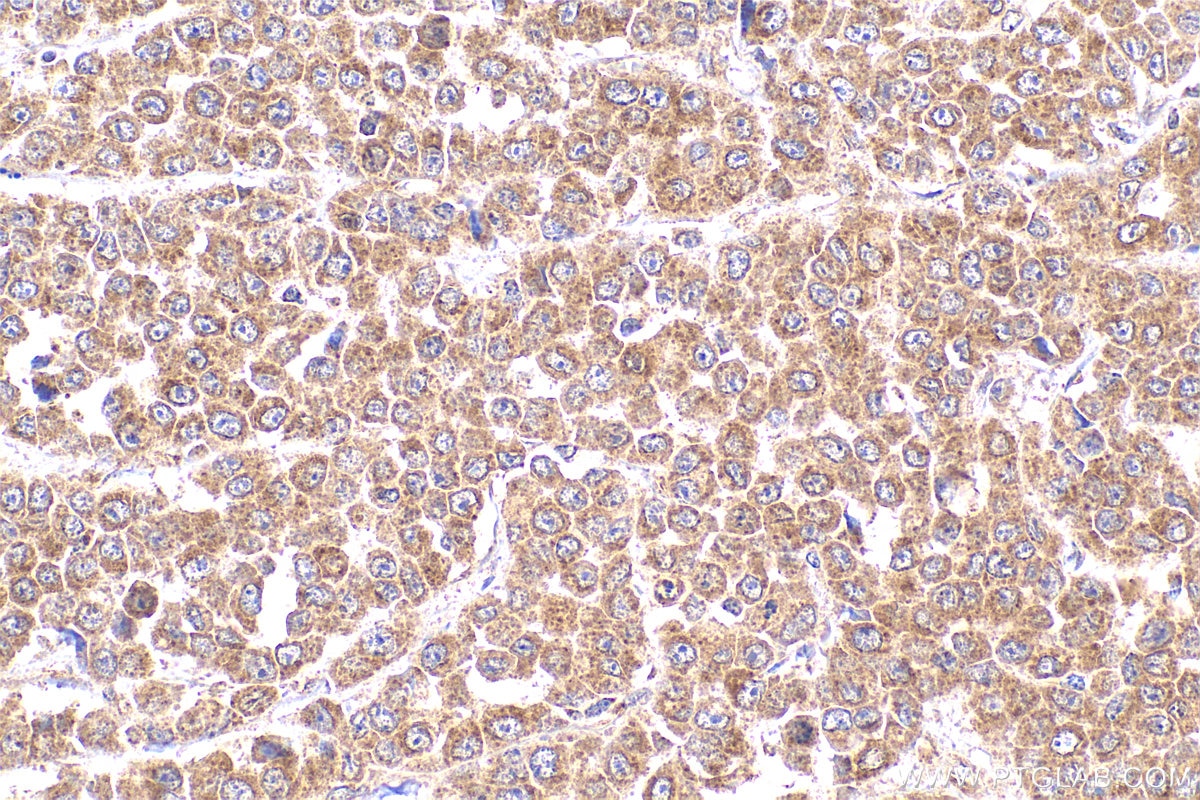
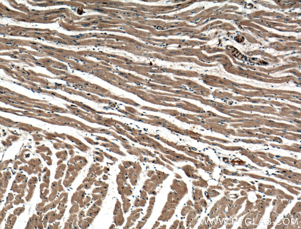
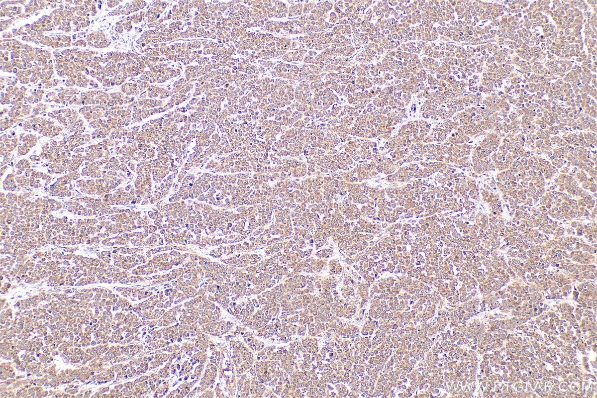
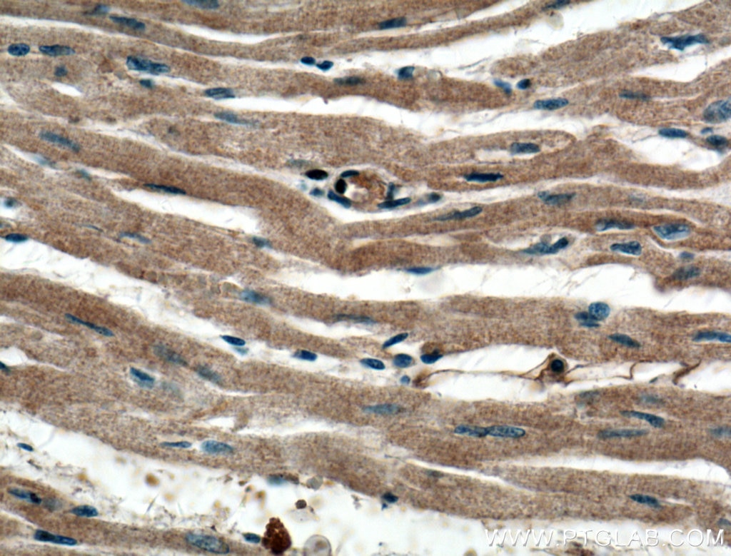
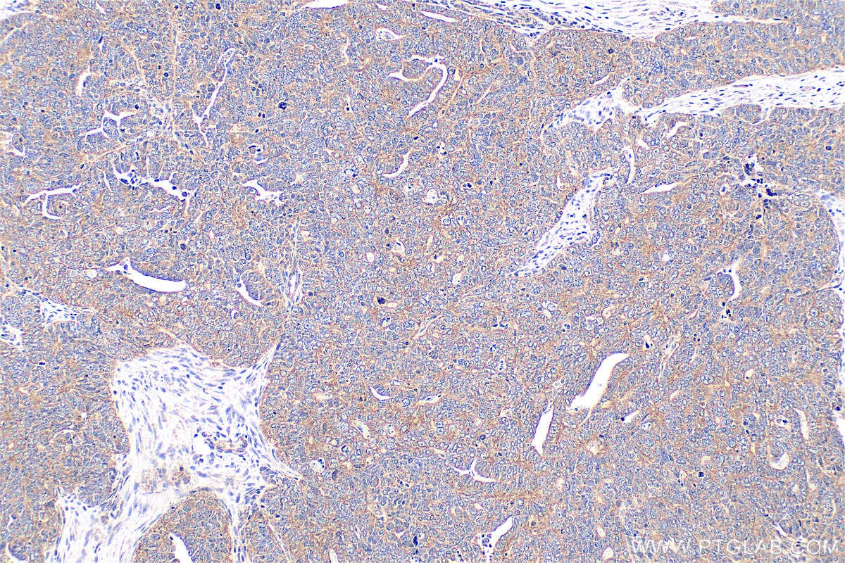
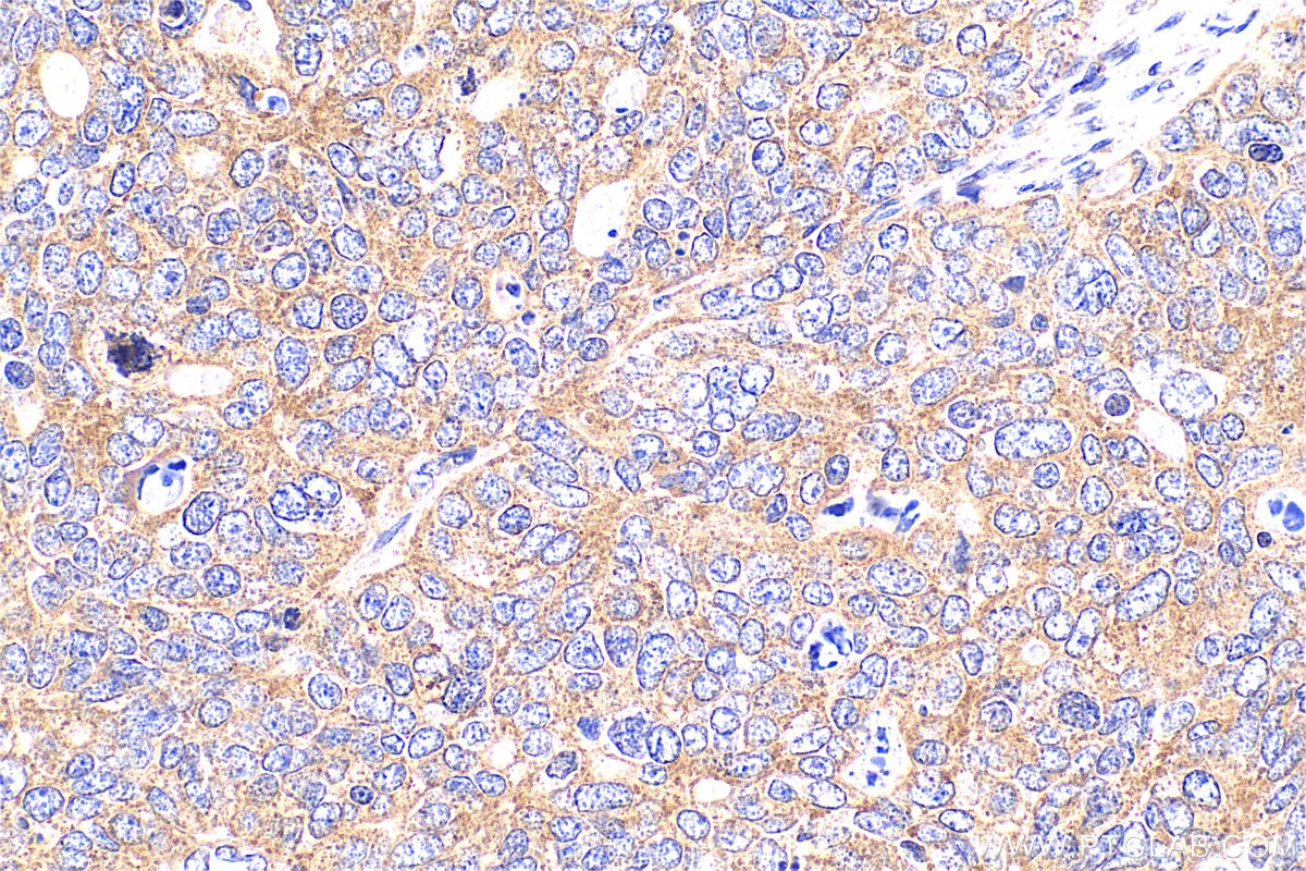
| Positive IHC detected in | human colon cancer tissue, human heart tissue, human ovary tumor tissue Note: suggested antigen retrieval with TE buffer pH 9.0; (*) Alternatively, antigen retrieval may be performed with citrate buffer pH 6.0 |
Recommended dilution
| Application | Dilution |
|---|---|
| Western Blot (WB) | WB : 1:3000-1:10000 |
| Immunoprecipitation (IP) | IP : 0.5-4.0 ug for 1.0-3.0 mg of total protein lysate |
| Immunohistochemistry (IHC) | IHC : 1:50-1:500 |
| It is recommended that this reagent should be titrated in each testing system to obtain optimal results. | |
| Sample-dependent, Check data in validation data gallery. | |
Product Information
22117-1-AP targets MYPT1 in WB, IHC, IF, IP, CoIP, ELISA applications and shows reactivity with human, mouse, rat samples.
| Tested Reactivity | human, mouse, rat |
| Cited Reactivity | human, mouse, rat, pig |
| Host / Isotype | Rabbit / IgG |
| Class | Polyclonal |
| Type | Antibody |
| Immunogen |
CatNo: Ag17496 Product name: Recombinant human MYPT1 protein Source: e coli.-derived, PET28a Tag: 6*His Domain: 707-981 aa of BC111752 Sequence: AEKTIGRSRSTRTREQENEEKEKEEKEKQDKEKQEEKKESETSREDEYKQKYSRTYDETYQRYRPVSTSSSTTPSSSLSTMSSSLYASSQLNRPNSLVGITSAYSRGITKENEREGEKREEEKEGEDKSQPKSIRERRRPREKRRSTGVSFWTQDSDENEQEQQSDTEEGSNKKETQTDSISRYETSSTSAGDRYDSLLGRSGSYSYLEERKPYSSRLEKDDSTDFKKLYEQILAENEKLKAQLHDTNMELTDLKLQLEKATQRQERFADRSLLE Predict reactive species |
| Full Name | protein phosphatase 1, regulatory (inhibitor) subunit 12A |
| Calculated Molecular Weight | 1030 aa, 115 kDa |
| Observed Molecular Weight | 130 kDa |
| GenBank Accession Number | BC111752 |
| Gene Symbol | MYPT1 |
| Gene ID (NCBI) | 4659 |
| RRID | AB_11183753 |
| Conjugate | Unconjugated |
| Form | Liquid |
| Purification Method | Antigen affinity purification |
| UNIPROT ID | O14974 |
| Storage Buffer | PBS with 0.02% sodium azide and 50% glycerol, pH 7.3. |
| Storage Conditions | Store at -20°C. Stable for one year after shipment. Aliquoting is unnecessary for -20oC storage. 20ul sizes contain 0.1% BSA. |
Background Information
Myosin phosphatase target subunit 1(MYPT1), which is also called PPP1R12A, is one of the subunits of myosin phosphatase. Myosin phosphatase regulates the interaction of actin and myosin downstream of the guanosine triphosphatase Rho. The small guanosine triphosphatase Rho is implicated in myosin light chain (MLC) phosphorylation, which results in contraction of smooth muscle and interaction of actin and myosin in nonmuscle cells. The guanosine triphosphate (GTP)-bound, active form of RhoA (GTP.RhoA) specifically interacted with the myosin-binding subunit (MBS) of myosin phosphatase, which regulates the extent of phosphorylation of MLC. Rho-associated kinase (Rho-kinase), which is activated by GTP. RhoA, phosphorylated MBS and consequently inactivated myosin phosphatase. Overexpression of RhoA or activated RhoA in NIH3T3 cells increased phosphorylation of MBS and MLC. Thus, Rho appears to inhibit myosin phosphatase through the action of Rho-kinase. Phosphorylation of MYPT1 at Thr696 and Thr853 results in phosphatase inhibition and cytoskeletal reorganization. Several transcript variants encoding different isoforms have been found for this gene.
Protocols
| Product Specific Protocols | |
|---|---|
| IHC protocol for MYPT1 antibody 22117-1-AP | Download protocol |
| IP protocol for MYPT1 antibody 22117-1-AP | Download protocol |
| WB protocol for MYPT1 antibody 22117-1-AP | Download protocol |
| Standard Protocols | |
|---|---|
| Click here to view our Standard Protocols |
Publications
What published studies show
The MYPT1 antibody (Cat# 22117-1-AP) has 26 citations across journals including EMBO Molecular Medicine, Journal of Experimental & Clinical Cancer Research, Cell Molecular Life Sciences, Communications Biology, Scientific Reports, International Journal of Molecular Science, and ACS Chemical Neuroscience. Research fields span oncology (glioblastoma, hepatocellular carcinoma, ovarian cancer), cardiovascular biology (pulmonary hypertension, vascular remodeling), fibrosis, neurology (ischemic stroke), smooth muscle physiology, and developmental biology. Applications are predominantly Western blot, with IF, IHC, CoIP, and IP also represented.
| Species | Application | Title |
|---|---|---|
J Exp Clin Cancer Res Non-canonical activation of MAPK signaling by the lncRNA ASH1L-AS1-encoded microprotein APPLE through inhibition of PP1/PP2A-mediated ERK1/2 dephosphorylation in hepatocellular carcinoma | ||
Thyroid Morphological and Functional Colonic Defects Caused by a Mutated Thyroid Hormone Receptor α | ||
Cell Mol Life Sci Apurinic/apyrimidinic endodeoxyribonuclease 1 (APE1) promotes stress granule formation via YBX1 phosphorylation in ovarian cancer | ||
Food Funct Chlorogenic acid improves the intestinal barrier by relieving endoplasmic reticulum stress and inhibiting ROCK/MLCK signaling pathways. | ||
Cells Mitotic Acetylation of Microtubules Promotes Centrosomal PLK1 Recruitment and Is Required to Maintain Bipolar Spindle Homeostasis.
|
Reviews
What customers say
Both reviews rate the antibody 5 out of 5 stars. One user successfully used it for Western Blot on mouse bone marrow stromal cells (M2-10B4) at a 1:1000 dilution, detecting a clear band around 120 KDa. Another user applied it for Immunohistochemistry on paraffin-embedded porcine coronary artery sections, also at 1:1000 dilution, and shared their microwave antigen retrieval protocol. Overall, customers report reliable performance across both applications with consistent results.
The reviews below have been submitted by verified Proteintech customers who received an incentive for providing their feedback.
Siddharth (Verified Customer) (07-28-2023) | Good for western blotting. Band detected around 120 KDa.
|
Claudia (Verified Customer) (10-19-2021) | Unmasking microwave: potencia medium, ph6, 10 min., cool 35 min., room temperature.
|












