

Tested Applications
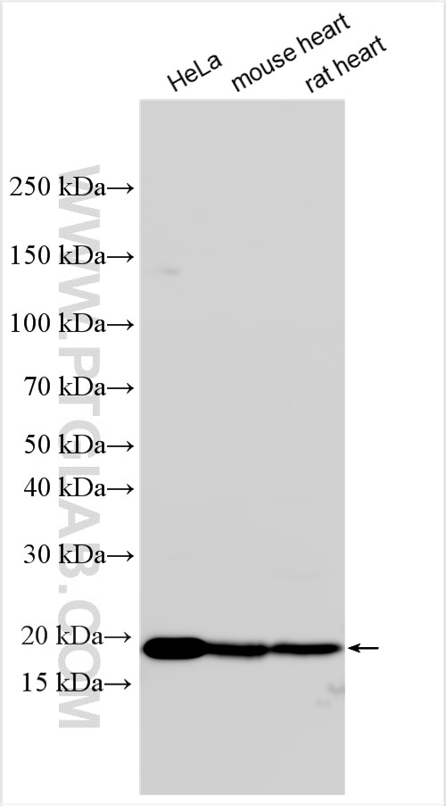
| Positive WB detected in | HeLa cells, mouse heart tissue, rat heart tissue |
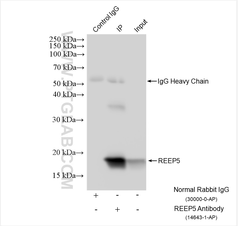
| Positive IP detected in | HeLa cells |
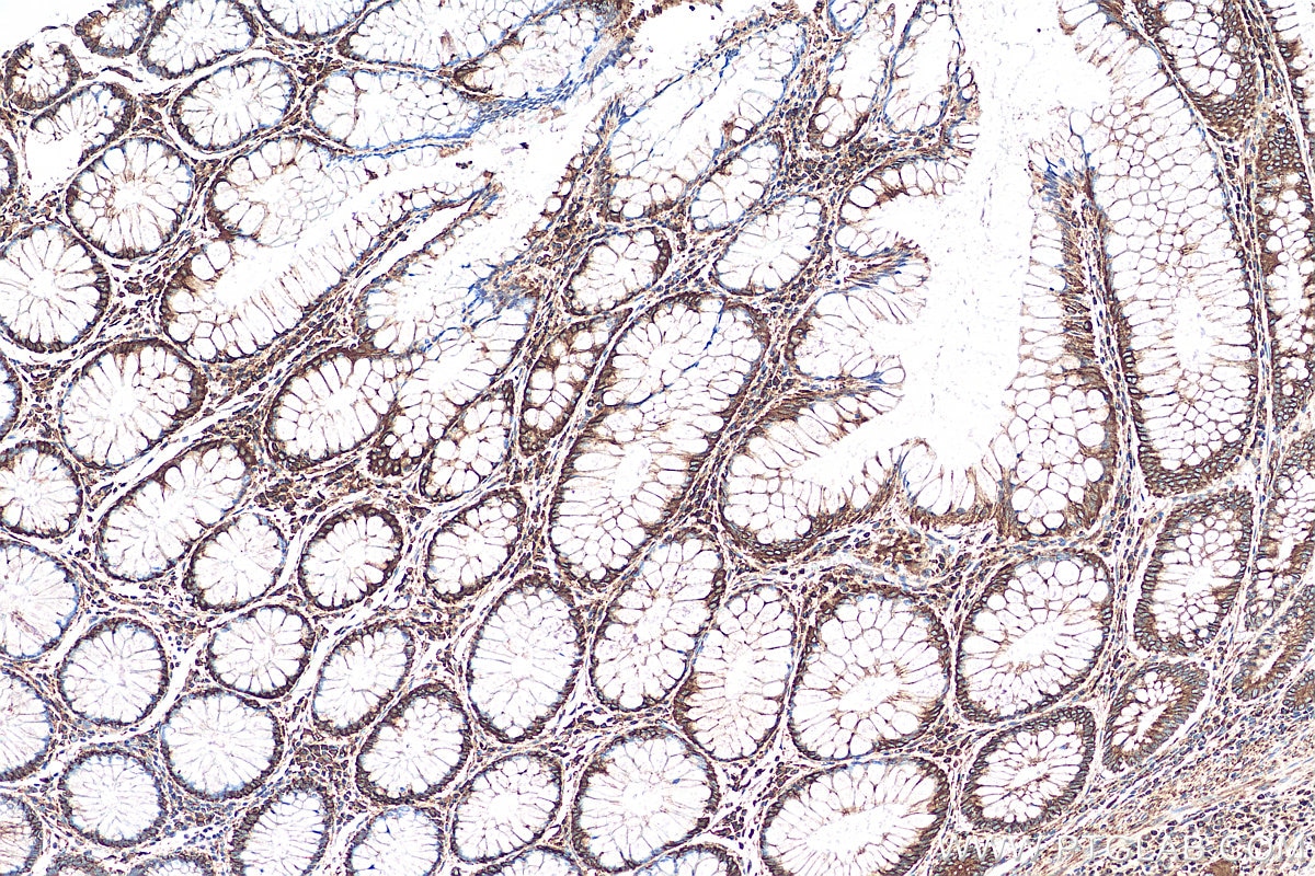
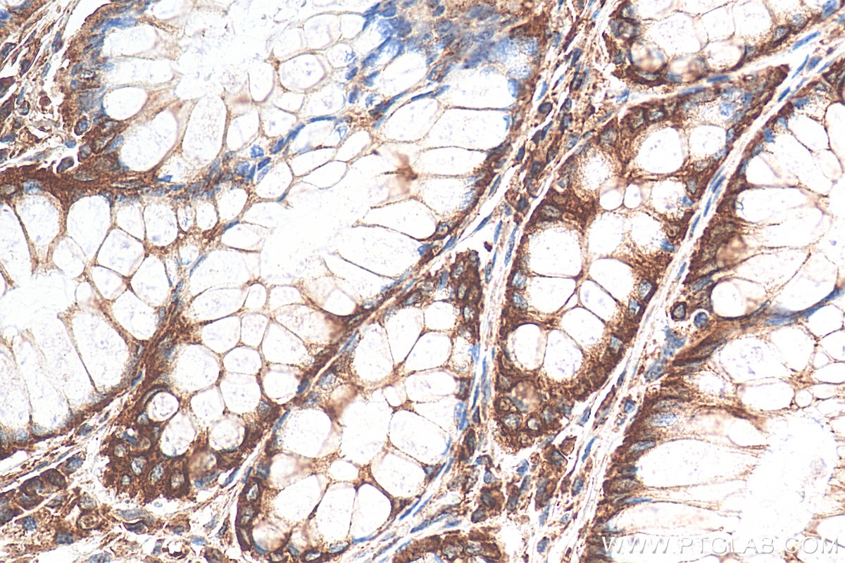
| Positive IHC detected in | human colon cancer tissue Note: suggested antigen retrieval with TE buffer pH 9.0; (*) Alternatively, antigen retrieval may be performed with citrate buffer pH 6.0 |
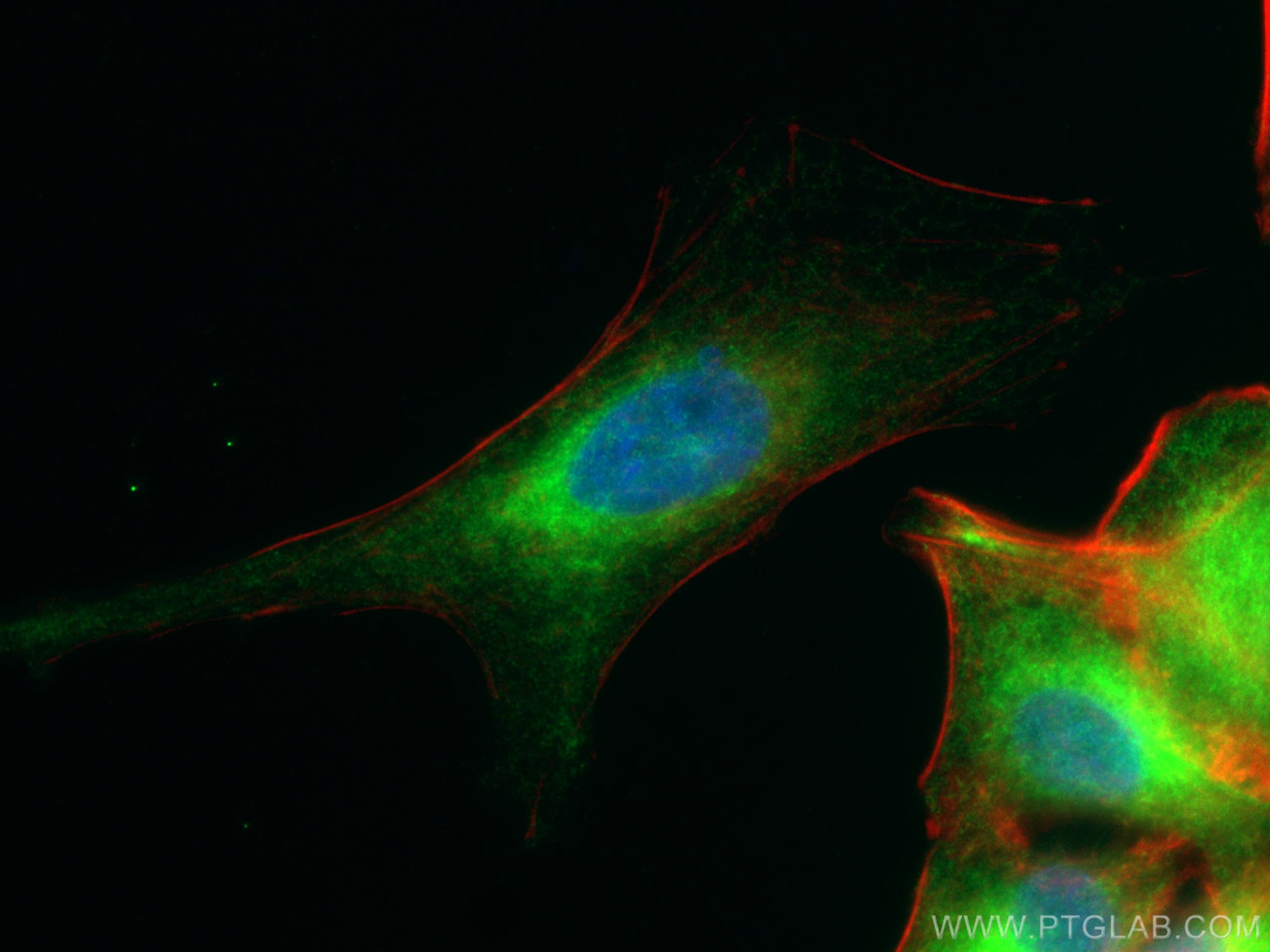
| Positive IF/ICC detected in | A549 cells |
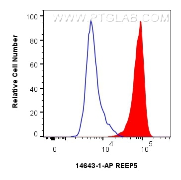
| Positive FC (Intra) detected in | HeLa cells |
Recommended dilution
| Application | Dilution |
|---|---|
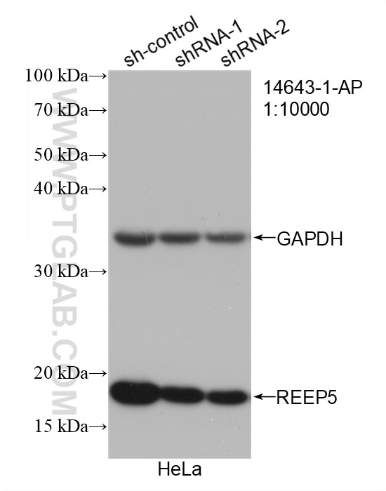
| Western Blot (WB) | WB : 1:5000-1:50000 |
| Immunoprecipitation (IP) | IP : 0.5-4.0 ug for 1.0-3.0 mg of total protein lysate |
| Immunohistochemistry (IHC) | IHC : 1:300-1:1200 |
| Immunofluorescence (IF)/ICC | IF/ICC : 1:50-1:500 |
| Flow Cytometry (FC) (INTRA) | FC (INTRA) : 0.25 ug per 10^6 cells in a 100 ul suspension |
| It is recommended that this reagent should be titrated in each testing system to obtain optimal results. | |
| Sample-dependent, Check data in validation data gallery. | |
Product Information
14643-1-AP targets REEP5 in WB, IHC, IF/ICC, FC (Intra), IP, ELISA applications and shows reactivity with human, mouse, rat samples.
| Tested Reactivity | human, mouse, rat |
| Cited Reactivity | human, mouse, monkey, xenopus |
| Host / Isotype | Rabbit / IgG |
| Class | Polyclonal |
| Type | Antibody |
| Immunogen |
CatNo: Ag6229 Product name: Recombinant human REEP5 protein Source: e coli.-derived, PET28a Tag: 6*His Domain: 1-189 aa of BC065926 Sequence: MSAAMRERFDRFLHEKNCMTDLLAKLEAKTGVNRSFIALGVIGLVALYLVFGYGASLLCNLIGFGYPAYISIKAIESPNKEDDTQWLTYWVVYGVFSIAEFFSDIFLSWFPFYYMLKCGFLLWCMAPSPSNGAELLYKRIIRPFFLKHESQMDSVVKDLKDKAKETADAITKEAKKATVNLLGEEKKST Predict reactive species |
| Full Name | receptor accessory protein 5 |
| Calculated Molecular Weight | 21 kDa |
| Observed Molecular Weight | 18-21 kDa |
| GenBank Accession Number | BC065926 |
| Gene Symbol | REEP5 |
| Gene ID (NCBI) | 7905 |
| RRID | AB_2178440 |
| Conjugate | Unconjugated |
| Form | Liquid |
| Purification Method | Antigen affinity purification |
| UNIPROT ID | Q00765 |
| Storage Buffer | PBS with 0.02% sodium azide and 50% glycerol, pH 7.3. |
| Storage Conditions | Store at -20°C. Stable for one year after shipment. Aliquoting is unnecessary for -20oC storage. 20ul sizes contain 0.1% BSA. |
Background Information
REEP5 (receptor expression-enhancing protein 5), also named as C5orf18, DP1 or TB2, is a membrane protein belonging to DP1/Yop1p protein family. All REEP proteins contain a conserved TB2/DP1, HVA22 domain which is involved in intracellular trafficking and secretion (PMID: 19123125). REEP5 is expressed in circumvallate papillae and testis, and may have the function to influence functional cell surface expression of taste receptors (PMID: 16720576). REEP5 can interact with atlastins (ATL1 and ATL2) which are involved in modulating the structure of ER network (PMID: 19665976).
Protocols
| Product Specific Protocols | |
|---|---|
| FC protocol for REEP5 antibody 14643-1-AP | Download protocol |
| IF protocol for REEP5 antibody 14643-1-AP | Download protocol |
| IHC protocol for REEP5 antibody 14643-1-AP | Download protocol |
| IP protocol for REEP5 antibody 14643-1-AP | Download protocol |
| WB protocol for REEP5 antibody 14643-1-AP | Download protocol |
| Standard Protocols | |
|---|---|
| Click here to view our Standard Protocols |
Publications
What published studies show
The REEP5 Polyclonal antibody (Cat# 14643-1-AP) has 63 citations. These appear in high-impact journals including Nature, Cell, Cell Metabolism, Nature Communications, Nature Cell Biology, Autophagy, Molecular Cell, Developmental Cell, EMBO Journal, Current Biology, Molecular Psychiatry, and eLife. Research fields span ER-phagy and selective autophagy, endoplasmic reticulum remodeling and tubule formation, lipid and lipoprotein metabolism, membrane curvature, and neurodegeneration (Alzheimer's disease, hereditary spastic paraplegia, Parkinson's), as well as cardiomyopathy and ER-mitochondria signaling.
| Species | Application | Title |
|---|---|---|
Signal Transduct Target Ther Targeting intracellular cholesterol imbalance rescues sarcomere-ER contact site signaling and ER remodeling in dilated cardiomyopathy. | ||
Reviews
What customers say
Able is busy answering your previous question. Please try again later.
The reviews below have been submitted by verified Proteintech customers who received an incentive for providing their feedback.
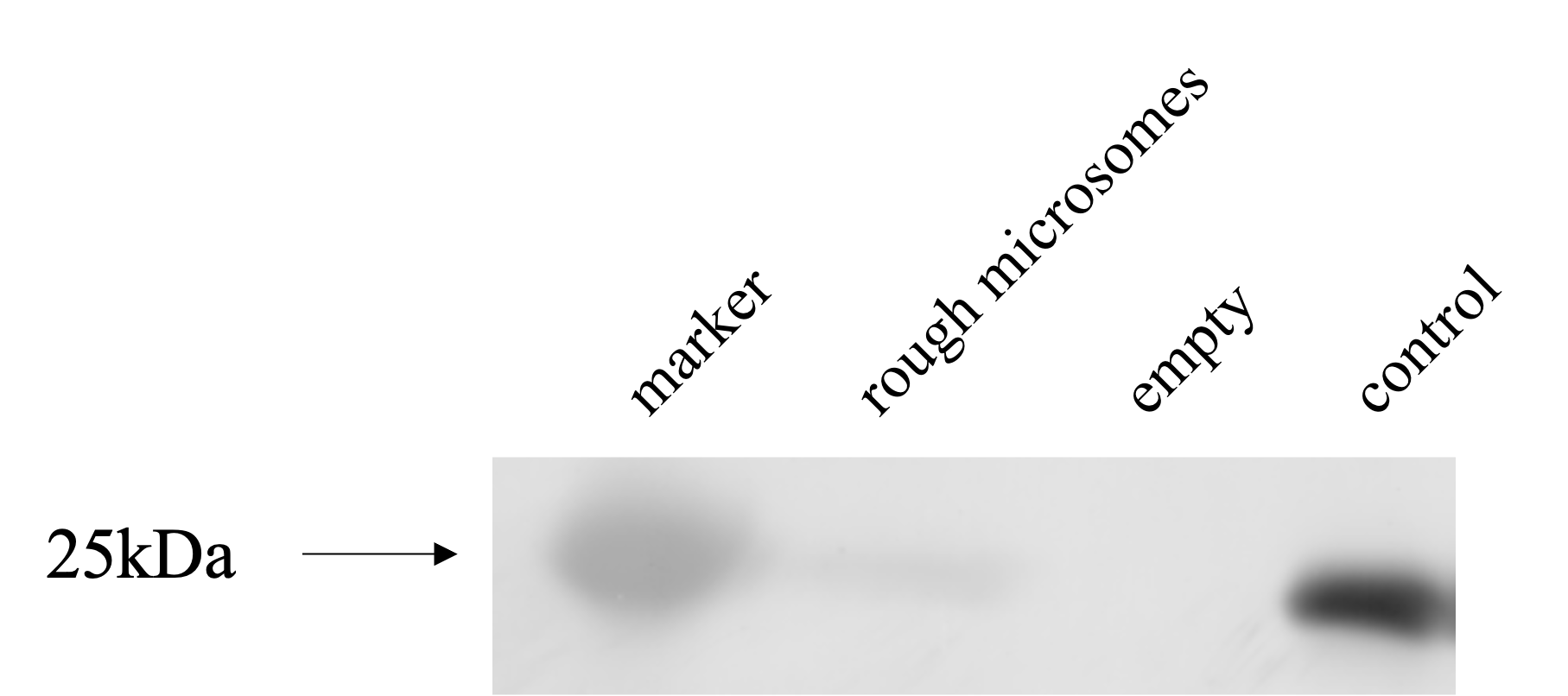
Andrea (Verified Customer) (05-10-2022) | Sharp and strong signal at 21 kDa with clear background.
 |
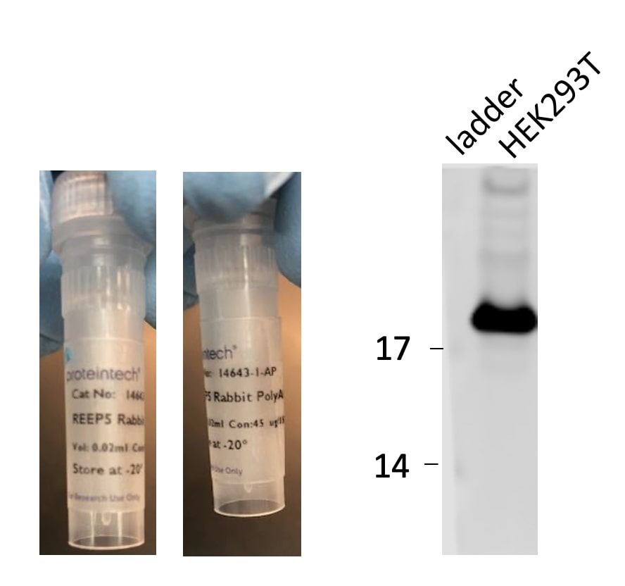
Christine (Verified Customer) (11-10-2021) | Very good detection of endogenous REEP5 protein in human HEK293T and HeLa cell lines. Band at about 20 kDa (expected size for REEP5)
|
Wei (Verified Customer) (09-22-2020) | The antibody gets sharp, single band on western blot.
|
WEI (Verified Customer) (06-29-2020) | The staining pattern is consistent with cardiomyocytes like ER localization.
|
H (Verified Customer) (04-05-2020) | The antibody is very sensitive, such that 1 hour incubation using 1:3000 dilution worked perfectly.
 |








