- Featured Product
- KD/KO Validated
ATP1A1-Specific Polyklonaler Antikörper
ATP1A1-Specific Polyklonal Antikörper für WB, IHC, IF/ICC, FC (Intra), ELISA
Wirt / Isotyp
Kaninchen / IgG
Getestete Reaktivität
human, Maus und mehr (1)
Anwendung
WB, IHC, IF/ICC, FC (Intra), ELISA
Konjugation
Unkonjugiert
Kat-Nr. : 55187-1-AP
Synonyme
 with sh-Control and sh-ATP1A1-Specific transfected HEK-293 cells.")
 at dilution of 1:40000 incubated at room temperature for 1.5 hours.")
 at dilution of 1:4000 incubated at room temperature for 1.5 hours.")
 at dilution of 1:1500 incubated at room temperature for 1.5 hours.")
 at dilution of 1:2000 incubated at room temperature for 1.5 hours.")
 at dilution of 1:500 incubated at room temperature for 1.5 hours.")
 at dilution of 1:800 incubated at room temperature for 1.5 hours.")
 at dilution of 1:2000 incubated at room temperature for 1.5 hours.")
 at dilution of 1:200 (under 40x lens. Heat mediated antigen retrieval with Tris-EDTA buffer (pH 9.0).")
 at dilution of 1:200 (under 10x lens). Heat mediated antigen retrieval with Tris-EDTA buffer (pH 9.0).")
 at dilution of 1:400 (under 10x lens). Heat mediated antigen retrieval with Tris-EDTA buffer (pH 9.0).")
 at dilution of 1:1000 (under 40x lens). Heat mediated antigen retrieval with Tris-EDTA buffer (pH 9.0).")
 at dilution of 1:800 (under 20x lens). Heat mediated antigen retrieval with Tris-EDTA buffer (pH 9.0).")
 at dilution of 1:200 (under 10x lens. Heat mediated antigen retrieval with Tris-EDTA buffer (pH 9.0).")
 at dilution of 1:200 (under 10x lens. Heat mediated antigen retrieval with Tris-EDTA buffer (pH 9.0).")
 at dilution of 1:200 (under 40x lens. Heat mediated antigen retrieval with Tris-EDTA buffer (pH 9.0).")
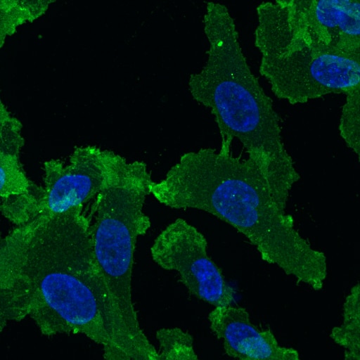
 fixed HEK-293 cells using ATP1A1-Specific antibody (55187-1-AP) at dilution of 1:1000 and CoraLite®488-Conjugated AffiniPure Goat Anti-Rabbit IgG(H+L).")
 fixed Caco-2 cells using 55187-1-AP (ATP1A1-Specific antibody) at dilution of 1:100 and CoraLite488-Conjugated AffiniPure Goat Anti-Rabbit IgG(H+L).")
 and CoraLite®488-Conjugated AffiniPure Goat Anti-Rabbit IgG(H+L) (SA00013-2)(red), or 0.4 ug rabbit IgG isotype control (blue). Cells were fixed with 4% PFA and permeabilized with Flow Cytometry Perm Buffer (PF00011-C).")
"ATP1A1-Specific Antibodies" Comparison
View side-by-side comparison of ATP1A1-Specific antibodies from other vendors to find the one that best suits your research needs.
Geprüfte Anwendungen
| Erfolgreiche Detektion in WB | bei 37°C inkubiertes Maushirngewebe, bei 37°C inkubiertes Mausherzgewebe, HEK-293-Zellen, HepG2-Zellen, MCF-7-Zellen, Neuro-2a-Zellen |
| Erfolgreiche Detektion in IHC | human ovary cancer tissue, humanes Kolongewebe, humanes Nierengewebe, humanes Leberkarzinomgewebe, Mausnierengewebe Hinweis: Antigendemaskierung mit TE-Puffer pH 9,0 empfohlen. (*) Wahlweise kann die Antigendemaskierung auch mit Citratpuffer pH 6,0 erfolgen. |
| Erfolgreiche Detektion in IF/ICC | HEK-293-Zellen, Caco-2-Zellen |
| Erfolgreiche Detektion in FC (Intra) | HEK-293-Zellen |
Empfohlene Verdünnung
| Anwendung | Verdünnung |
|---|---|
| Western Blot (WB) | WB : 1:5000-1:50000 |
| Immunhistochemie (IHC) | IHC : 1:500-1:2000 |
| Immunfluoreszenz (IF)/ICC | IF/ICC : 1:500-1:2000 |
| Durchflusszytometrie (FC) (INTRA) | FC (INTRA) : 0.40 ug per 10^6 cells in a 100 µl suspension |
| It is recommended that this reagent should be titrated in each testing system to obtain optimal results. | |
| Sample-dependent, check data in validation data gallery | |
Veröffentlichte Anwendungen
| WB | See 15 publications below |
| IF | See 7 publications below |
Produktinformation
55187-1-AP bindet in WB, IHC, IF/ICC, FC (Intra), ELISA ATP1A1-Specific und zeigt Reaktivität mit human, Maus
| Getestete Reaktivität | human, Maus |
| In Publikationen genannte Reaktivität | human, Maus, Ratte |
| Wirt / Isotyp | Kaninchen / IgG |
| Klonalität | Polyklonal |
| Typ | Antikörper |
| Immunogen | Peptid |
| Vollständiger Name | ATPase, Na+/K+ transporting, alpha 1 polypeptide |
| Berechnetes Molekulargewicht | 113 kDa |
| Beobachtetes Molekulargewicht | 100-110 kDa |
| GenBank-Zugangsnummer | NM_000701 |
| Gene symbol | ATP1A1 |
| Gene ID (NCBI) | 476 |
| Konjugation | Unkonjugiert |
| Form | Liquid |
| Reinigungsmethode | Antigen-Affinitätsreinigung |
| Lagerungspuffer | PBS with 0.02% sodium azide and 50% glycerol |
| Lagerungsbedingungen | Bei -20°C lagern. Nach dem Versand ein Jahr lang stabil Aliquotieren ist bei -20oC Lagerung nicht notwendig. 20ul Größen enthalten 0,1% BSA. |
Hintergrundinformationen
ATP1A1 is the catalytic component of Na+/K+-ATPase which is a membrane bound enzyme primarily involved in generation of Na+ and K+ gradients across plasma membranes and in determination of cytoplasmic Na+ levels. ATP1A1 is a ubiquitously expressed membrane protein and often used as the marker or internal control for plasma membrane protein. This antibody is specific to ATP1A1.
Protokolle
| PRODUKTSPEZIFISCHE PROTOKOLLE | |
|---|---|
| WB protocol for ATP1A1-Specific antibody 55187-1-AP | Protokoll herunterladen |
| IHC protocol for ATP1A1-Specific antibody 55187-1-AP | Protokoll herunterladenl |
| IF protocol for ATP1A1-Specific antibody 55187-1-AP | Protokoll herunterladen |
| FC protocol for ATP1A1-Specific antibody 55187-1-AP | Download protocol |
| STANDARD-PROTOKOLLE | |
|---|---|
| Klicken Sie hier, um unsere Standardprotokolle anzuzeigen |
Publikationen
| Species | Application | Title |
|---|---|---|
Autophagy Hepatocyte CD36 modulates UBQLN1-mediated proteasomal degradation of autophagic SNARE proteins contributing to septic liver injury | ||
Nat Commun CLICs-dependent chloride efflux is an essential and proximal upstream event for NLRP3 inflammasome activation. | ||
Adv Healthc Mater Effect of Nanoparticle Rigidity on the Interaction of Stromal Membrane Particles with Leukemia Cells | ||
Hypertension Renal Natriuretic Peptide Receptor-C Deficiency Attenuates NaCl Cotransporter Activity in Angiotensin II-Induced Hypertension. | ||
Sci Rep Fluorescence- and magnetic-activated cell sorting strategies to separate spermatozoa involving plural contributors from biological mixtures for human identification. |
Rezensionen
The reviews below have been submitted by verified Proteintech customers who received an incentive for providing their feedback.
FH Paulina (Verified Customer) (03-26-2025) | Fixation: 2% PFA for 20min. Permeabilization/Antibodies dilution solution: 0.05% saponin, 5% horse serum in PBS. ATP1A1 antibody was diluted 1:100 in saponin solution and incubated on cells at room temperature for 1 hour, followed by 1 hour incubation with anti-rabbit conjugated to AlexaFluor 488 (1:200).
 |

