- Phare
- Validé par KD/KO
Anticorps Polyclonal de lapin anti-ATP1A1-Specific
ATP1A1-Specific Polyclonal Antibody for WB, IHC, IF/ICC, FC (Intra), ELISA
Hôte / Isotype
Lapin / IgG
Réactivité testée
Humain, souris et plus (1)
Applications
WB, IHC, IF/ICC, FC (Intra), ELISA
Conjugaison
Non conjugué
N° de cat : 55187-1-AP
Synonymes
 with sh-Control and sh-ATP1A1-Specific transfected HEK-293 cells.")
 at dilution of 1:40000 incubated at room temperature for 1.5 hours.")
 at dilution of 1:4000 incubated at room temperature for 1.5 hours.")
 at dilution of 1:1500 incubated at room temperature for 1.5 hours.")
 at dilution of 1:2000 incubated at room temperature for 1.5 hours.")
 at dilution of 1:500 incubated at room temperature for 1.5 hours.")
 at dilution of 1:800 incubated at room temperature for 1.5 hours.")
 at dilution of 1:2000 incubated at room temperature for 1.5 hours.")
 at dilution of 1:200 (under 40x lens. Heat mediated antigen retrieval with Tris-EDTA buffer (pH 9.0).")
 at dilution of 1:200 (under 10x lens). Heat mediated antigen retrieval with Tris-EDTA buffer (pH 9.0).")
 at dilution of 1:400 (under 10x lens). Heat mediated antigen retrieval with Tris-EDTA buffer (pH 9.0).")
 at dilution of 1:1000 (under 40x lens). Heat mediated antigen retrieval with Tris-EDTA buffer (pH 9.0).")
 at dilution of 1:800 (under 20x lens). Heat mediated antigen retrieval with Tris-EDTA buffer (pH 9.0).")
 at dilution of 1:200 (under 10x lens. Heat mediated antigen retrieval with Tris-EDTA buffer (pH 9.0).")
 at dilution of 1:200 (under 10x lens. Heat mediated antigen retrieval with Tris-EDTA buffer (pH 9.0).")
 at dilution of 1:200 (under 40x lens. Heat mediated antigen retrieval with Tris-EDTA buffer (pH 9.0).")
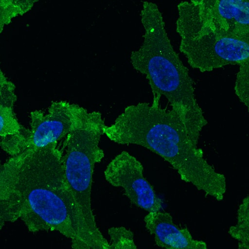
 fixed HEK-293 cells using ATP1A1-Specific antibody (55187-1-AP) at dilution of 1:1000 and CoraLite®488-Conjugated AffiniPure Goat Anti-Rabbit IgG(H+L).")
 fixed Caco-2 cells using 55187-1-AP (ATP1A1-Specific antibody) at dilution of 1:100 and CoraLite488-Conjugated AffiniPure Goat Anti-Rabbit IgG(H+L).")
 and CoraLite®488-Conjugated AffiniPure Goat Anti-Rabbit IgG(H+L) (SA00013-2)(red), or 0.4 ug rabbit IgG isotype control (blue). Cells were fixed with 4% PFA and permeabilized with Flow Cytometry Perm Buffer (PF00011-C).")
"ATP1A1-Specific Antibodies" Comparison
View side-by-side comparison of ATP1A1-Specific antibodies from other vendors to find the one that best suits your research needs.
Applications testées
| Résultats positifs en WB | tissu cérébral de souris incubé à 37 °C, cellules HEK-293, cellules HepG2, cellules MCF-7, cellules Neuro-2a, tissu cardiaque de souris incubé à 37 °C |
| Résultats positifs en IHC | human ovary cancer tissue, tissu de cancer du foie humain, tissu de côlon humain, tissu rénal de souris, tissu rénal humain il est suggéré de démasquer l'antigène avec un tampon de TE buffer pH 9.0; (*) À défaut, 'le démasquage de l'antigène peut être 'effectué avec un tampon citrate pH 6,0. |
| Résultats positifs en IF/ICC | cellules HEK-293, cellules Caco-2 |
| Résultats positifs en FC (Intra) | cellules HEK-293, |
Dilution recommandée
| Application | Dilution |
|---|---|
| Western Blot (WB) | WB : 1:5000-1:50000 |
| Immunohistochimie (IHC) | IHC : 1:500-1:2000 |
| Immunofluorescence (IF)/ICC | IF/ICC : 1:500-1:2000 |
| Flow Cytometry (FC) (INTRA) | FC (INTRA) : 0.40 ug per 10^6 cells in a 100 µl suspension |
| It is recommended that this reagent should be titrated in each testing system to obtain optimal results. | |
| Sample-dependent, check data in validation data gallery | |
Applications publiées
| WB | See 15 publications below |
| IF | See 7 publications below |
Informations sur le produit
55187-1-AP cible ATP1A1-Specific dans les applications de WB, IHC, IF/ICC, FC (Intra), ELISA et montre une réactivité avec des échantillons Humain, souris
| Réactivité | Humain, souris |
| Réactivité citée | rat, Humain, souris |
| Hôte / Isotype | Lapin / IgG |
| Clonalité | Polyclonal |
| Type | Anticorps |
| Immunogène | Peptide |
| Nom complet | ATPase, Na+/K+ transporting, alpha 1 polypeptide |
| Masse moléculaire calculée | 113 kDa |
| Poids moléculaire observé | 100-110 kDa |
| Numéro d’acquisition GenBank | NM_000701 |
| Symbole du gène | ATP1A1 |
| Identification du gène (NCBI) | 476 |
| Conjugaison | Non conjugué |
| Forme | Liquide |
| Méthode de purification | Purification par affinité contre l'antigène |
| Tampon de stockage | PBS with 0.02% sodium azide and 50% glycerol |
| Conditions de stockage | Stocker à -20°C. Stable pendant un an après l'expédition. L'aliquotage n'est pas nécessaire pour le stockage à -20oC Les 20ul contiennent 0,1% de BSA. |
Informations générales
ATP1A1 is the catalytic component of Na+/K+-ATPase which is a membrane bound enzyme primarily involved in generation of Na+ and K+ gradients across plasma membranes and in determination of cytoplasmic Na+ levels. ATP1A1 is a ubiquitously expressed membrane protein and often used as the marker or internal control for plasma membrane protein. This antibody is specific to ATP1A1.
Protocole
| Product Specific Protocols | |
|---|---|
| WB protocol for ATP1A1-Specific antibody 55187-1-AP | Download protocol |
| IHC protocol for ATP1A1-Specific antibody 55187-1-AP | Download protocol |
| IF protocol for ATP1A1-Specific antibody 55187-1-AP | Download protocol |
| FC protocol for ATP1A1-Specific antibody 55187-1-AP | Download protocol |
| Standard Protocols | |
|---|---|
| Click here to view our Standard Protocols |
Publications
| Species | Application | Title |
|---|---|---|
Autophagy Hepatocyte CD36 modulates UBQLN1-mediated proteasomal degradation of autophagic SNARE proteins contributing to septic liver injury | ||
Nat Commun CLICs-dependent chloride efflux is an essential and proximal upstream event for NLRP3 inflammasome activation. | ||
Adv Healthc Mater Effect of Nanoparticle Rigidity on the Interaction of Stromal Membrane Particles with Leukemia Cells | ||
Hypertension Renal Natriuretic Peptide Receptor-C Deficiency Attenuates NaCl Cotransporter Activity in Angiotensin II-Induced Hypertension. | ||
Sci Rep Fluorescence- and magnetic-activated cell sorting strategies to separate spermatozoa involving plural contributors from biological mixtures for human identification. |
Avis
The reviews below have been submitted by verified Proteintech customers who received an incentive for providing their feedback.
FH Paulina (Verified Customer) (03-26-2025) | Fixation: 2% PFA for 20min. Permeabilization/Antibodies dilution solution: 0.05% saponin, 5% horse serum in PBS. ATP1A1 antibody was diluted 1:100 in saponin solution and incubated on cells at room temperature for 1 hour, followed by 1 hour incubation with anti-rabbit conjugated to AlexaFluor 488 (1:200).
 |

