Recombinant Human L-selectin/CD62L protein (rFc Tag)
Species
Human
Purity
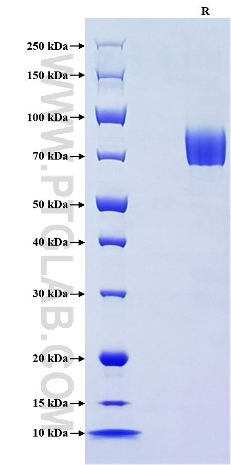
>90 %, SDS-PAGE
Tag
rFc Tag
Activity
not tested
Cat no : Eg3006
Validation Data Gallery
Product Information
| Purity | >90 %, SDS-PAGE |
| Endotoxin | <0.1 EU/μg protein, LAL method |
| Activity |
Not tested |
| Expression | HEK293-derived Human L-selectin protein Trp39-Asn332 (Accession# P14151-1) with a rabbit IgG Fc tag at the C-terminus. |
| GeneID | 6402 |
| Accession | P14151-1 |
| PredictedSize | 59.1 kDa |
| SDS-PAGE | 65-90 kDa, reducing (R) conditions |
| Formulation | Lyophilized from 0.22 μm filtered solution in PBS, pH 7.4. Normally 5% trehalose and 5% mannitol are added as protectants before lyophilization. |
| Reconstitution | Briefly centrifuge the tube before opening. Reconstitute at 0.1-0.5 mg/mL in sterile water. |
| Storage Conditions |
It is recommended that the protein be aliquoted for optimal storage. Avoid repeated freeze-thaw cycles.
|
| Shipping | The product is shipped at ambient temperature. Upon receipt, store it immediately at the recommended temperature. |
Background
CD62L, also known as L-selectin or SELL, is a member of the selectin family of adhesion molecules that also include CD62E (E-selectin) and CD62P (P-selectin). CD62L is a highly glycosylated protein of 95-105 kDa on neutrophils and 74 kDa on lymphocytes. CD62L is expressed on the surface of most leukocytes, including lymphocytes, neutrophils, monocytes, eosinophils, hematopoietic progenitor cells, and immature thymocytes. It mediates the binding of lymphocytes to high endothelial venules (HEV) of peripheral lymph nodes through interactions with a constitutively expressed ligand, and is also involved in lymphocyte, neutrophil, and monocyte attachment to endothelium at sites of inflammation.
References:
1.Bowen BR. et al. (1989) J Cell Biol. 109(1):421-427.
2.Tedder TF. et al. (1989) J Exp Med. 170(1):123-133.
3.Griffin JD. et al. (1990) J Immunol. 145(2):576-584.
4.Tedder TF. et al. (1990) Eur J Immunol. 20(6):1351-1355.
5.Tedder TF. et al. (1990) J Immunol. 144(2):532-540.
6.Schleiffenbaum B. et al. (1992) J Cell Biol. 119(1):229-238.

