Recombinant Human SPINT2/HAI-2 protein (rFc Tag)
Species
Human
Purity
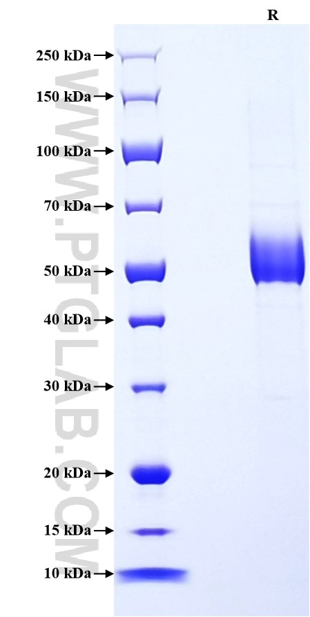
>90 %, SDS-PAGE
Tag
rFc Tag
Activity
not tested
Cat no : Eg3130
Validation Data Gallery
Product Information
| Purity | >90 %, SDS-PAGE |
| Endotoxin | <0.1 EU/μg protein, LAL method |
| Activity |
Not tested |
| Expression | HEK293-derived Human SPINT2 protein Ala28-Lys197 (Accession# O43291-1) with a rabbit IgG Fc tag at the C-terminus. |
| GeneID | 10653 |
| Accession | O43291-1 |
| PredictedSize | 45.2 kDa |
| SDS-PAGE | 48-60 kDa, reducing (R) conditions |
| Formulation | Lyophilized from 0.22 μm filtered solution in PBS, pH 7.4. Normally 5% trehalose and 5% mannitol are added as protectants before lyophilization. |
| Reconstitution | Briefly centrifuge the tube before opening. Reconstitute at 0.1-0.5 mg/mL in sterile water. |
| Storage Conditions |
It is recommended that the protein be aliquoted for optimal storage. Avoid repeated freeze-thaw cycles.
|
| Shipping | The product is shipped at ambient temperature. Upon receipt, store it immediately at the recommended temperature. |
Background
SPINT2 (serine peptidase inhibitor Kunitz type 2), also known as hepatocyte growth factor activator inhibitor type-2(10), belongs to the Kunitz family of serine protease inhibitors, which is a plasma membrane-localized serine protease inhibitor found in epithelial cells of various tissues including the respiratory tract and all major organs. SPINT2 possesses one transmembrane domain and two kunitz-type inhibitor domains that are exposed to the extracellular space and which are believed to facilitate a potent inhibition of target proteases. SPINT2 regulates cell proliferation, migration and invasion via a reduction in HGFA, leading to apoptosis and necrosis of uterine leiomyosarcoma cells.
References:
1. Li J, et al. (2023). Exp Ther Med. Oct 9;26(6):546.
2. Straus, M.R., et al. (2020). Virology. Apr;543:43-53.
3. Heinz-Erian P, et al. (2009). Am J Hum Genet. 84(2):188-196.
4. Pereira, M.S., et al. (2016). J Histochem Cytochem. Jan;64(1):32-41.

