Tested Applications
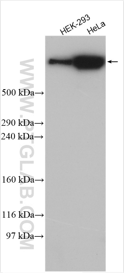
| Positive WB detected in | HEK-293 cells, HeLa cells |
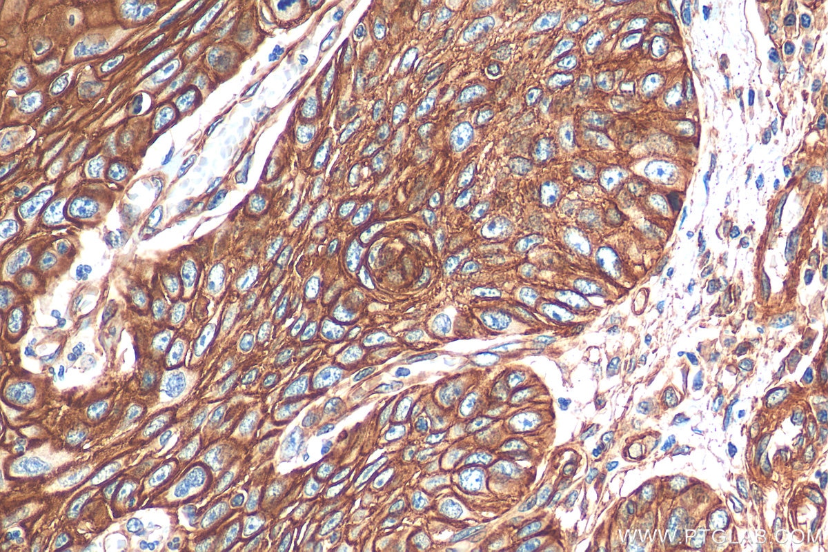
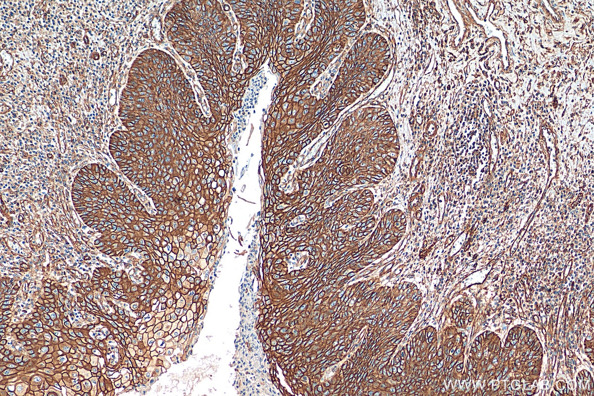
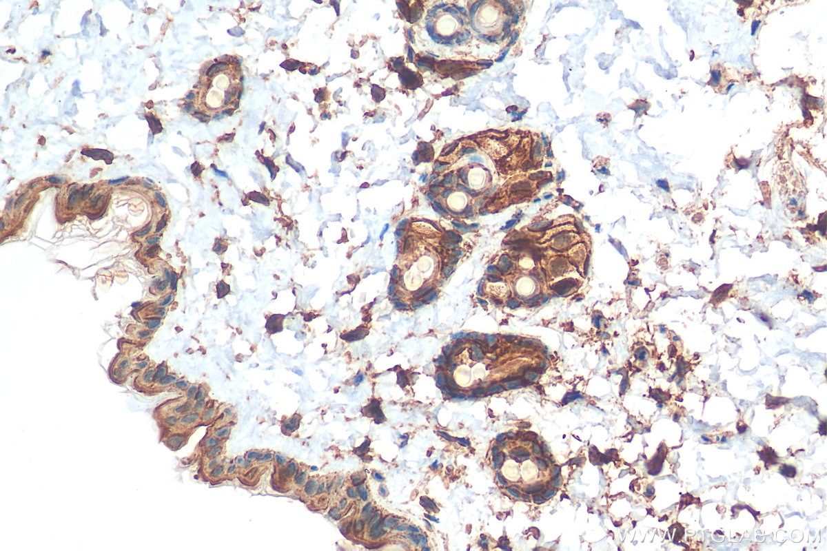
| Positive IHC detected in | human oesophagus cancer tissue, mouse skin tissue Note: suggested antigen retrieval with TE buffer pH 9.0; (*) Alternatively, antigen retrieval may be performed with citrate buffer pH 6.0 |
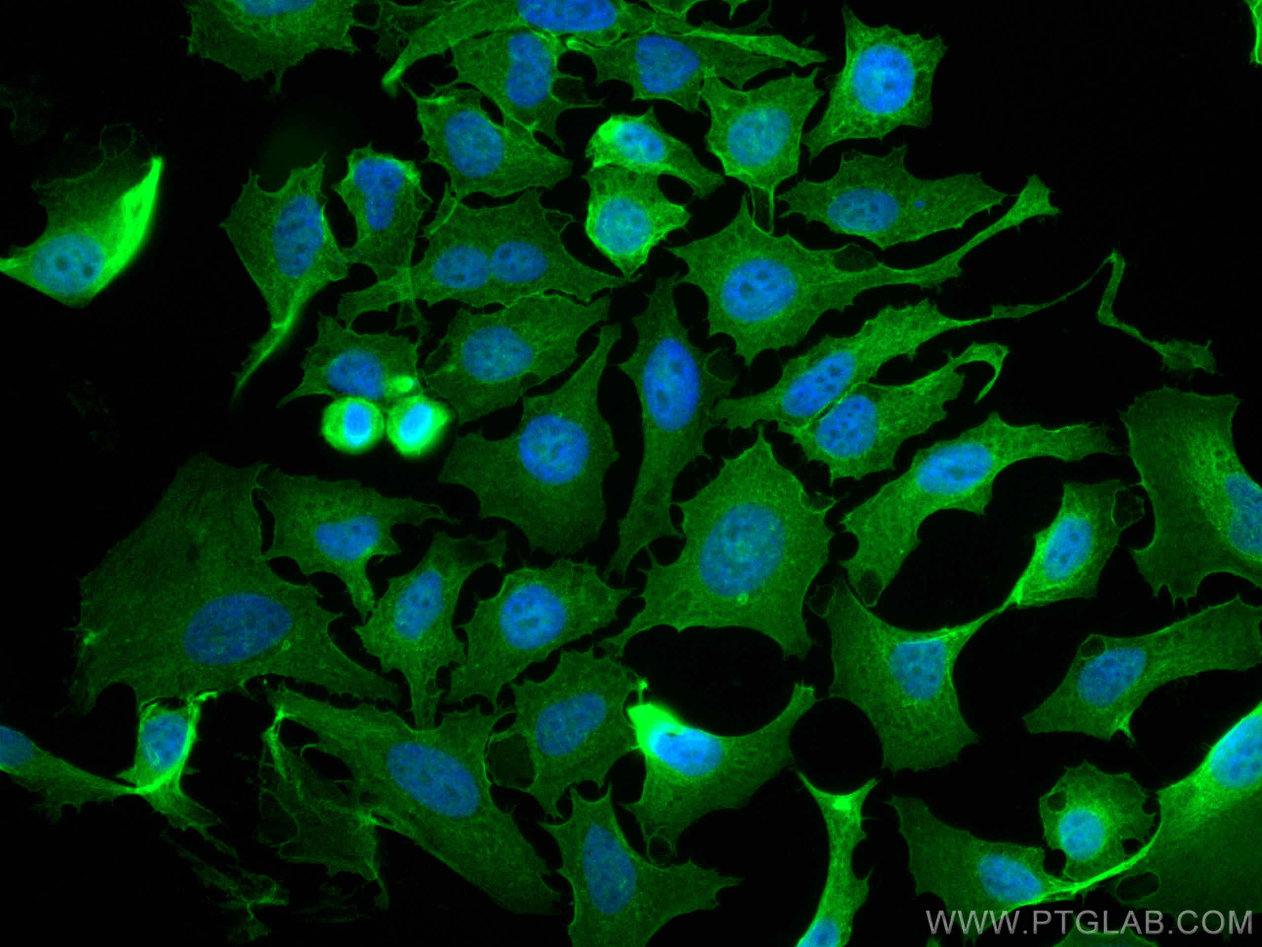
| Positive IF/ICC detected in | HeLa cells |
Recommended dilution
| Application | Dilution |
|---|---|
| Western Blot (WB) | WB : 1:1000-1:4000 |
| Immunohistochemistry (IHC) | IHC : 1:50-1:500 |
| Immunofluorescence (IF)/ICC | IF/ICC : 1:50-1:500 |
| It is recommended that this reagent should be titrated in each testing system to obtain optimal results. | |
| Sample-dependent, Check data in validation data gallery. | |
Published Applications
| IF | See 3 publications below |
| FC | See 1 publications below |
Product Information
16637-1-AP targets AHNAK in WB, IHC, IF/ICC, ELISA applications and shows reactivity with human, mouse samples.
| Tested Reactivity | human, mouse |
| Cited Reactivity | human |
| Host / Isotype | Rabbit / IgG |
| Class | Polyclonal |
| Type | Antibody |
| Immunogen |
CatNo: Ag9986 Product name: Recombinant human AHNAK protein Source: e coli.-derived, PGEX-4T Tag: GST Domain: 1-149 aa of BC000926 Sequence: MEKEEETTRELLLPNWQGSGSHGLTIAQRDDGVFVQEVTQNSPAARTGVVKEGDQIVGATIYFDNLQSGEVTQLLNTMGHHTVGLKLHRKGDRSPEPGQTWTREVFSSCSSEVVLNTPQPSALECKDQNKQKEASSQAGAVSVSTPNAG Predict reactive species |
| Full Name | AHNAK nucleoprotein |
| Calculated Molecular Weight | 629 kDa |
| GenBank Accession Number | BC000926 |
| Gene Symbol | AHNAK |
| Gene ID (NCBI) | 79026 |
| ENSEMBL Gene ID | ENSG00000124942 |
| RRID | AB_2878291 |
| Conjugate | Unconjugated |
| Form | Liquid |
| Purification Method | Antigen affinity purification |
| UNIPROT ID | Q09666 |
| Storage Buffer | PBS with 0.02% sodium azide and 50% glycerol, pH 7.3. |
| Storage Conditions | Store at -20°C. Stable for one year after shipment. Aliquoting is unnecessary for -20oC storage. 20ul sizes contain 0.1% BSA. |
Background Information
AHNAK, also known as desmoyokin, is described as a giant scaffold protein based on its large size (629 kDa) and ability to interact with different proteins to form multi-protein complexes. The bulk of the protein is assembled in 128-residue repetitive elements known as the central repeated unit (CRU). Its proposed functions are quite diverse, ranging from a role in the formation of the blood brain barrier, in cell architecture and migration, to the regulation of cardiac channels and muscle membrane repair. AHNAK is differentially expressed in some cancer cell lines. The expression of AHNAK is subsequently localized to the plasma membrane of keratinocytes in human epidermis. AHNAK has been reported in many intracellular locations including the nucleus, cytoplasm and plasma membrane.
Protocols
| Product Specific Protocols | |
|---|---|
| IF protocol for AHNAK antibody 16637-1-AP | Download protocol |
| IHC protocol for AHNAK antibody 16637-1-AP | Download protocol |
| WB protocol for AHNAK antibody 16637-1-AP | Download protocol |
| Standard Protocols | |
|---|---|
| Click here to view our Standard Protocols |
Publications
| Species | Application | Title |
|---|---|---|
Nat Commun Innate-like self-reactive B cells infiltrate human renal allografts during transplant rejection. | ||
EMBO Rep Microautophagy regulated by STK38 and GABARAPs is essential to repair lysosomes and prevent aging | ||
Clin Exp Immunol The AHNAK induces increased IL-6 production in CD4+T cells and serves as a potential diagnostic biomarker for recurrent pregnancy loss. | ||
Transl Lung Cancer Res Tumor-educated platelet RNA as a diagnostic biomarker for ground-glass opacity-related lung adenocarcinoma | ||
J Thromb Haemost Single-cell transcriptomic analysis reveals distinct plasma cell populations in chronic thromboembolic pulmonary hypertension | ||
JCI Insight Lactate programs CRIP1 protein lactylation to drive synovial proliferation in rheumatoid arthritis. |