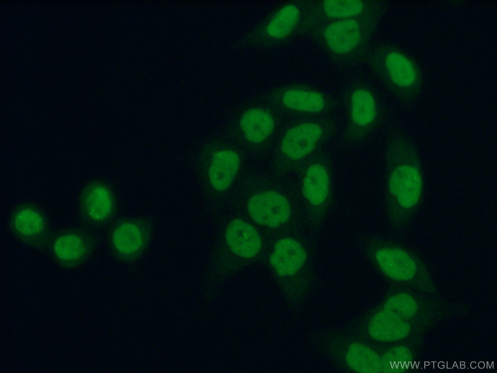
Tested Applications
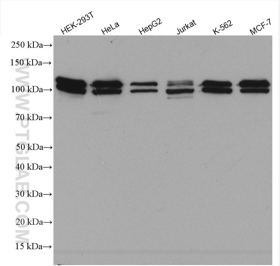
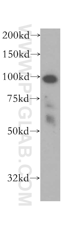
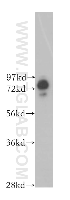
| Positive WB detected in | HEK-293T cells, HeLa cells, COLO 320 cells, HepG2 cells, Jurkat cells, K-562 cells, MCF-7 cells |
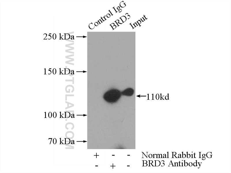
| Positive IP detected in | HeLa cells |
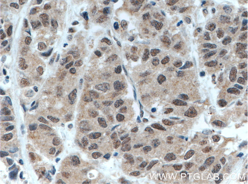
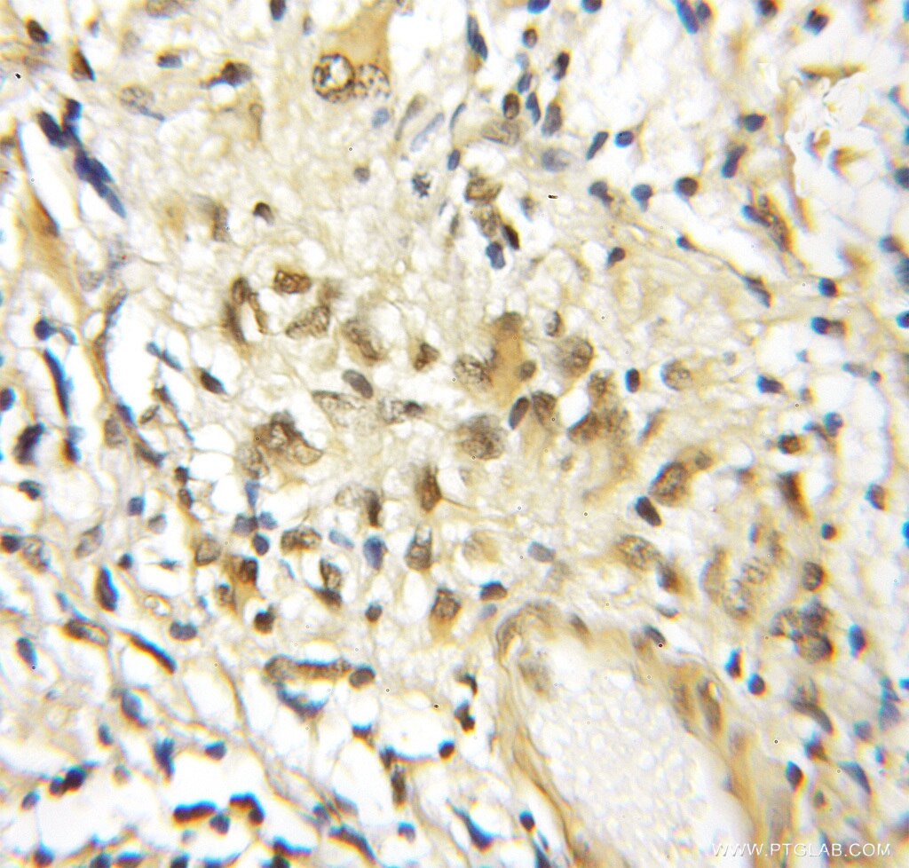
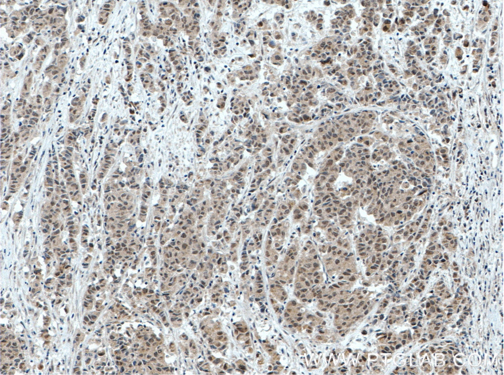
| Positive IHC detected in | human prostate cancer tissue, human gliomas tissue Note: suggested antigen retrieval with TE buffer pH 9.0; (*) Alternatively, antigen retrieval may be performed with citrate buffer pH 6.0 |
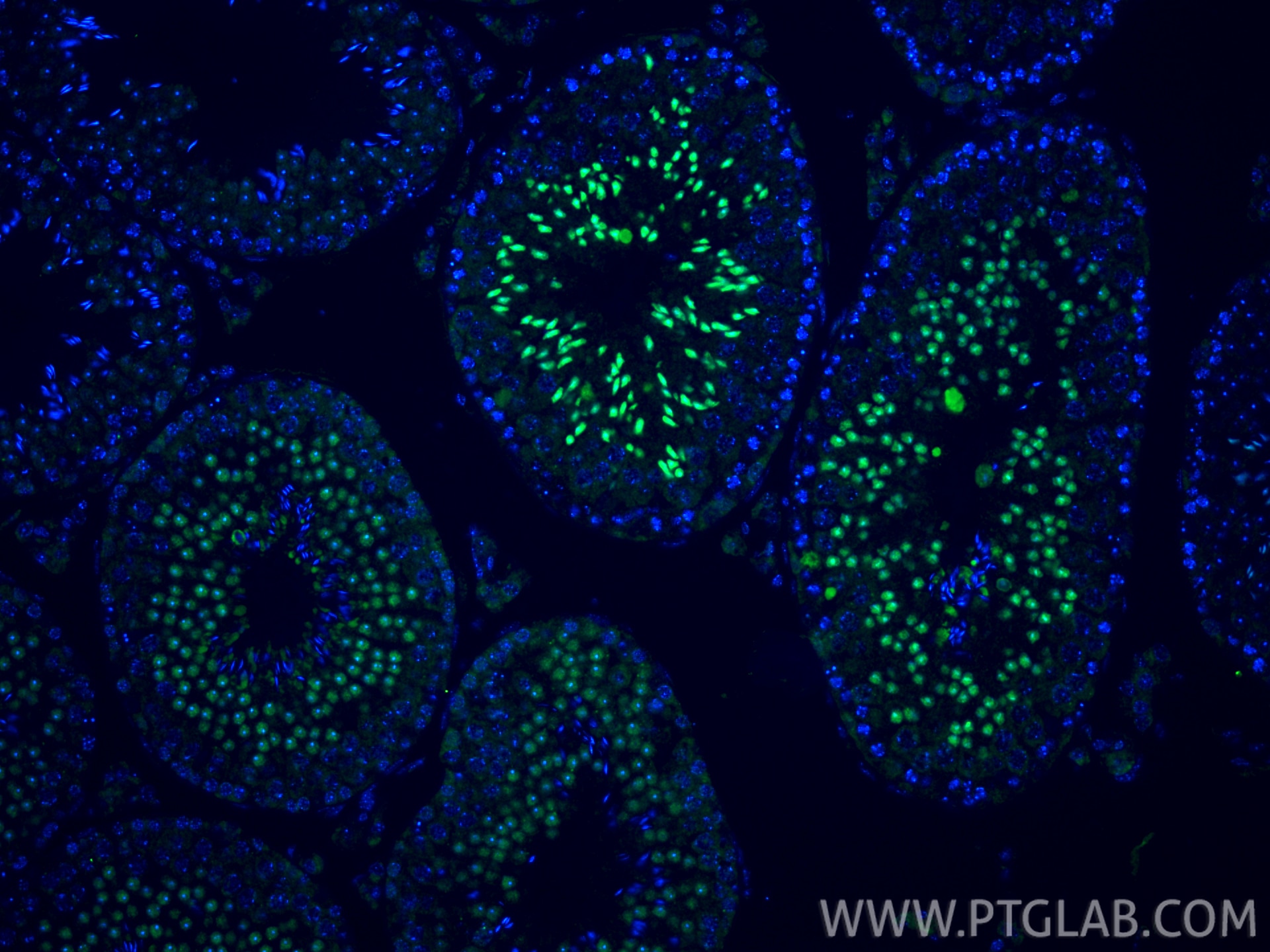
| Positive IF-P detected in | mouse testis tissue |
| Positive IF/ICC detected in | HeLa cells |
Recommended dilution
| Application | Dilution |
|---|---|
| Western Blot (WB) | WB : 1:500-1:1000 |
| Immunoprecipitation (IP) | IP : 0.5-4.0 ug for 1.0-3.0 mg of total protein lysate |
| Immunohistochemistry (IHC) | IHC : 1:50-1:500 |
| Immunofluorescence (IF)-P | IF-P : 1:200-1:800 |
| Immunofluorescence (IF)/ICC | IF/ICC : 1:50-1:500 |
| It is recommended that this reagent should be titrated in each testing system to obtain optimal results. | |
| Sample-dependent, Check data in validation data gallery. | |
Published Applications
| KD/KO | See 2 publications below |
| WB | See 28 publications below |
| IHC | See 2 publications below |
Product Information
11859-1-AP targets BRD3 in WB, IHC, IF/ICC, IF-P, IP, ELISA applications and shows reactivity with human samples.
| Tested Reactivity | human |
| Cited Reactivity | human, mouse |
| Host / Isotype | Rabbit / IgG |
| Class | Polyclonal |
| Type | Antibody |
| Immunogen |
CatNo: Ag2433 Product name: Recombinant human BRD3 protein Source: e coli.-derived, PGEX-4T Tag: GST Domain: 1-400 aa of BC032124 Sequence: MSTATTVAPAGIPATPGPVNPPPPEVSNPSKPGRKTNQLQYMQNVVVKTLWKHQFAWPFYQPVDAIKLNLPDYHKIIKNPMDMGTIKKRLENNYYWSASECMQDFNTMFTNCYIYNKPTDDIVLMAQALEKIFLQKVAQMPQEEVELLPPAPKGKGRKPAAGAQSAGTQQVAAVSSVSPATPFQSVPPTVSQTPVIAATPVPTITANVTSVPVPPAAAPPPPATPIVPVVPPTPPVVKKKGVKRKADTTTPTTSAITASRSESPPPLSDPKQAKVVARRESGGRPIKPPKKDLEDGEVPQHAGKKGKLSEHLRYCDSILREMLSKKHAAYAWPFYKPVDAEALELHDYHDIIKHPMDLSTVKRKMDGREYPDAQGFAADVRLMFSNCYKYNPPDHEVVAM Predict reactive species |
| Full Name | bromodomain containing 3 |
| Calculated Molecular Weight | 556 aa, 61 kDa |
| Observed Molecular Weight | 95-105 kDa |
| GenBank Accession Number | BC032124 |
| Gene Symbol | BRD3 |
| Gene ID (NCBI) | 8019 |
| RRID | AB_2065902 |
| Conjugate | Unconjugated |
| Form | Liquid |
| Purification Method | Antigen affinity purification |
| UNIPROT ID | Q15059 |
| Storage Buffer | PBS with 0.02% sodium azide and 50% glycerol, pH 7.3. |
| Storage Conditions | Store at -20°C. Stable for one year after shipment. Aliquoting is unnecessary for -20oC storage. 20ul sizes contain 0.1% BSA. |
Background Information
BRD3 was belongs to the BET family that a small group of unique, highly conserved bromodomain-containing genes with four members in mouse and human. There are BRD2, BRD3, BRD4 and BRDT. And BET family proteins all localize in the nucleus, and contain two tandems N-terminal BRDs, an extra-terminal (ET) domain and a more divergent C-terminal recruitment domain (PMID:25849938). BRD2, BRD3, and BRD4 play key roles in transcription of genes required for both inflammation and cancer. Northern hybridization revealed that BRD3 is most abundant in testis, ovary, placenta, uterus, and brain(PMID:15261828; PMID:27451123). The antibody could recognize the common about 100 kDa protein in applications as the papers (PMID:29437854; 22595521).
Protocols
| Product Specific Protocols | |
|---|---|
| IF protocol for BRD3 antibody 11859-1-AP | Download protocol |
| IHC protocol for BRD3 antibody 11859-1-AP | Download protocol |
| IP protocol for BRD3 antibody 11859-1-AP | Download protocol |
| WB protocol for BRD3 antibody 11859-1-AP | Download protocol |
| Standard Protocols | |
|---|---|
| Click here to view our Standard Protocols |
Publications
| Species | Application | Title |
|---|---|---|
Nat Med Prostate cancer-associated SPOP mutations confer resistance to BET inhibitors through stabilization of BRD4. | ||
Mol Cell A di-acetyl-decorated chromatin signature couples liquid condensation to suppress DNA end synapsis | ||
Nat Commun A proximity biotinylation-based approach to identify protein-E3 ligase interactions induced by PROTACs and molecular glues. | ||
Nat Commun Stromal induction of BRD4 phosphorylation Results in Chromatin Remodeling and BET inhibitor Resistance in Colorectal Cancer. |
Reviews
The reviews below have been submitted by verified Proteintech customers who received an incentive for providing their feedback.
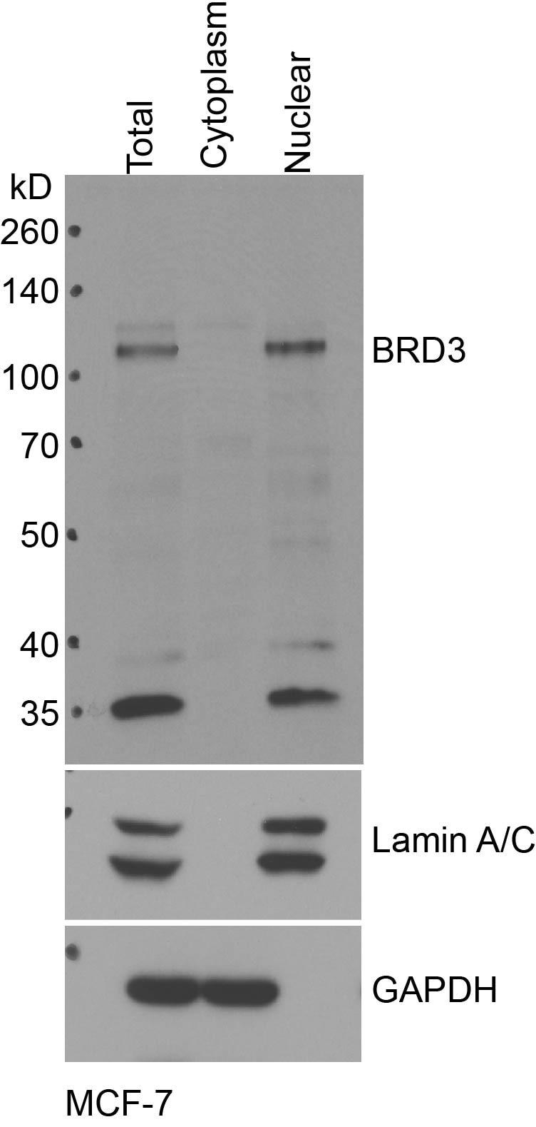
FH Zehua (Verified Customer) (02-06-2019) | Works well on fractionation lysates of MCF-7 cells.
 |

