Tested Applications
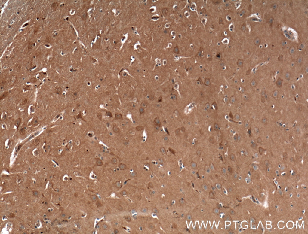
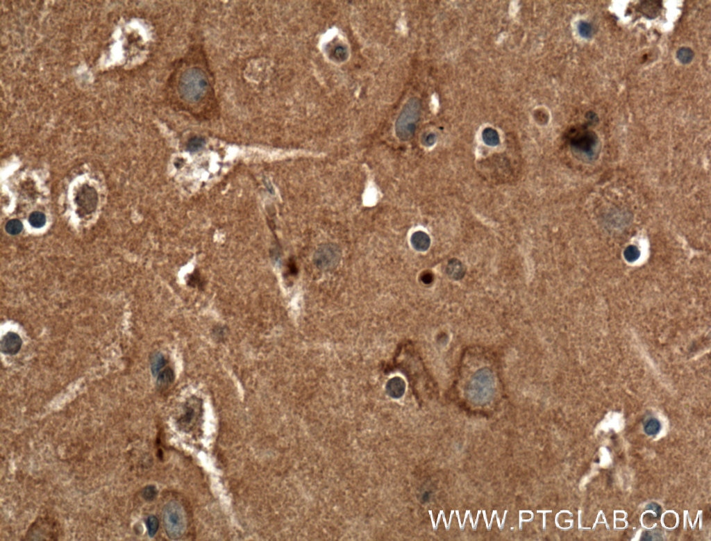
| Positive IHC detected in | human brain tissue Note: suggested antigen retrieval with TE buffer pH 9.0; (*) Alternatively, antigen retrieval may be performed with citrate buffer pH 6.0 |
Recommended dilution
| Application | Dilution |
|---|---|
| Immunohistochemistry (IHC) | IHC : 1:50-1:500 |
| It is recommended that this reagent should be titrated in each testing system to obtain optimal results. | |
| Sample-dependent, Check data in validation data gallery. | |
Product Information
27227-1-AP targets CACNA1A in IHC, ELISA applications and shows reactivity with human samples.
| Tested Reactivity | human |
| Host / Isotype | Rabbit / IgG |
| Class | Polyclonal |
| Type | Antibody |
| Immunogen |
CatNo: Ag25943 Product name: Recombinant human CACNA1A protein Source: e coli.-derived, PGEX-4T Tag: GST Domain: 821-920 aa of NM_000068 Sequence: MDRPLVVDPQENRNNNTNKSRAAEPTVDQRLGQQRAEDFLRKQARYHDRARDPSGSAGLDARRPWAGSQEAELSREGPYGRESDHHAREGSLEQPGFWEGE Predict reactive species |
| Full Name | calcium channel, voltage-dependent, P/Q type, alpha 1A subunit |
| Calculated Molecular Weight | 282 kDa |
| GenBank Accession Number | NM_000068 |
| Gene Symbol | CACNA1A |
| Gene ID (NCBI) | 773 |
| RRID | AB_2880810 |
| Conjugate | Unconjugated |
| Form | Liquid |
| Purification Method | Antigen affinity purification |
| UNIPROT ID | O00555 |
| Storage Buffer | PBS with 0.02% sodium azide and 50% glycerol, pH 7.3. |
| Storage Conditions | Store at -20°C. Stable for one year after shipment. Aliquoting is unnecessary for -20oC storage. 20ul sizes contain 0.1% BSA. |
Protocols
| Product Specific Protocols | |
|---|---|
| IHC protocol for CACNA1A antibody 27227-1-AP | Download protocol |
| Standard Protocols | |
|---|---|
| Click here to view our Standard Protocols |
Reviews
The reviews below have been submitted by verified Proteintech customers who received an incentive for providing their feedback.
FH Lea (Verified Customer) (10-08-2024) | I transfected HEK 293T cells with 2 different CACNA1A constructs (12Q and 27Q, 2 replicates each) and an empty vector as a negative control. The expected band at ~280 kDa is clearly visible in the western blot and no unspecific bands are visible in the negative control. I was very happy with the results.
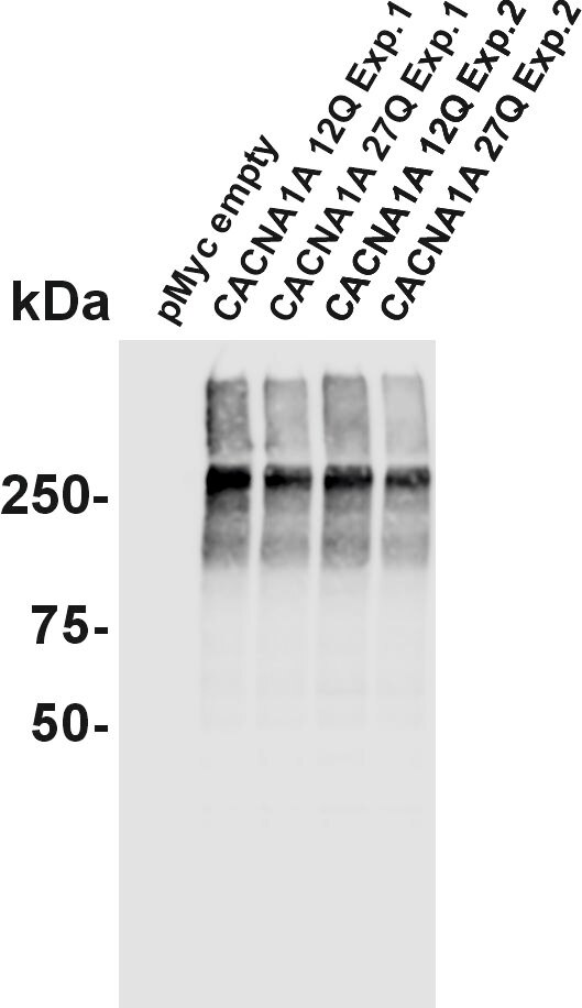 |
FH Ümit (Verified Customer) (08-11-2022) | Cav2.1 (black) staining of two rhesus monkey brainstem sections (5µm &7µm) visualized with immunoperoxidase method (DAB-Nickel).
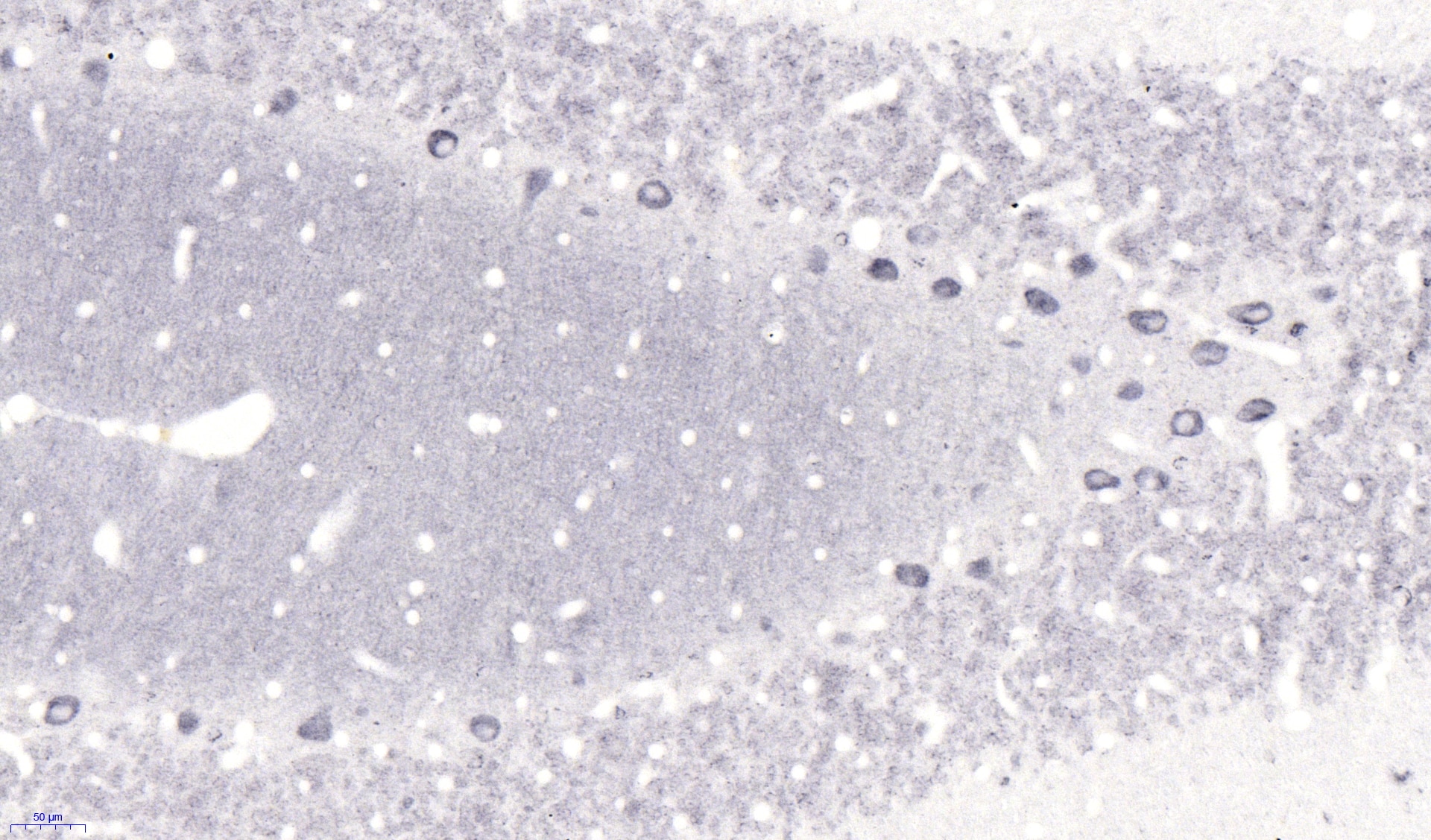 |