Tested Applications
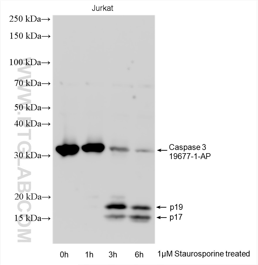
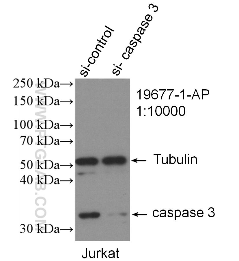
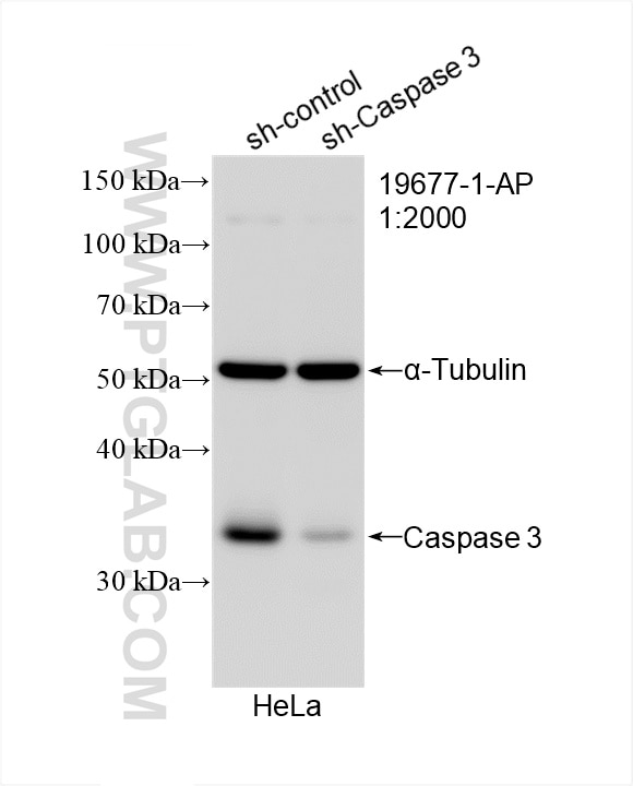
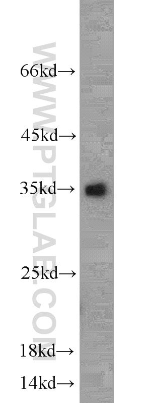
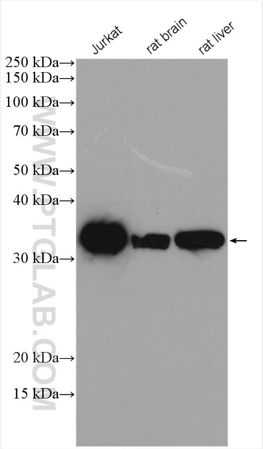
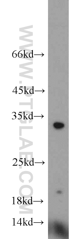
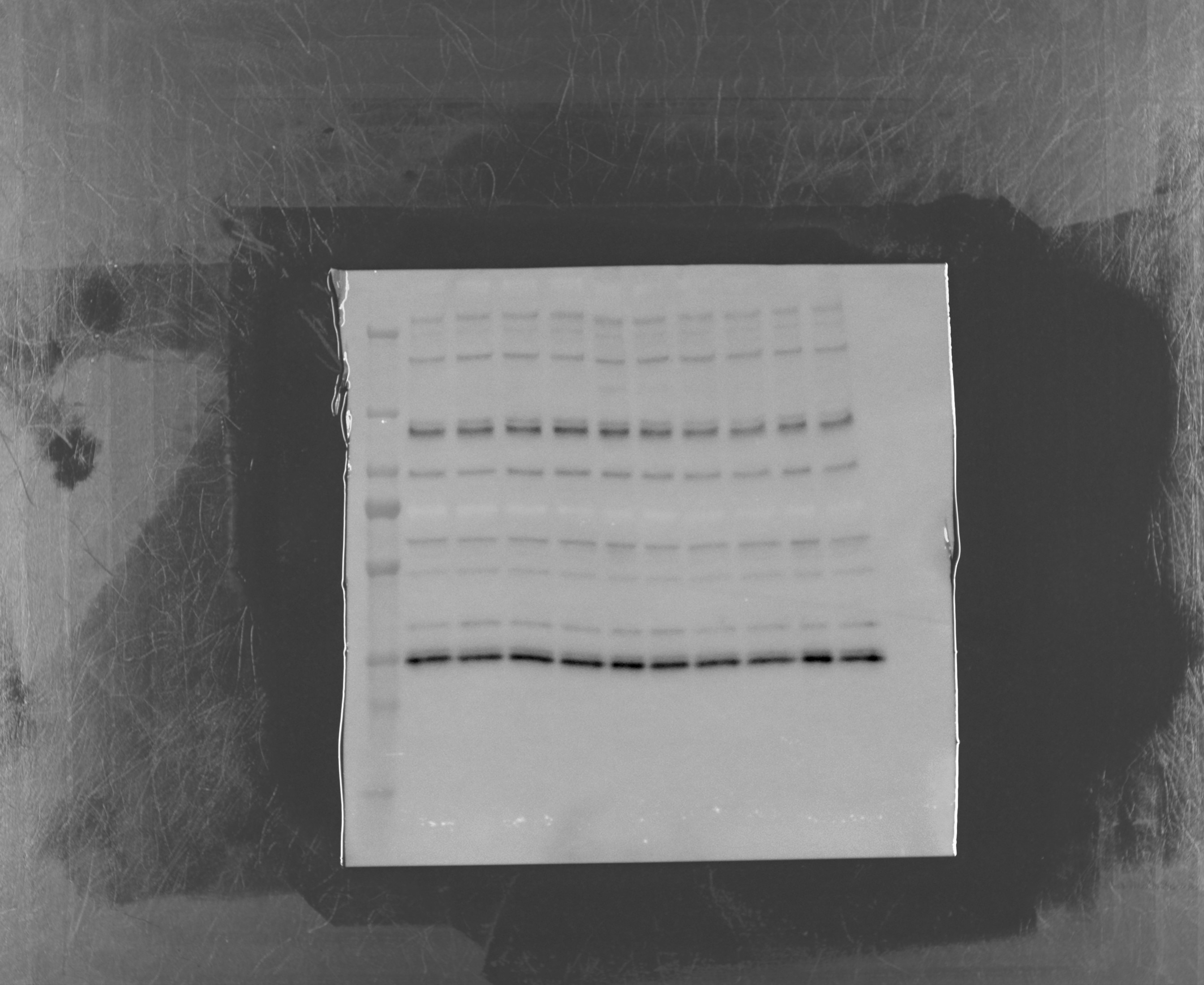
| Positive WB detected in | Jurkat cells, HeLa cells, mouse spleen tissue, Staurosporine treated Jurkat cells, rat brain tissue, rat liver tissue |
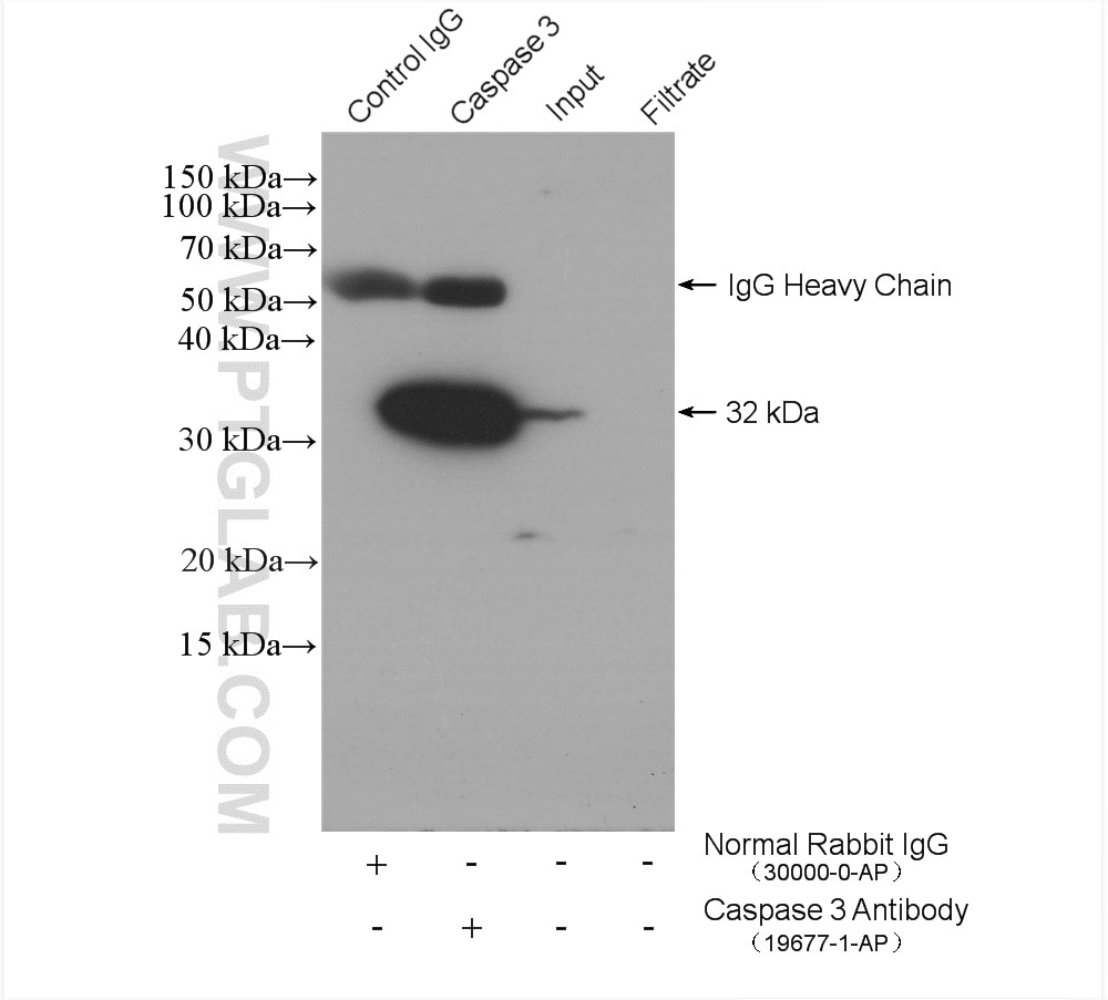
| Positive IP detected in | NIH/3T3 cells |
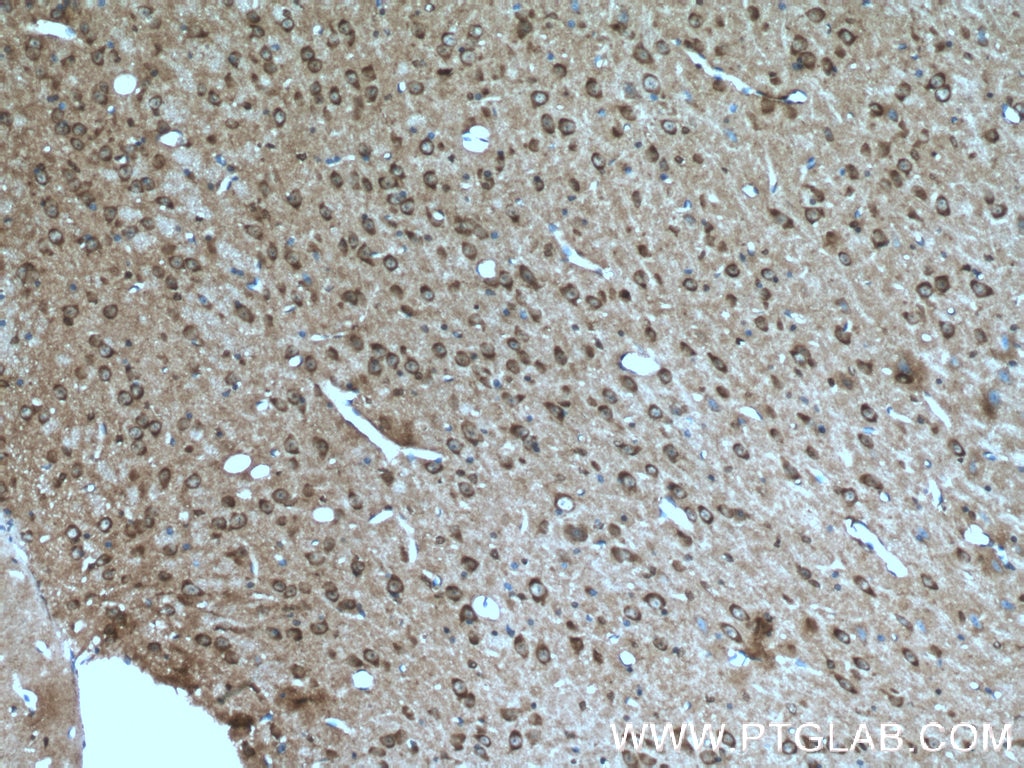
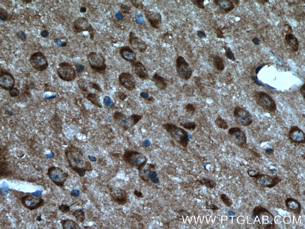
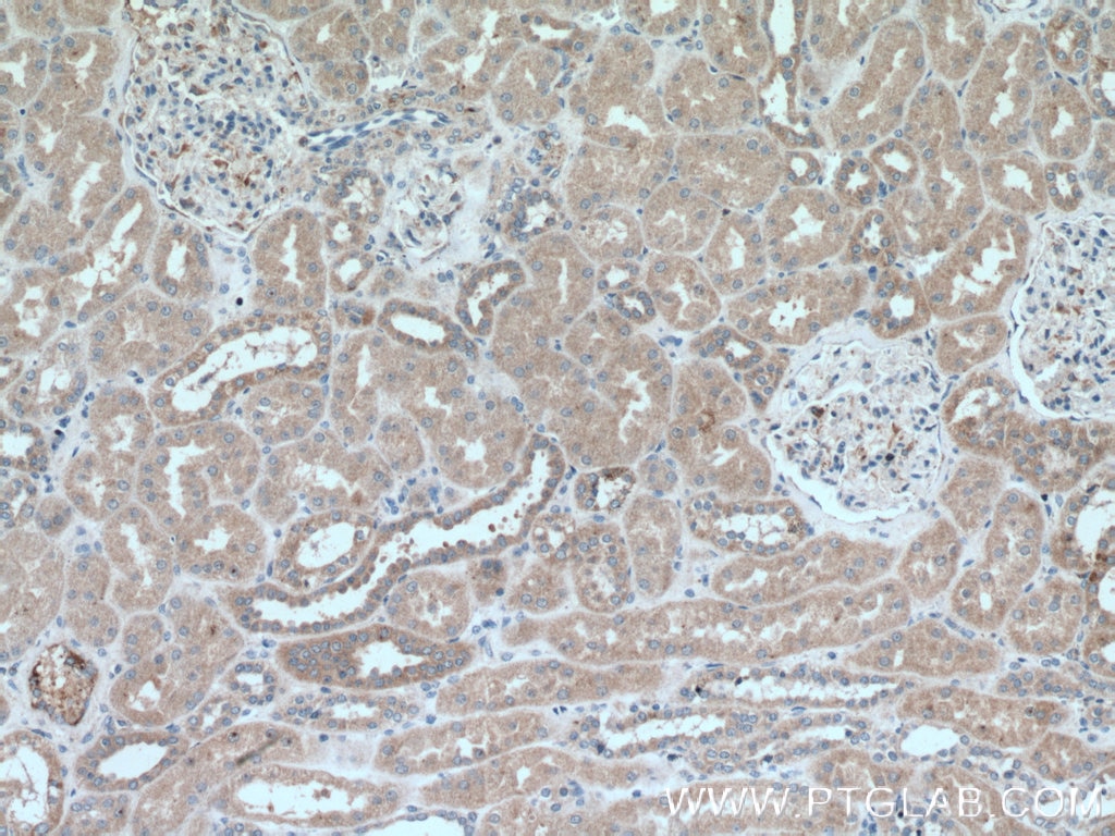
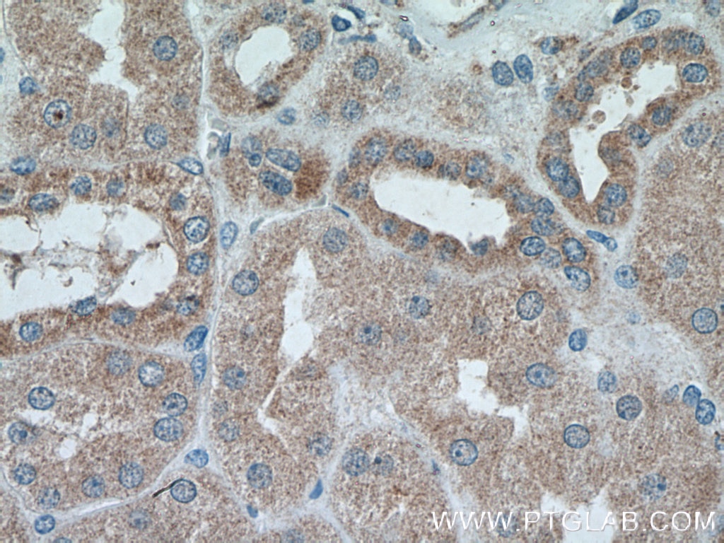
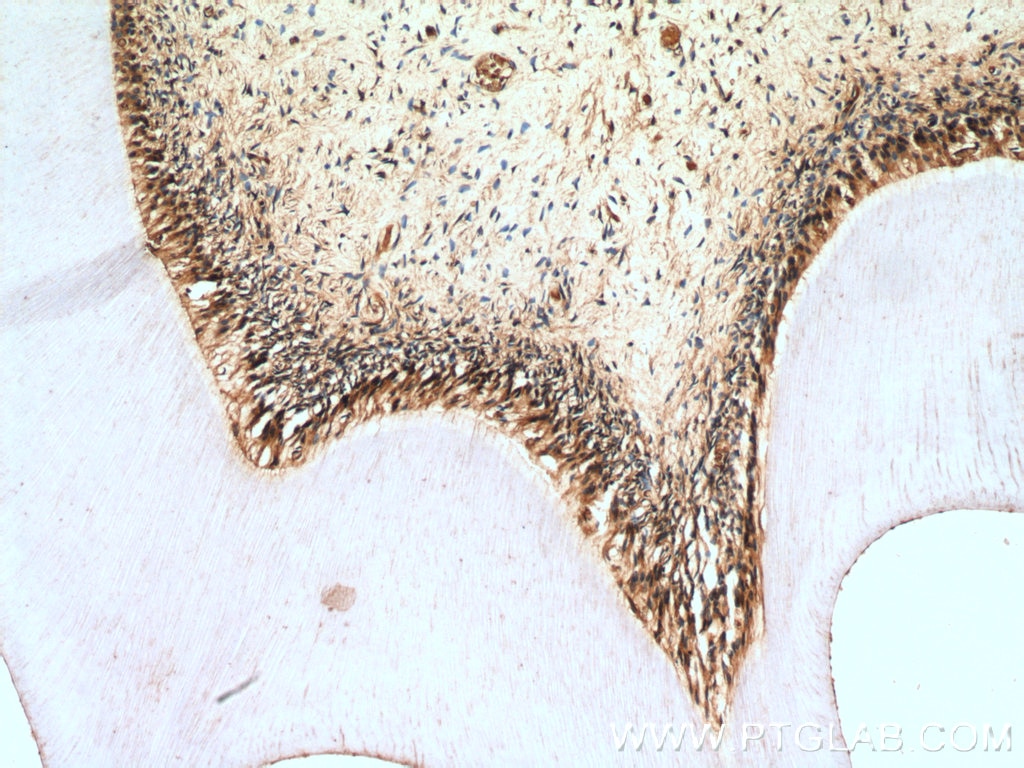
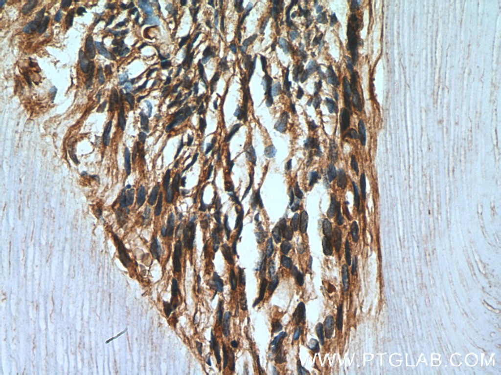
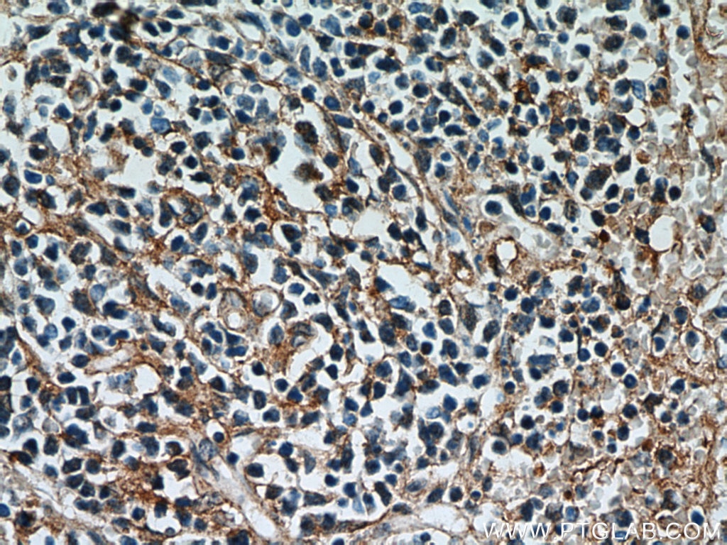
| Positive IHC detected in | mouse brain tissue, human teeth tissue, human spleen tissue, human kidney tissue Note: suggested antigen retrieval with TE buffer pH 9.0; (*) Alternatively, antigen retrieval may be performed with citrate buffer pH 6.0 |
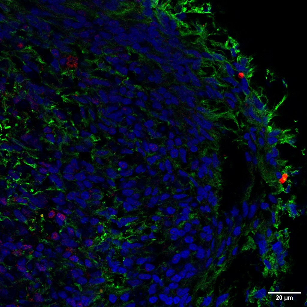
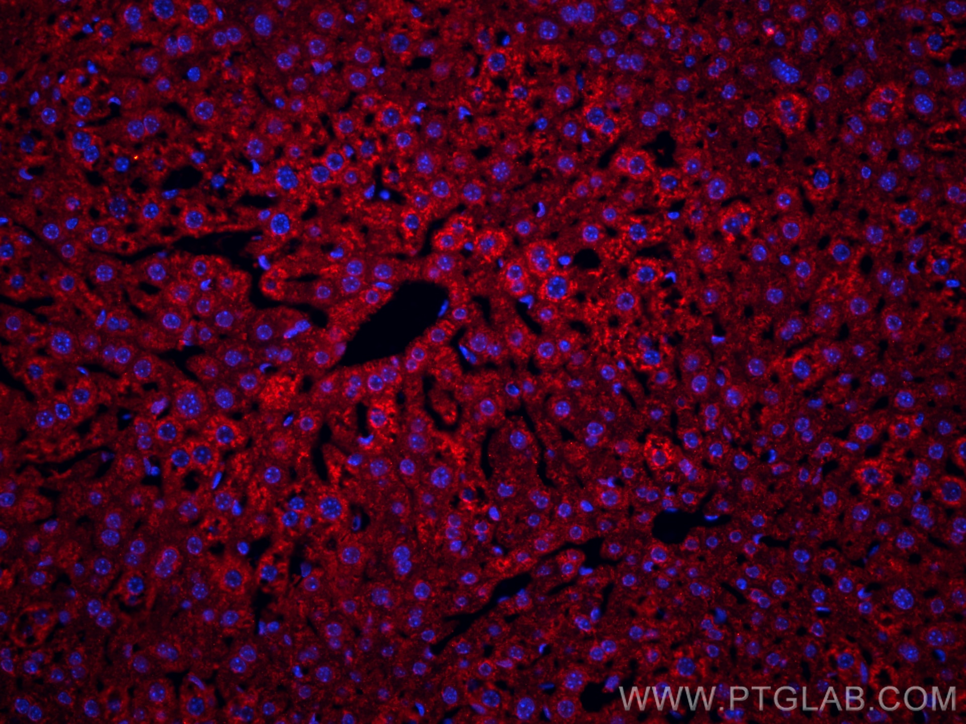
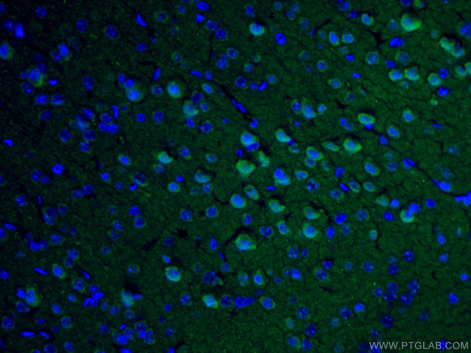
| Positive IF-P detected in | mouse liver tissue, mouse brain tissue, mouse eye tissue |
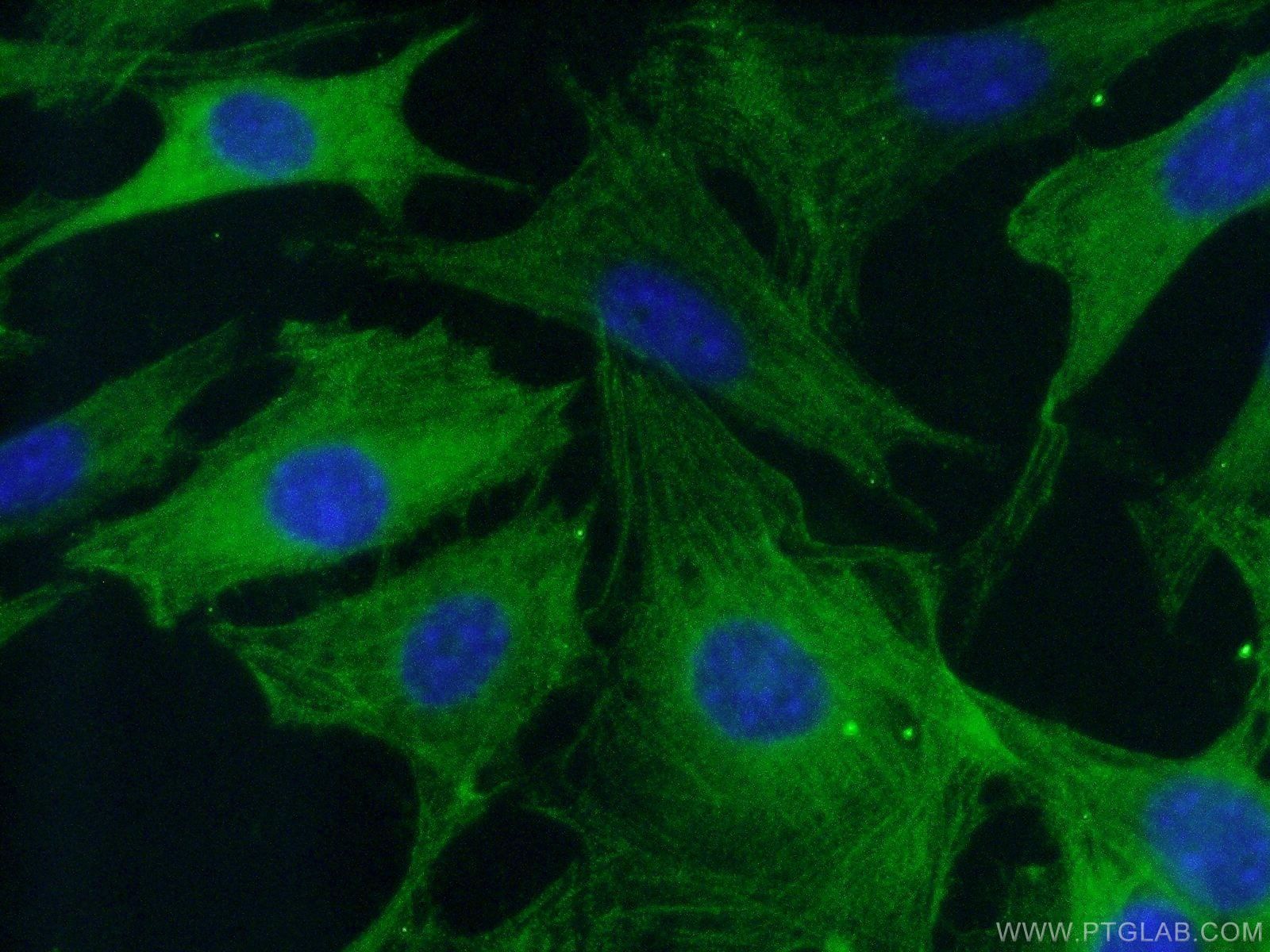
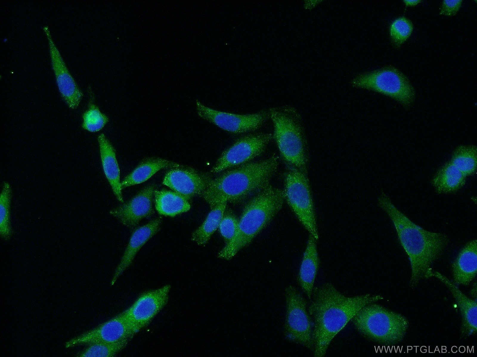
| Positive IF/ICC detected in | NIH/3T3 cells, HeLa cells |
Recommended dilution
| Application | Dilution |
|---|---|
| Western Blot (WB) | WB : 1:500-1:2000 |
| Immunoprecipitation (IP) | IP : 0.5-4.0 ug for 1.0-3.0 mg of total protein lysate |
| Immunohistochemistry (IHC) | IHC : 1:50-1:500 |
| Immunofluorescence (IF)-P | IF-P : 1:200-1:800 |
| Immunofluorescence (IF)/ICC | IF/ICC : 1:50-1:500 |
| It is recommended that this reagent should be titrated in each testing system to obtain optimal results. | |
| Sample-dependent, Check data in validation data gallery. | |
Published Applications
| KD/KO | See 7 publications below |
| WB | See 2625 publications below |
| IHC | See 259 publications below |
| IF | See 158 publications below |
| IP | See 5 publications below |
| ELISA | See 2 publications below |
| RIP | See 1 publications below |
Product Information
19677-1-AP targets Caspase 3/P17/P19 in WB, IHC, IF/ICC, IF-P, IP, RIP, ELISA applications and shows reactivity with human, mouse, rat samples.
| Tested Reactivity | human, mouse, rat |
| Cited Reactivity | human, mouse, canine, monkey, chicken, bovine, hamster, goat, duck |
| Host / Isotype | Rabbit / IgG |
| Class | Polyclonal |
| Type | Antibody |
| Immunogen |
Peptide Predict reactive species |
| Full Name | caspase 3, apoptosis-related cysteine peptidase |
| Calculated Molecular Weight | 32 kDa |
| Observed Molecular Weight | 32-35 kDa, 17 kDa, 19 kDa |
| GenBank Accession Number | NM_004346 |
| Gene Symbol | Caspase 3 |
| Gene ID (NCBI) | 836 |
| RRID | AB_10733244 |
| Conjugate | Unconjugated |
| Form | Liquid |
| Purification Method | Antigen affinity purification |
| UNIPROT ID | P42574 |
| Storage Buffer | PBS with 0.02% sodium azide and 50% glycerol, pH 7.3. |
| Storage Conditions | Store at -20°C. Stable for one year after shipment. Aliquoting is unnecessary for -20oC storage. 20ul sizes contain 0.1% BSA. |
Background Information
Caspases, a family of endoproteases, are critical players in cell regulatory networks controlling inflammation and cell death. Initiator caspases (caspase-2, -8, -9, -10, -11, and -12) cleave and activate downstream effector caspases (caspase-3, -6, and -7), which in turn execute apoptosis by cleaving targeted cellular proteins. Caspase 3 (also named CPP32, SCA-1, and Apopain) proteolytically cleaves poly(ADP-ribose) polymerase (PARP) at the beginning of apoptosis. Caspase 3 plays a key role in the activation of sterol regulatory element binding proteins (SREBPs) between the basic helix-loop-helix leucine zipper domain and the membrane attachment domain. Caspase 3 can also form heterocomplex with other proteins and performs the molecular mass of 50-70 kDa(PMID:9747872). This antibody can recognize p17, p19 and p32 of Caspase 3.
Protocols
| Product Specific Protocols | |
|---|---|
| IF protocol for Caspase 3/P17/P19 antibody 19677-1-AP | Download protocol |
| IHC protocol for Caspase 3/P17/P19 antibody 19677-1-AP | Download protocol |
| IP protocol for Caspase 3/P17/P19 antibody 19677-1-AP | Download protocol |
| WB protocol for Caspase 3/P17/P19 antibody 19677-1-AP | Download protocol |
| Standard Protocols | |
|---|---|
| Click here to view our Standard Protocols |
Publications
| Species | Application | Title |
|---|---|---|
Mol Cancer hsa_circ_0007919 induces LIG1 transcription by binding to FOXA1/TET1 to enhance the DNA damage response and promote gemcitabine resistance in pancreatic ductal adenocarcinoma | ||
Nat Microbiol Yersinia infection induces glucose depletion and AMPK-dependent inhibition of pyroptosis in mice | ||
Bioact Mater Silicate ions as soluble form of bioactive ceramics alleviate aortic aneurysm and dissection | ||
Nat Commun Macrophage lineage cells-derived migrasomes activate complement-dependent blood-brain barrier damage in cerebral amyloid angiopathy mouse model | ||
Nat Commun Protective effects of Pt-N-C single-atom nanozymes against myocardial ischemia-reperfusion injury | ||
Sci Transl Med PTEN status determines chemosensitivity to proteasome inhibition in cholangiocarcinoma. |
Reviews
The reviews below have been submitted by verified Proteintech customers who received an incentive for providing their feedback.
FH Vignesh (Verified Customer) (01-28-2026) | Excellent
|
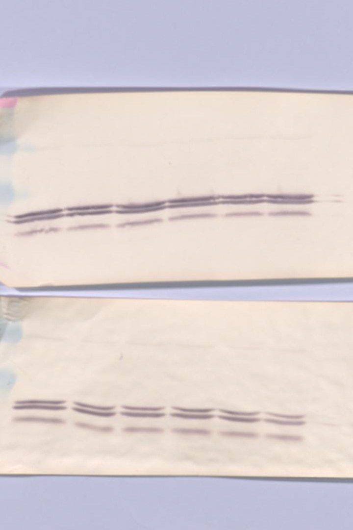
FH Charlotte (Verified Customer) (08-15-2024) | Done in milk. Many bands but the most intense one matches the p32 of the caspase 3
 |
FH Alessandro (Verified Customer) (12-09-2023) | Reliable apoptosis Ab for IF
|
FH Azita (Verified Customer) (06-02-2021) | Western blot analysis using LC3B-Specific Polyclonal antibody in NSC34 cell line at dilution of 1:1000.
|
FH Hala (Verified Customer) (04-12-2021) | works very well
|
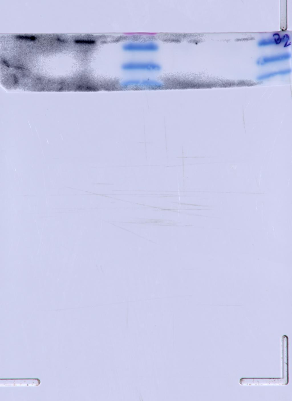
FH Isha (Verified Customer) (02-03-2021) | Gave me crystal clear bands in kidney lysates. really happy with the product
|
FH Diane (Verified Customer) (02-02-2021) | Incubated overnight 4 degrees C. Secondary 1:2500. Excellent bands. Used Opti-4CN substrate kit for visualization.
 |
FH Chao (Verified Customer) (03-12-2020) | Full-length but not cleaved isoform is detected by western blot
|
FH Kishor (Verified Customer) (01-30-2019) | It is an excellent antibody, worked every time when I used and got satisfactory results.
 |