Tested Applications
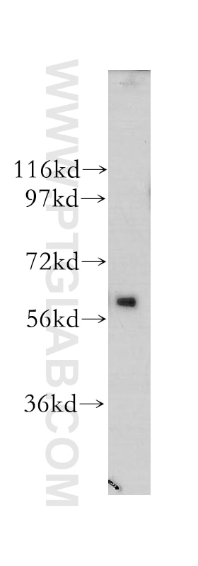
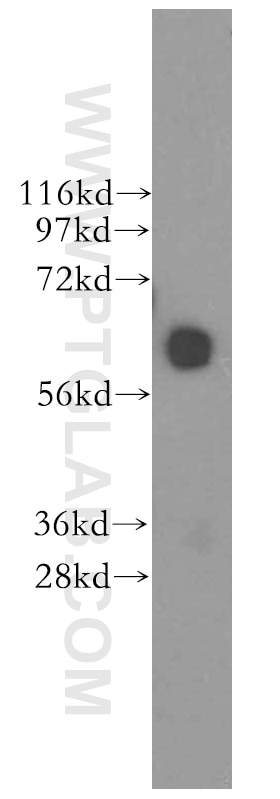
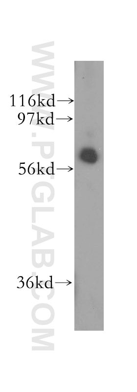
| Positive WB detected in | human brain tissue, Y79 cells |
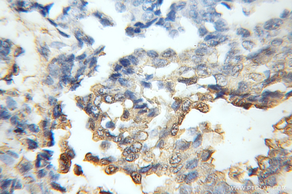
| Positive IHC detected in | human breast cancer tissue Note: suggested antigen retrieval with TE buffer pH 9.0; (*) Alternatively, antigen retrieval may be performed with citrate buffer pH 6.0 |
Recommended dilution
| Application | Dilution |
|---|---|
| Western Blot (WB) | WB : 1:500-1:2000 |
| Immunohistochemistry (IHC) | IHC : 1:20-1:200 |
| It is recommended that this reagent should be titrated in each testing system to obtain optimal results. | |
| Sample-dependent, Check data in validation data gallery. | |
Published Applications
| WB | See 1 publications below |
| IP | See 1 publications below |
Product Information
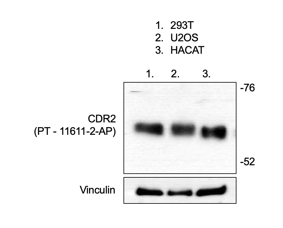
11611-2-AP targets CDR2 in WB, IP, IHC, ELISA applications and shows reactivity with human, mouse samples.
| Tested Reactivity | human, mouse |
| Cited Reactivity | human |
| Host / Isotype | Rabbit / IgG |
| Class | Polyclonal |
| Type | Antibody |
| Immunogen |
CatNo: Ag2183 Product name: Recombinant human CDR2 protein Source: e coli.-derived, PGEX-4T Tag: GST Domain: 105-454 aa of BC017503 Sequence: SKASQQKILSLTETIECLQTNIDHLQSQVEELKSSGQGRRSPGKCDQEKPAPSFACLKELYDLRQHFVYDHVFAEKITSLQGQPSPDEEENEHLKKTVTMLQAQLSLERQKRVTMEEEYGLVLKENSELEQQLGATGAYRARALELEAEVAEMRQMLQSEHPFVNGVEKLVPDSLYVPFKEPSQSLLEEMFLTVPESHRKPLKRSSSETILSSLAGSDIVKGHEETCIRRAKAVKQRGISLLHEVDTQYSALKVKYEELLKKCQEEQDSLSHKAVQTSRAAAKDLTGVNAQSEPVASGWELASVNPEPVSSPTTPPEYKALFKEIFSCIKKTKQEIDEQRTKYRSLSSHS Predict reactive species |
| Full Name | cerebellar degeneration-related protein 2, 62kDa |
| Calculated Molecular Weight | 454 aa, 52 kDa |
| Observed Molecular Weight | 62 kDa |
| GenBank Accession Number | BC017503 |
| Gene Symbol | CDR2 |
| Gene ID (NCBI) | 1039 |
| RRID | AB_2076734 |
| Conjugate | Unconjugated |
| Form | Liquid |
| Purification Method | Antigen affinity purification |
| UNIPROT ID | Q01850 |
| Storage Buffer | PBS with 0.02% sodium azide and 50% glycerol, pH 7.3. |
| Storage Conditions | Store at -20°C. Stable for one year after shipment. Aliquoting is unnecessary for -20oC storage. 20ul sizes contain 0.1% BSA. |
Background Information
Patients with paraneoplastic cerebellar degeneration (PCD) carry a characteristic antibody called anti-Yo. On Western blot analysis of Purkinje cells and tumor tissue from CD patient, the anti-Yo sera react with at least 2 antigens, a major species of 62 kD called CDR62 or CDR2 [PMID:18045792]. CDR2 is partly characterized, as CDR2 through its leucine zipper motif has been demonstrated to interact with c-myc, with cell cycle-related proteins and with a protein kinase, indicating that CDR2 is involved in signal transduction and gene transcription. Furthermore, CDR2 has been found to attenuate hypoxic response in renal cell carcinoma, and recent data suggest a role for CDR2 and c-myc in mitosis in cycling cells [PMID:21080165].
Protocols
| Product Specific Protocols | |
|---|---|
| IHC protocol for CDR2 antibody 11611-2-AP | Download protocol |
| WB protocol for CDR2 antibody 11611-2-AP | Download protocol |
| Standard Protocols | |
|---|---|
| Click here to view our Standard Protocols |
Reviews
The reviews below have been submitted by verified Proteintech customers who received an incentive for providing their feedback.
FH anwesh (Verified Customer) (11-11-2019) | CDR2 was successfully IP'ed from A549 cells. The antibody had not background bands.
|
FH anwesh (Verified Customer) (07-29-2019) | The protein was found at the right size and no additional background bands were observed around the size range. Perfect!
 |