Tested Applications
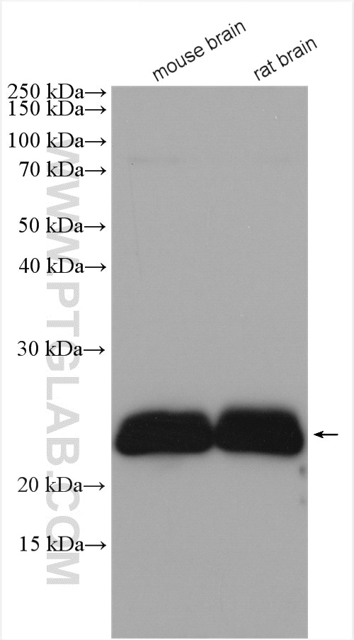
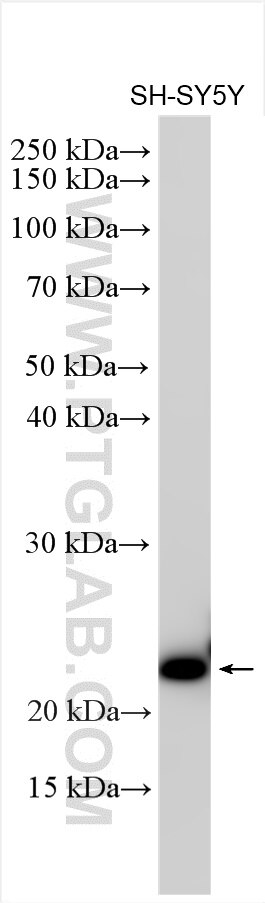
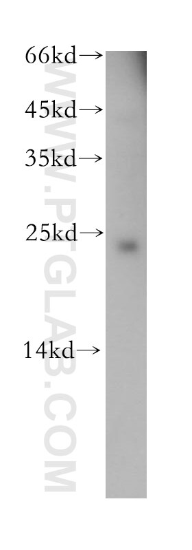
| Positive WB detected in | mouse brain tissue, SH-SY5Y cells, human brain tissue, rat brain tissue |
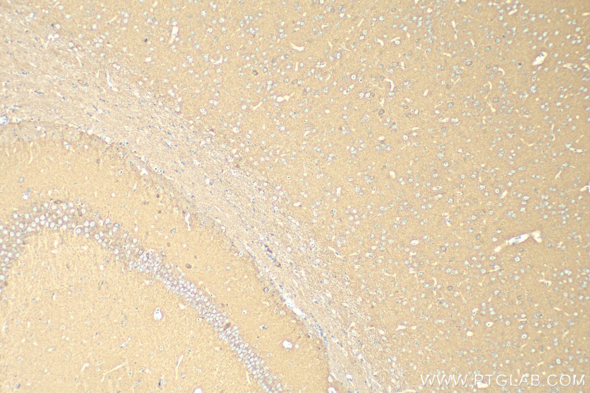
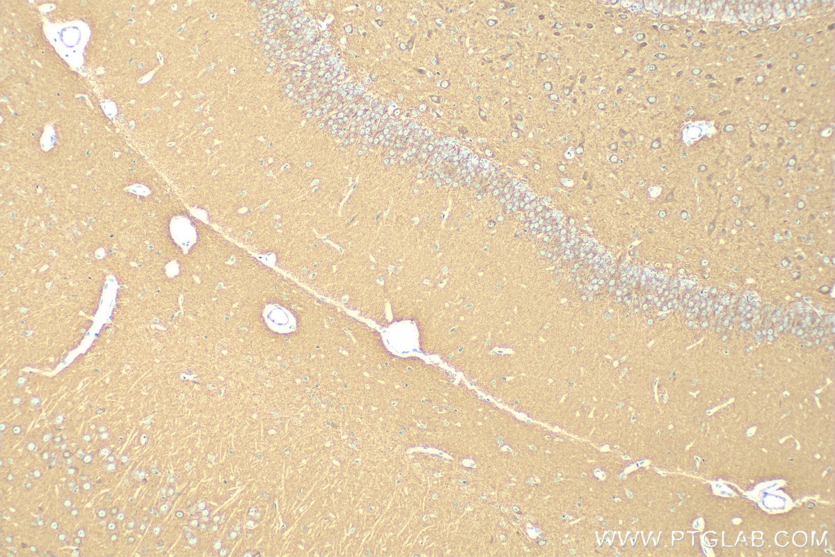
| Positive IHC detected in | mouse brain tissue, rat brain tissue Note: suggested antigen retrieval with TE buffer pH 9.0; (*) Alternatively, antigen retrieval may be performed with citrate buffer pH 6.0 |
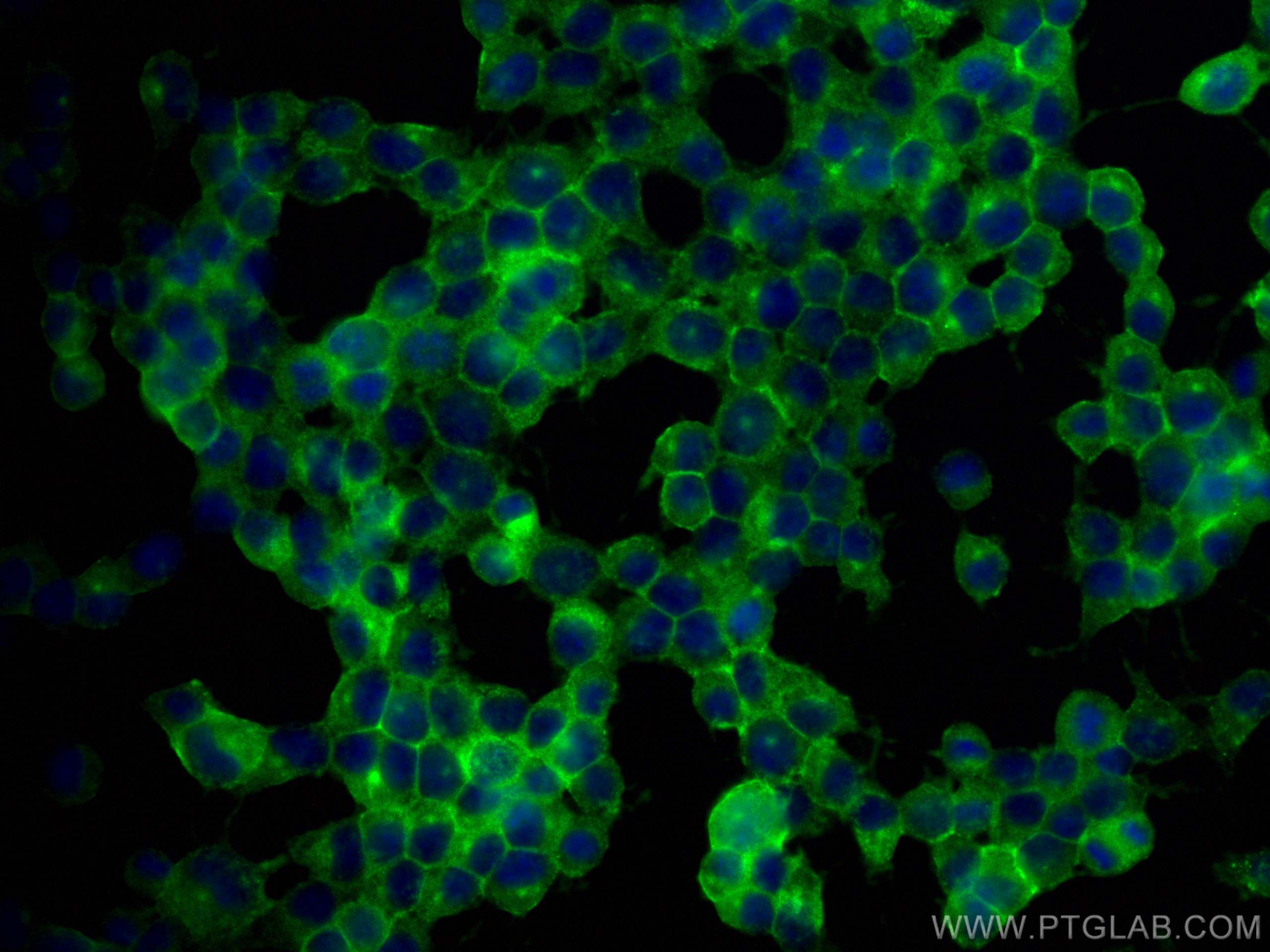
| Positive IF/ICC detected in | Neuro-2a cells |
Recommended dilution
| Application | Dilution |
|---|---|
| Western Blot (WB) | WB : 1:500-1:1000 |
| Immunohistochemistry (IHC) | IHC : 1:50-1:500 |
| Immunofluorescence (IF)/ICC | IF/ICC : 1:50-1:500 |
| It is recommended that this reagent should be titrated in each testing system to obtain optimal results. | |
| Sample-dependent, Check data in validation data gallery. | |
Published Applications
| WB | See 2 publications below |
Product Information
13280-1-AP targets CEND1 in WB, IHC, IF/ICC, ELISA applications and shows reactivity with human, mouse, rat samples.
| Tested Reactivity | human, mouse, rat |
| Cited Reactivity | rat, goat |
| Host / Isotype | Rabbit / IgG |
| Class | Polyclonal |
| Type | Antibody |
| Immunogen |
CatNo: Ag4024 Product name: Recombinant human CEND1 protein Source: e coli.-derived, PGEX-4T Tag: GST Domain: 1-123 aa of BC034732 Sequence: MESRGKSASSPKPDTKVPQVTTEAKVPPAADGKAPLTKPSKKEAPAEKQQPPAAPTTAPAKKTSAKADPALLNNHSNLKPAPTVPSSPDATPEPKGPGDGAEEDEAASGGPGGRGPWSCENFN Predict reactive species |
| Full Name | cell cycle exit and neuronal differentiation 1 |
| Calculated Molecular Weight | 149 aa, 15 kDa |
| Observed Molecular Weight | 23 kDa |
| GenBank Accession Number | BC034732 |
| Gene Symbol | CEND1 |
| Gene ID (NCBI) | 51286 |
| RRID | AB_2291632 |
| Conjugate | Unconjugated |
| Form | Liquid |
| Purification Method | Antigen affinity purification |
| UNIPROT ID | Q8N111 |
| Storage Buffer | PBS with 0.02% sodium azide and 50% glycerol, pH 7.3. |
| Storage Conditions | Store at -20°C. Stable for one year after shipment. Aliquoting is unnecessary for -20oC storage. 20ul sizes contain 0.1% BSA. |
Background Information
Cell cycle exit and neuronal differentiation 1 (CEND1) also known as BM88, is a neuronal protein associated in vivo with terminal neuron-generating divisions, marking the exit of proliferative cells from the cell cycle (PMID: 17971443). CEND1 expression is critical for the NEUROG2-driven reprogramming of astrocytes, suggesting the existence of a reciprocal feedback loop leading to neurogenesis (PMID: 26321141). CEND1 is an integral membrane protein composed of two 23-kDa polypeptide chains linked together by disulfide bridges.
Protocols
| Product Specific Protocols | |
|---|---|
| IF protocol for CEND1 antibody 13280-1-AP | Download protocol |
| IHC protocol for CEND1 antibody 13280-1-AP | Download protocol |
| WB protocol for CEND1 antibody 13280-1-AP | Download protocol |
| Standard Protocols | |
|---|---|
| Click here to view our Standard Protocols |
Publications
| Species | Application | Title |
|---|---|---|
Cell Death Discov Molecular mechanism underlying miR-204-5p regulation of adipose-derived stem cells differentiation into cells from three germ layers | ||
J Ethnopharmacol The Protective Effects and Mechanisms of Essential oil from Chimonanthus nitens Oliv. Leaves in Allergic Rhinitis Based on the NF-κB and T-bet/GATA-3 signaling pathways |