Tested Applications
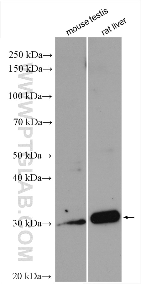
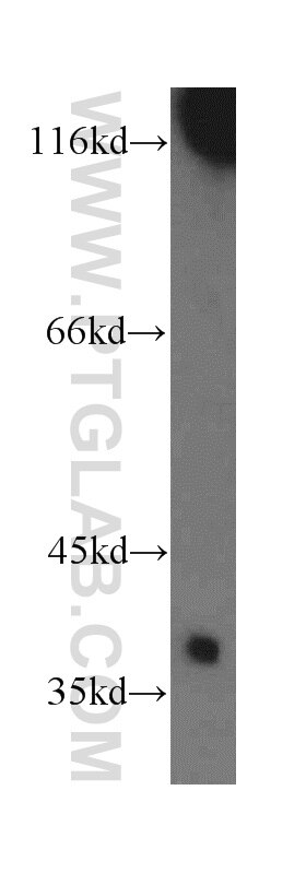
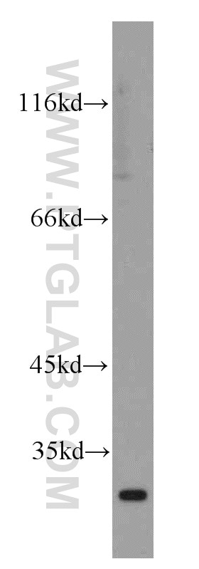
| Positive WB detected in | mouse testis tissue, human testis tissue, rat liver tissue |
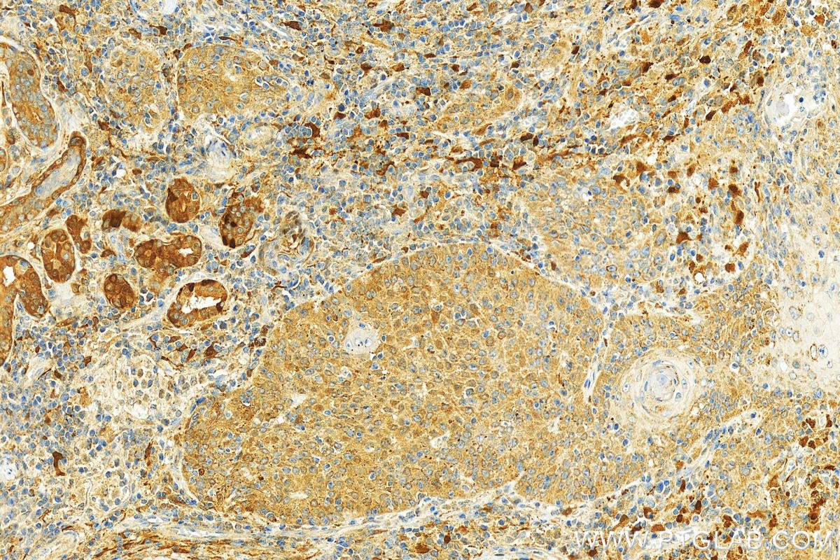
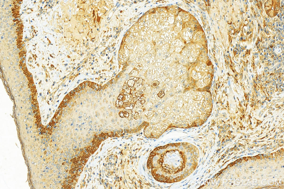
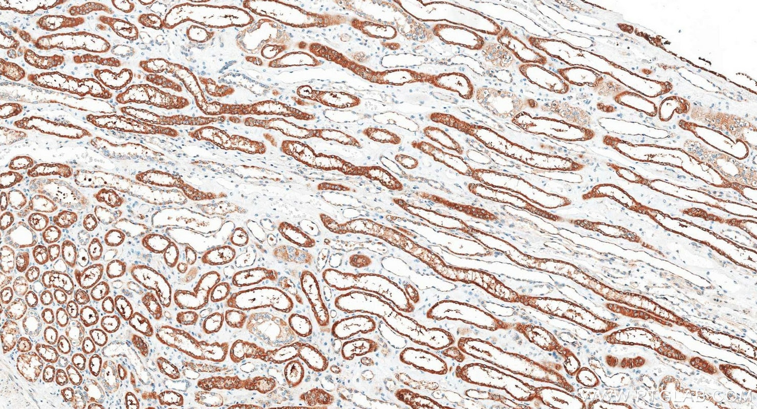
| Positive IHC detected in | human skin cancer tissue, human kidney tissue Note: suggested antigen retrieval with TE buffer pH 9.0; (*) Alternatively, antigen retrieval may be performed with citrate buffer pH 6.0 |
Recommended dilution
| Application | Dilution |
|---|---|
| Western Blot (WB) | WB : 1:500-1:1000 |
| Immunohistochemistry (IHC) | IHC : 1:50-1:500 |
| It is recommended that this reagent should be titrated in each testing system to obtain optimal results. | |
| Sample-dependent, Check data in validation data gallery. | |
Published Applications
| WB | See 1 publications below |
| IHC | See 1 publications below |
Product Information
18442-1-AP targets Cathepsin V in WB, IHC, ELISA applications and shows reactivity with human, mouse, rat samples.
| Tested Reactivity | human, mouse, rat |
| Cited Reactivity | human |
| Host / Isotype | Rabbit / IgG |
| Class | Polyclonal |
| Type | Antibody |
| Immunogen |
CatNo: Ag13284 Product name: Recombinant human CTSL2 protein Source: e coli.-derived, PGEX-4T Tag: GST Domain: 1-334 aa of BC023504 Sequence: MNLSLVLAAFCLGIASAVPKFDQNLDTKWYQWKATHRRLYGANEEGWRRAVWEKNMKMIELHNGEYSQGKHGFTMAMNAFGDMTNEEFRQMMGCFRNQKFRKGKVFREPLFLDLPKSVDWRKKGYVTPVKNQKQCGSCWAFSATGALEGQMFRKTGKLVSLSEQNLVDCSRPQGNQGCNGGFMARAFQYVKENGGLDSEESYPYVAVDEICKYRPENSVANDTGFTVVAPGKEKALMKAVATVGPISVAMDAGHSSFQFYKSGIYFEPDCSSKNLDHGVLVVGYGFEGANSNNSKYWLVKNSWGPEWGSNGYVKIAKDKNNHCGIATAASYPNV Predict reactive species |
| Full Name | cathepsin L2 |
| Calculated Molecular Weight | 37 kDa |
| Observed Molecular Weight | 26-36 kDa |
| GenBank Accession Number | BC023504 |
| Gene Symbol | Cathepsin V |
| Gene ID (NCBI) | 1515 |
| RRID | AB_10644340 |
| Conjugate | Unconjugated |
| Form | Liquid |
| Purification Method | Antigen affinity purification |
| UNIPROT ID | O60911 |
| Storage Buffer | PBS with 0.02% sodium azide and 50% glycerol, pH 7.3. |
| Storage Conditions | Store at -20°C. Stable for one year after shipment. Aliquoting is unnecessary for -20oC storage. 20ul sizes contain 0.1% BSA. |
Background Information
Cathepsin V is a lysosomal cysteine protease that has an important role in corneal physiology. Cathepsin V was found to be differentially expressed in relation to pigmentary skin backgrounds, and was about 7.5-folds higher in keratinocytes from lightly pigmented, relative to darkly pigmented skins. It is expressed in primary keratinocytes, melanocytes, and HaCaT keratinocytes, but not in primary fibroblasts. The purified cathepsin L2 was a high molecular weight proteinase of about 78 kDa, but it was unstable to SDS and reducing conditions, which dissociated parts of the proteinase into 66 kDa, 31 kDa and 26 kDa fragments(Food Chemistry,Volume11,Issue4,879-886) and it can form a complex of 70 kDa with an unidentified subunit(PMID:1930136).
Protocols
| Product Specific Protocols | |
|---|---|
| IHC protocol for Cathepsin V antibody 18442-1-AP | Download protocol |
| WB protocol for Cathepsin V antibody 18442-1-AP | Download protocol |
| Standard Protocols | |
|---|---|
| Click here to view our Standard Protocols |
Publications
| Species | Application | Title |
|---|---|---|
Mol Carcinog Cathepsin V is correlated with the prognosis and tumor microenvironment in liver cancer | ||
Mol Cell The RNA-stability-independent role of the RNA m6A reader YTHDF2 in promoting protein translation to confer tumor chemotherapy resistance |