- Home
- Products
- Primary Antibodies
- Cytokeratin 5 Monoclonal antibody, PBS Only (66727-1-PBS)
")
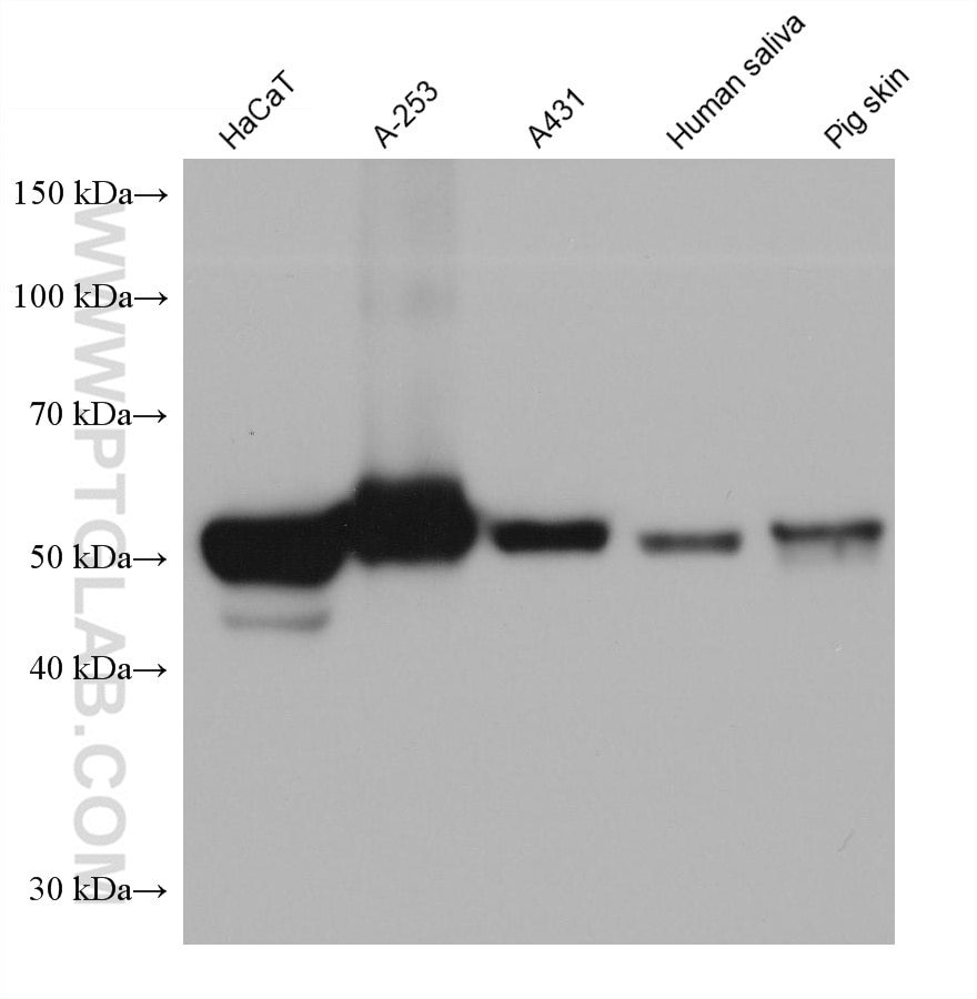
WB analysis using 66727-1-Ig (same clone as 66727-1-PBS)
Various lysates were subjected to SDS PAGE followed by western blot with 66727-1-Ig (Cytokeratin 5 antibody) at dilution of 1:100000 incubated at room temperature for 1.5 hours. This data was developed using the same antibody clone with 66727-1-PBS in a different storage buffer formulation.
× Various lysates were subjected to SDS PAGE followed by western blot with 66727-1-Ig (Cytokeratin 5 antibody) at dilution of 1:100000 incubated at room temperature for 1.5 hours. This data was developed using the same antibody clone with 66727-1-PBS in a different storage buffer formulation.
")
WB analysis of mouse skin using 66727-1-Ig (same clone as 66727-1-PBS)
mouse skin tissue were subjected to SDS PAGE followed by western blot with 66727-1-Ig (Cytokeratin 5 antibody) at dilution of 1:100000 incubated at room temperature for 1.5 hours. This data was developed using the same antibody clone with 66727-1-PBS in a different storage buffer formulation.
× mouse skin tissue were subjected to SDS PAGE followed by western blot with 66727-1-Ig (Cytokeratin 5 antibody) at dilution of 1:100000 incubated at room temperature for 1.5 hours. This data was developed using the same antibody clone with 66727-1-PBS in a different storage buffer formulation.
")
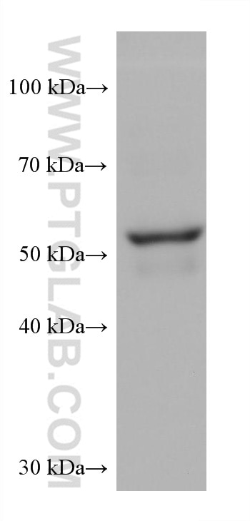
WB analysis of SKOV-3 using 66727-1-Ig (same clone as 66727-1-PBS)
SKOV-3 cells were subjected to SDS PAGE followed by western blot with 66727-1-Ig (Cytokeratin 5 antibody) at dilution of 1:100000 incubated at room temperature for 1.5 hours. This data was developed using the same antibody clone with 66727-1-PBS in a different storage buffer formulation.
× SKOV-3 cells were subjected to SDS PAGE followed by western blot with 66727-1-Ig (Cytokeratin 5 antibody) at dilution of 1:100000 incubated at room temperature for 1.5 hours. This data was developed using the same antibody clone with 66727-1-PBS in a different storage buffer formulation.
")
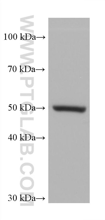
WB analysis of MCF-7 using 66727-1-Ig (same clone as 66727-1-PBS)
MCF-7 cells were subjected to SDS PAGE followed by western blot with 66727-1-Ig (Cytokeratin 5 antibody) at dilution of 1:100000 incubated at room temperature for 1.5 hours. This data was developed using the same antibody clone with 66727-1-PBS in a different storage buffer formulation.
× MCF-7 cells were subjected to SDS PAGE followed by western blot with 66727-1-Ig (Cytokeratin 5 antibody) at dilution of 1:100000 incubated at room temperature for 1.5 hours. This data was developed using the same antibody clone with 66727-1-PBS in a different storage buffer formulation.
")
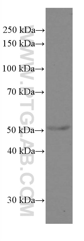
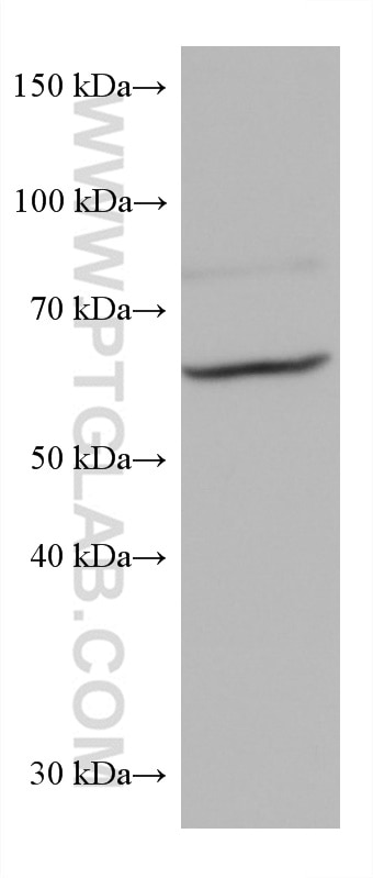
WB analysis of A549 using 66727-1-Ig (same clone as 66727-1-PBS)
A549 cells were subjected to SDS PAGE followed by western blot with 66727-1-Ig (Cytokeratin 5 antibody) at dilution of 1:100000 incubated at room temperature for 1.5 hours. This data was developed using the same antibody clone with 66727-1-PBS in a different storage buffer formulation.
× A549 cells were subjected to SDS PAGE followed by western blot with 66727-1-Ig (Cytokeratin 5 antibody) at dilution of 1:100000 incubated at room temperature for 1.5 hours. This data was developed using the same antibody clone with 66727-1-PBS in a different storage buffer formulation.
")
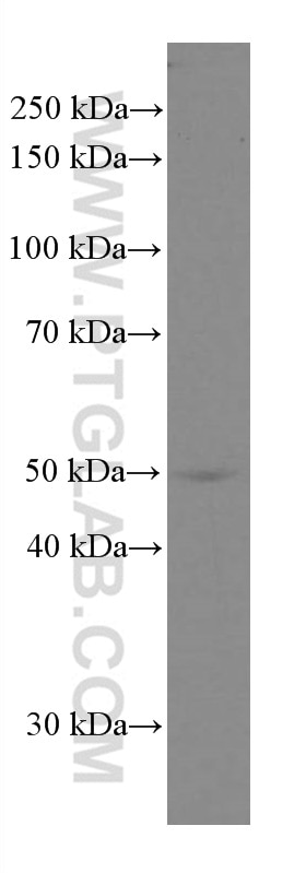
WB analysis of mouse skin using 66727-1-Ig (same clone as 66727-1-PBS)
mouse skin tissue were subjected to SDS PAGE followed by western blot with 66727-1-Ig (Cytokeratin 5 antibody) at dilution of 1:50000 incubated at room temperature for 1.5 hours. This data was developed using the same antibody clone with 66727-1-PBS in a different storage buffer formulation.
× mouse skin tissue were subjected to SDS PAGE followed by western blot with 66727-1-Ig (Cytokeratin 5 antibody) at dilution of 1:50000 incubated at room temperature for 1.5 hours. This data was developed using the same antibody clone with 66727-1-PBS in a different storage buffer formulation.
")
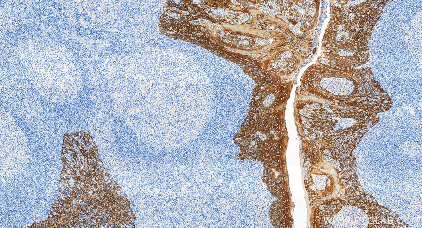
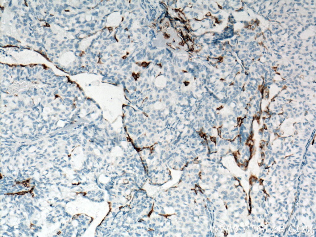
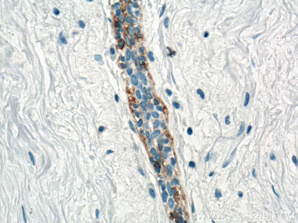
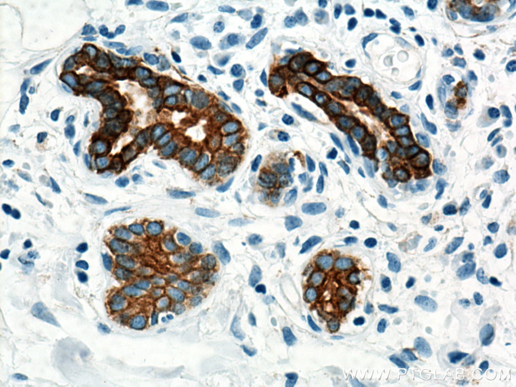
IHC staining of human tonsillitis using 66727-1-Ig (same clone as 66727-1-PBS)
Immunohistochemical analysis of paraffin-embedded human tonsillitis tissue slide using 66727-1-Ig (Cytokeratin 5 antibody) at dilution of 1:10000 (under 20x lens). Heat mediated antigen retrieval with Tris-EDTA buffer (pH 9.0). This data was developed using the same antibody clone with 66727-1-PBS in a different storage buffer formulation.
× Immunohistochemical analysis of paraffin-embedded human tonsillitis tissue slide using 66727-1-Ig (Cytokeratin 5 antibody) at dilution of 1:10000 (under 20x lens). Heat mediated antigen retrieval with Tris-EDTA buffer (pH 9.0). This data was developed using the same antibody clone with 66727-1-PBS in a different storage buffer formulation.
")
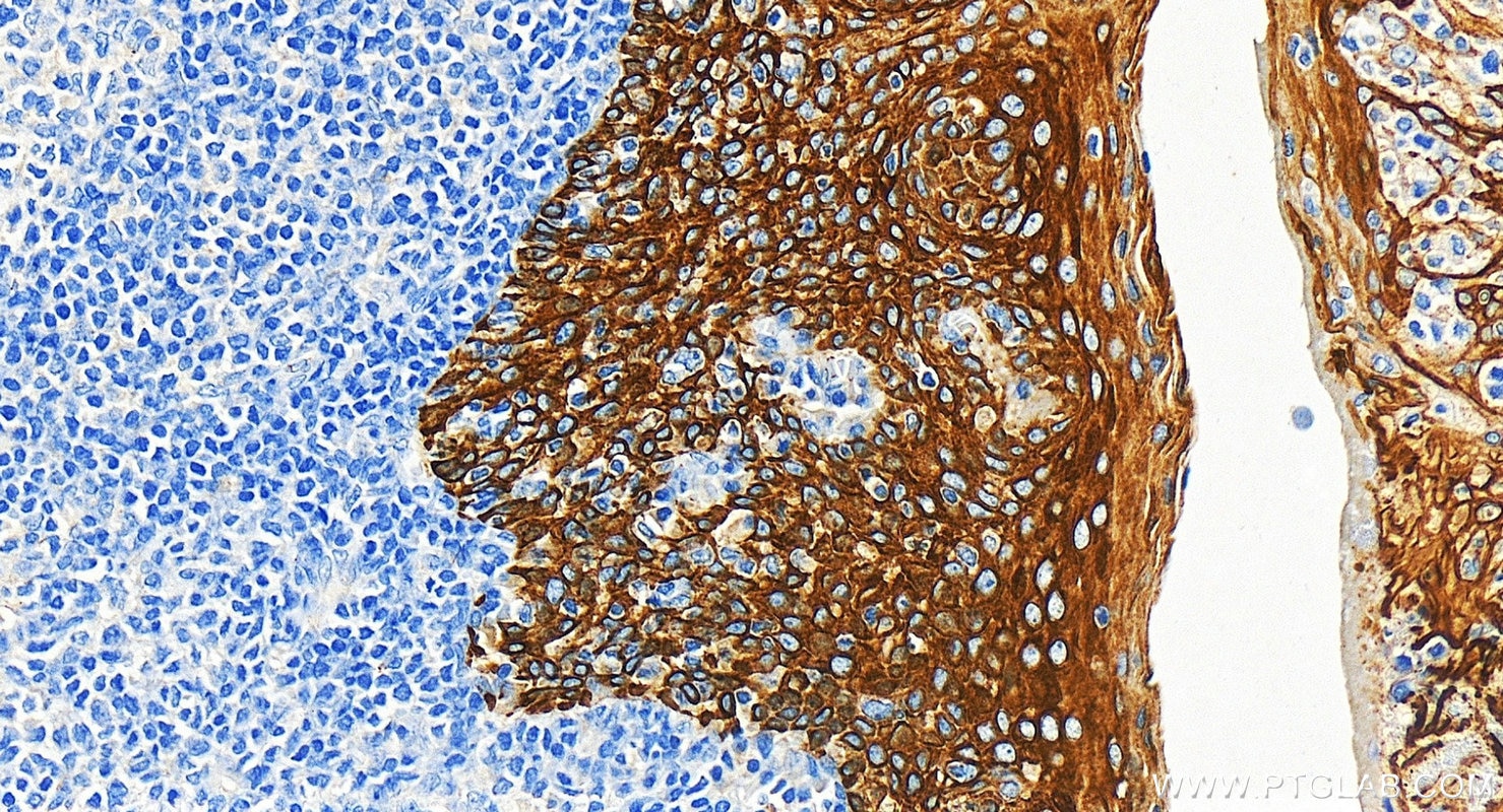
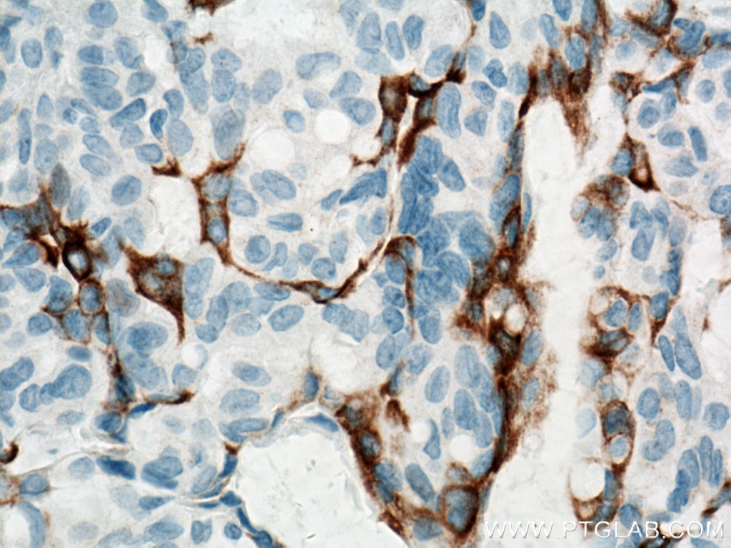
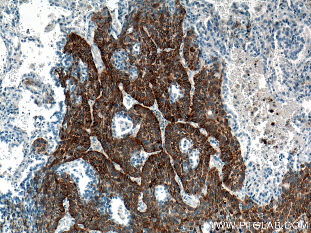
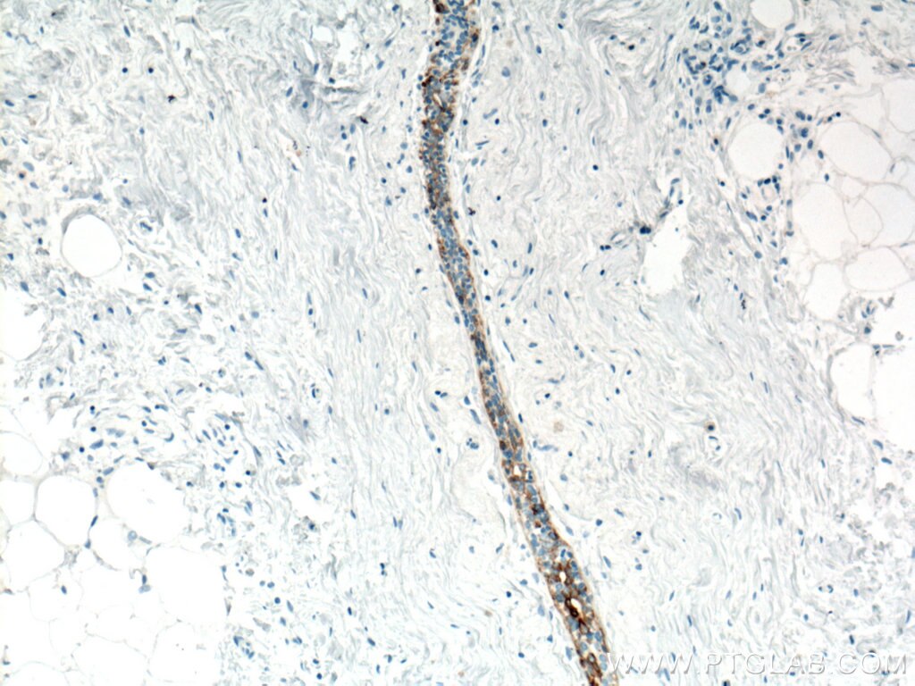
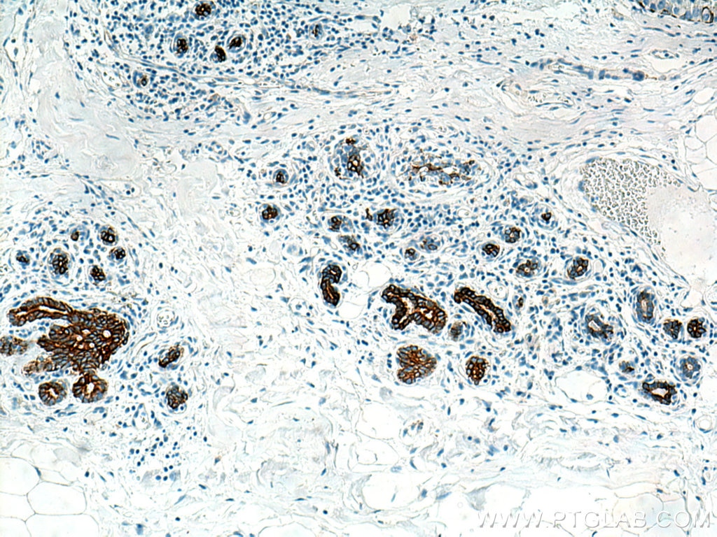
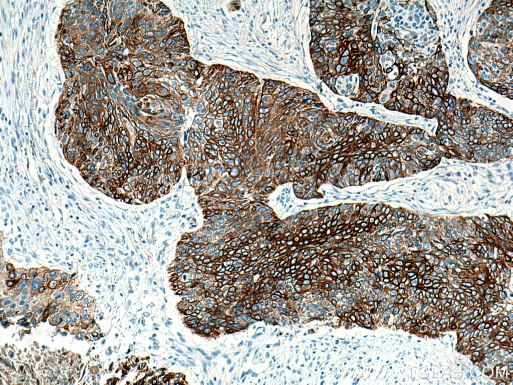
IHC staining of human oesophagus cancer using 66727-1-Ig (same clone as 66727-1-PBS)
Immunohistochemical analysis of paraffin-embedded human oesophagus cancer tissue slide using 66727-1-Ig (Cytokeratin 5 antibody) at dilution of 1:2000 (under 10x lens). Heat mediated antigen retrieval with Tris-EDTA buffer (pH 9.0). This data was developed using the same antibody clone with 66727-1-PBS in a different storage buffer formulation.
× Immunohistochemical analysis of paraffin-embedded human oesophagus cancer tissue slide using 66727-1-Ig (Cytokeratin 5 antibody) at dilution of 1:2000 (under 10x lens). Heat mediated antigen retrieval with Tris-EDTA buffer (pH 9.0). This data was developed using the same antibody clone with 66727-1-PBS in a different storage buffer formulation.
")
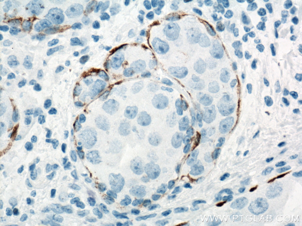
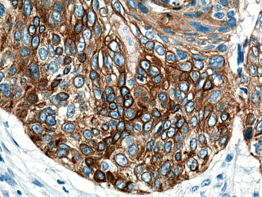
IHC staining of human cervical cancer using 66727-1-Ig (same clone as 66727-1-PBS)
Immunohistochemical analysis of paraffin-embedded human cervical cancer tissue slide using 66727-1-Ig (Cytokeratin 5 antibody) at dilution of 1:3000 (under 10x lens. Heat mediated antigen retrieval with Tris-EDTA buffer (pH 9.0). This data was developed using the same antibody clone with 66727-1-PBS in a different storage buffer formulation.
× Immunohistochemical analysis of paraffin-embedded human cervical cancer tissue slide using 66727-1-Ig (Cytokeratin 5 antibody) at dilution of 1:3000 (under 10x lens. Heat mediated antigen retrieval with Tris-EDTA buffer (pH 9.0). This data was developed using the same antibody clone with 66727-1-PBS in a different storage buffer formulation.
")
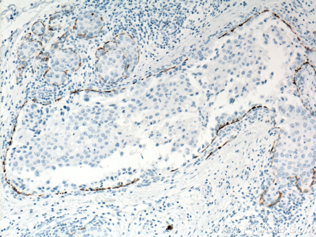
IHC staining of human cervical cancer using 66727-1-Ig (same clone as 66727-1-PBS)
Immunohistochemical analysis of paraffin-embedded human cervical cancer tissue slide using 66727-1-Ig (Cytokeratin 5 antibody) at dilution of 1:3000 (under 40x lens. Heat mediated antigen retrieval with Tris-EDTA buffer (pH 9.0). This data was developed using the same antibody clone with 66727-1-PBS in a different storage buffer formulation.
× Immunohistochemical analysis of paraffin-embedded human cervical cancer tissue slide using 66727-1-Ig (Cytokeratin 5 antibody) at dilution of 1:3000 (under 40x lens. Heat mediated antigen retrieval with Tris-EDTA buffer (pH 9.0). This data was developed using the same antibody clone with 66727-1-PBS in a different storage buffer formulation.
")
IHC staining of human breast cancer using 66727-1-Ig (same clone as 66727-1-PBS)
Immunohistochemical analysis of paraffin-embedded human breast cancer tissue slide using 66727-1-Ig (Cytokeratin 5 antibody) at dilution of 1:6000 (under 10x lens. Heat mediated antigen retrieval with Tris-EDTA buffer (pH 9.0). This data was developed using the same antibody clone with 66727-1-PBS in a different storage buffer formulation.
× Immunohistochemical analysis of paraffin-embedded human breast cancer tissue slide using 66727-1-Ig (Cytokeratin 5 antibody) at dilution of 1:6000 (under 10x lens. Heat mediated antigen retrieval with Tris-EDTA buffer (pH 9.0). This data was developed using the same antibody clone with 66727-1-PBS in a different storage buffer formulation.
")
IHC staining of human breast cancer using 66727-1-Ig (same clone as 66727-1-PBS)
Immunohistochemical analysis of paraffin-embedded human breast cancer tissue slide using 66727-1-Ig (Cytokeratin 5 antibody) at dilution of 1:6000 (under 40x lens. Heat mediated antigen retrieval with Tris-EDTA buffer (pH 9.0). This data was developed using the same antibody clone with 66727-1-PBS in a different storage buffer formulation.
× Immunohistochemical analysis of paraffin-embedded human breast cancer tissue slide using 66727-1-Ig (Cytokeratin 5 antibody) at dilution of 1:6000 (under 40x lens. Heat mediated antigen retrieval with Tris-EDTA buffer (pH 9.0). This data was developed using the same antibody clone with 66727-1-PBS in a different storage buffer formulation.
")
IHC staining of human breast hyperplasia using 66727-1-Ig (same clone as 66727-1-PBS)
Immunohistochemical analysis of paraffin-embedded human breast hyperplasia tissue slide using 66727-1-Ig (Cytokeratin 5 antibody) at dilution of 1:6000 (under 10x lens. Heat mediated antigen retrieval with Tris-EDTA buffer (pH 9.0). This data was developed using the same antibody clone with 66727-1-PBS in a different storage buffer formulation.
× Immunohistochemical analysis of paraffin-embedded human breast hyperplasia tissue slide using 66727-1-Ig (Cytokeratin 5 antibody) at dilution of 1:6000 (under 10x lens. Heat mediated antigen retrieval with Tris-EDTA buffer (pH 9.0). This data was developed using the same antibody clone with 66727-1-PBS in a different storage buffer formulation.
")
IHC staining of human breast hyperplasia using 66727-1-Ig (same clone as 66727-1-PBS)
Immunohistochemical analysis of paraffin-embedded human breast hyperplasia tissue slide using 66727-1-Ig (Cytokeratin 5 antibody) at dilution of 1:6000 (under 40x lens. Heat mediated antigen retrieval with Tris-EDTA buffer (pH 9.0). This data was developed using the same antibody clone with 66727-1-PBS in a different storage buffer formulation.
× Immunohistochemical analysis of paraffin-embedded human breast hyperplasia tissue slide using 66727-1-Ig (Cytokeratin 5 antibody) at dilution of 1:6000 (under 40x lens. Heat mediated antigen retrieval with Tris-EDTA buffer (pH 9.0). This data was developed using the same antibody clone with 66727-1-PBS in a different storage buffer formulation.
")
IHC staining of human lung cancer using 66727-1-Ig (same clone as 66727-1-PBS)
Immunohistochemical analysis of paraffin-embedded human lung cancer tissue slide using 66727-1-Ig (Cytokeratin 5 antibody) at dilution of 1:6000 (under 10x lens. Heat mediated antigen retrieval with Tris-EDTA buffer (pH 9.0). This data was developed using the same antibody clone with 66727-1-PBS in a different storage buffer formulation.
× Immunohistochemical analysis of paraffin-embedded human lung cancer tissue slide using 66727-1-Ig (Cytokeratin 5 antibody) at dilution of 1:6000 (under 10x lens. Heat mediated antigen retrieval with Tris-EDTA buffer (pH 9.0). This data was developed using the same antibody clone with 66727-1-PBS in a different storage buffer formulation.
")
IHC staining of human lung cancer using 66727-1-Ig (same clone as 66727-1-PBS)
Immunohistochemical analysis of paraffin-embedded human lung cancer tissue slide using 66727-1-Ig (Cytokeratin 5 antibody) at dilution of 1:6000 (under 40x lens. Heat mediated antigen retrieval with Tris-EDTA buffer (pH 9.0). This data was developed using the same antibody clone with 66727-1-PBS in a different storage buffer formulation.
× Immunohistochemical analysis of paraffin-embedded human lung cancer tissue slide using 66727-1-Ig (Cytokeratin 5 antibody) at dilution of 1:6000 (under 40x lens. Heat mediated antigen retrieval with Tris-EDTA buffer (pH 9.0). This data was developed using the same antibody clone with 66727-1-PBS in a different storage buffer formulation.
")
IHC staining of human breast hyperplasia using 66727-1-Ig (same clone as 66727-1-PBS)
Immunohistochemical analysis of paraffin-embedded human breast hyperplasia tissue slide using 66727-1-Ig (Cytokeratin 5 antibody) at dilution of 1:6000 (under 40x lens. Heat mediated antigen retrieval with Tris-EDTA buffer (pH 9.0). This data was developed using the same antibody clone with 66727-1-PBS in a different storage buffer formulation.
× Immunohistochemical analysis of paraffin-embedded human breast hyperplasia tissue slide using 66727-1-Ig (Cytokeratin 5 antibody) at dilution of 1:6000 (under 40x lens. Heat mediated antigen retrieval with Tris-EDTA buffer (pH 9.0). This data was developed using the same antibody clone with 66727-1-PBS in a different storage buffer formulation.
")
IHC staining of human breast hyperplasia using 66727-1-Ig (same clone as 66727-1-PBS)
Immunohistochemical analysis of paraffin-embedded human breast hyperplasia tissue slide using 66727-1-Ig (Cytokeratin 5 antibody) at dilution of 1:6000 (under 10x lens. Heat mediated antigen retrieval with Tris-EDTA buffer (pH 9.0). This data was developed using the same antibody clone with 66727-1-PBS in a different storage buffer formulation.
× Immunohistochemical analysis of paraffin-embedded human breast hyperplasia tissue slide using 66727-1-Ig (Cytokeratin 5 antibody) at dilution of 1:6000 (under 10x lens. Heat mediated antigen retrieval with Tris-EDTA buffer (pH 9.0). This data was developed using the same antibody clone with 66727-1-PBS in a different storage buffer formulation.
")
IHC staining of human breast cancer using 66727-1-Ig (same clone as 66727-1-PBS)
Immunohistochemical analysis of paraffin-embedded human breast cancer tissue slide using 66727-1-Ig (Cytokeratin 5 antibody) at dilution of 1:6000 (under 40x lens. Heat mediated antigen retrieval with Tris-EDTA buffer (pH 9.0). This data was developed using the same antibody clone with 66727-1-PBS in a different storage buffer formulation.
× Immunohistochemical analysis of paraffin-embedded human breast cancer tissue slide using 66727-1-Ig (Cytokeratin 5 antibody) at dilution of 1:6000 (under 40x lens. Heat mediated antigen retrieval with Tris-EDTA buffer (pH 9.0). This data was developed using the same antibody clone with 66727-1-PBS in a different storage buffer formulation.
")
IHC staining of human breast cancer using 66727-1-Ig (same clone as 66727-1-PBS)
Immunohistochemical analysis of paraffin-embedded human breast cancer tissue slide using 66727-1-Ig (Cytokeratin 5 antibody) at dilution of 1:6000 (under 10x lens. Heat mediated antigen retrieval with Tris-EDTA buffer (pH 9.0). This data was developed using the same antibody clone with 66727-1-PBS in a different storage buffer formulation.
× Immunohistochemical analysis of paraffin-embedded human breast cancer tissue slide using 66727-1-Ig (Cytokeratin 5 antibody) at dilution of 1:6000 (under 10x lens. Heat mediated antigen retrieval with Tris-EDTA buffer (pH 9.0). This data was developed using the same antibody clone with 66727-1-PBS in a different storage buffer formulation.
")
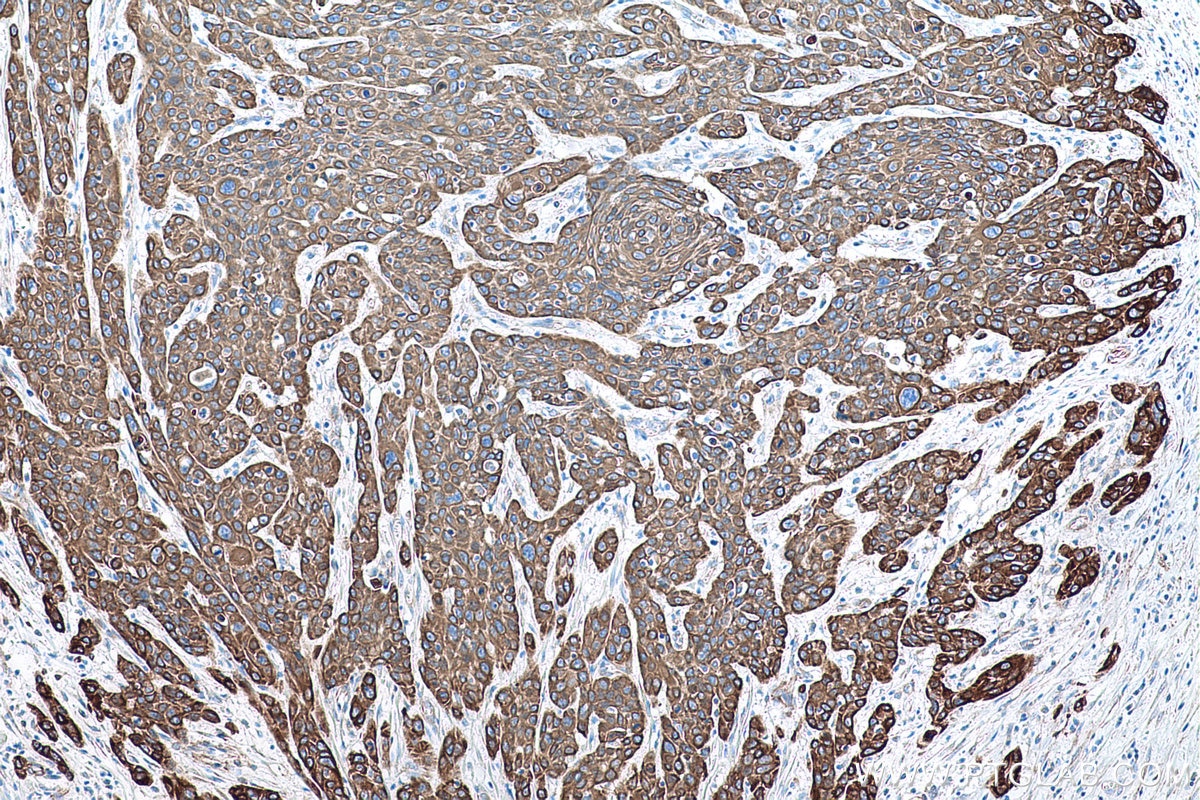
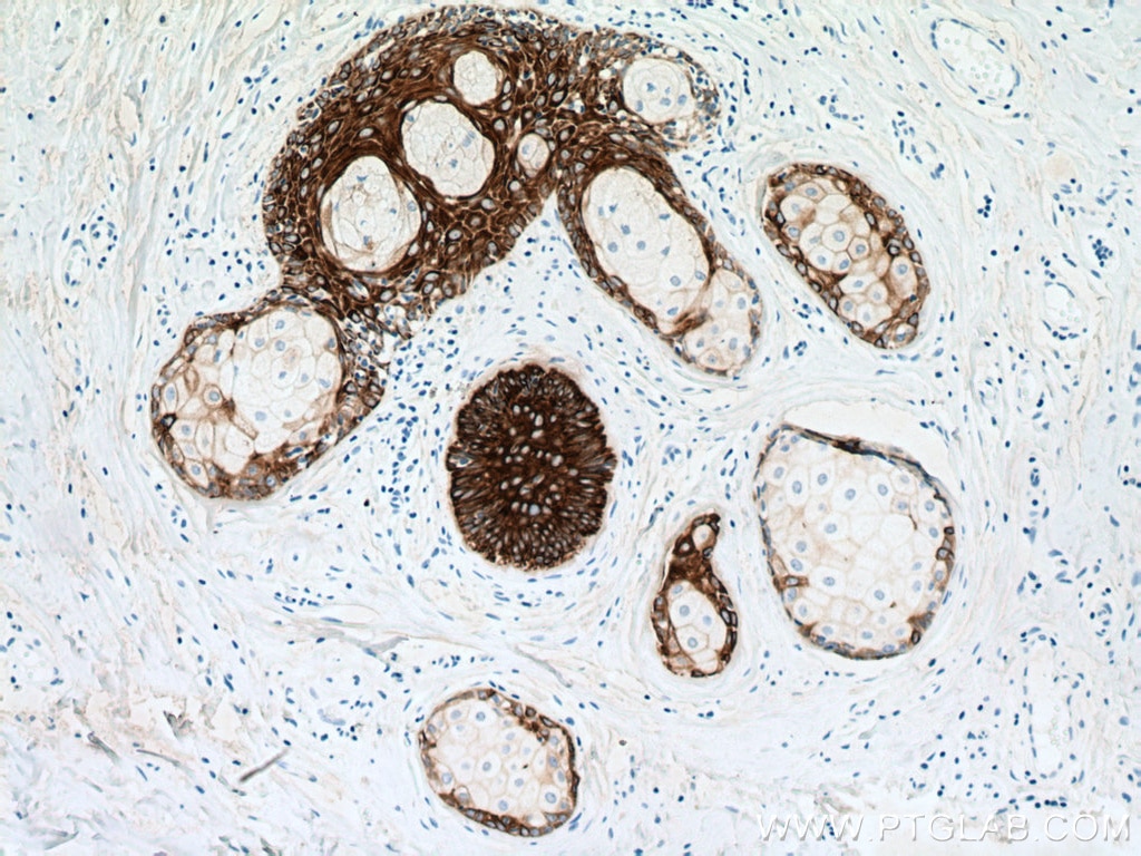
IHC staining of human skin cancer using 66727-1-Ig (same clone as 66727-1-PBS)
Immunohistochemical analysis of paraffin-embedded human skin cancer tissue slide using 66727-1-Ig (Cytokeratin 5 antibody) at dilution of 1:6000 (under 10x lens). Heat mediated antigen retrieval with Tris-EDTA buffer (pH 9.0). This data was developed using the same antibody clone with 66727-1-PBS in a different storage buffer formulation.
× Immunohistochemical analysis of paraffin-embedded human skin cancer tissue slide using 66727-1-Ig (Cytokeratin 5 antibody) at dilution of 1:6000 (under 10x lens). Heat mediated antigen retrieval with Tris-EDTA buffer (pH 9.0). This data was developed using the same antibody clone with 66727-1-PBS in a different storage buffer formulation.
")
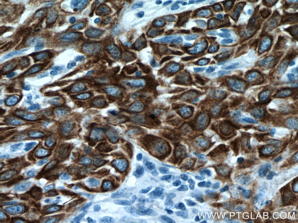
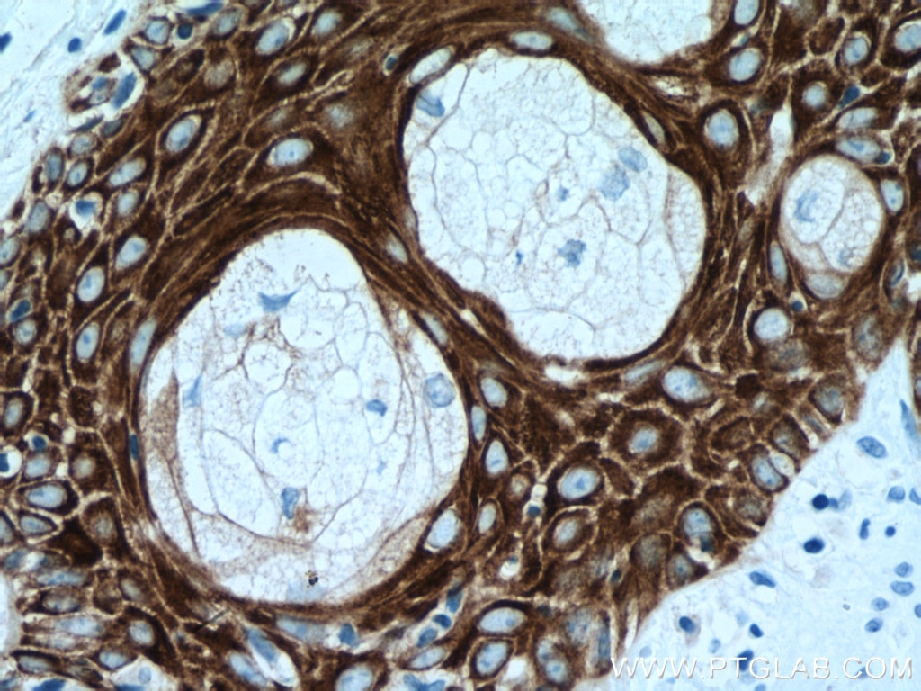
IHC staining of human skin cancer using 66727-1-Ig (same clone as 66727-1-PBS)
Immunohistochemical analysis of paraffin-embedded human skin cancer tissue slide using 66727-1-Ig (Cytokeratin 5 antibody) at dilution of 1:6000 (under 40x lens). Heat mediated antigen retrieval with Tris-EDTA buffer (pH 9.0). This data was developed using the same antibody clone with 66727-1-PBS in a different storage buffer formulation.
× Immunohistochemical analysis of paraffin-embedded human skin cancer tissue slide using 66727-1-Ig (Cytokeratin 5 antibody) at dilution of 1:6000 (under 40x lens). Heat mediated antigen retrieval with Tris-EDTA buffer (pH 9.0). This data was developed using the same antibody clone with 66727-1-PBS in a different storage buffer formulation.
")
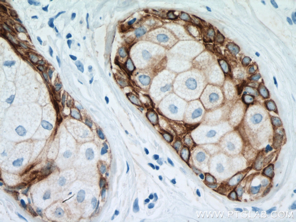
IHC staining of human breast cancer using 66727-1-Ig (same clone as 66727-1-PBS)
Immunohistochemical analysis of paraffin-embedded human breast cancer tissue slide using 66727-1-Ig (Cytokeratin 5 antibody) at dilution of 1:10000 (under 10x lens). Heat mediated antigen retrieval with Tris-EDTA buffer (pH 9.0). This data was developed using the same antibody clone with 66727-1-PBS in a different storage buffer formulation.
× Immunohistochemical analysis of paraffin-embedded human breast cancer tissue slide using 66727-1-Ig (Cytokeratin 5 antibody) at dilution of 1:10000 (under 10x lens). Heat mediated antigen retrieval with Tris-EDTA buffer (pH 9.0). This data was developed using the same antibody clone with 66727-1-PBS in a different storage buffer formulation.
")
IHC staining of human breast cancer using 66727-1-Ig (same clone as 66727-1-PBS)
Immunohistochemical analysis of paraffin-embedded human breast cancer tissue slide using 66727-1-Ig (Cytokeratin 5 antibody) at dilution of 1:10000 (under 40x lens). Heat mediated antigen retrieval with Tris-EDTA buffer (pH 9.0). This data was developed using the same antibody clone with 66727-1-PBS in a different storage buffer formulation.
× Immunohistochemical analysis of paraffin-embedded human breast cancer tissue slide using 66727-1-Ig (Cytokeratin 5 antibody) at dilution of 1:10000 (under 40x lens). Heat mediated antigen retrieval with Tris-EDTA buffer (pH 9.0). This data was developed using the same antibody clone with 66727-1-PBS in a different storage buffer formulation.
")
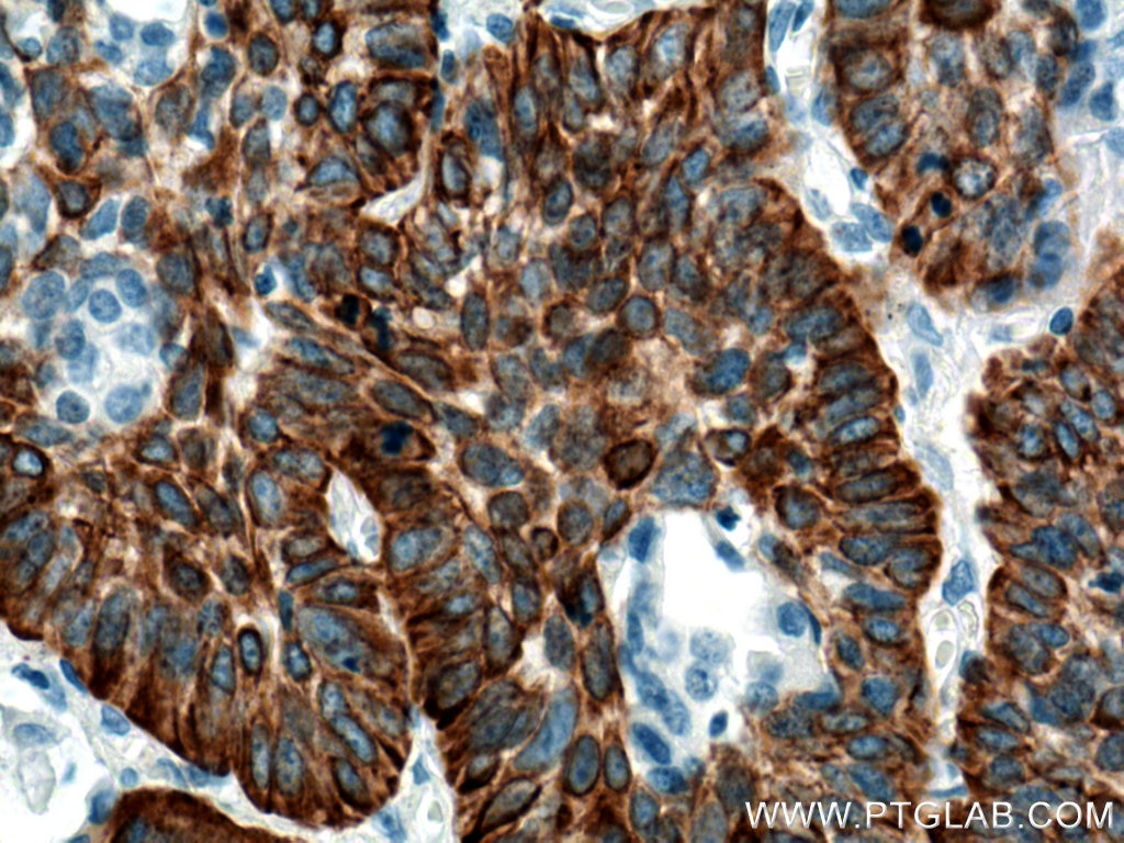
IHC staining of human oesophagus cancer using 66727-1-Ig (same clone as 66727-1-PBS)
Immunohistochemical analysis of paraffin-embedded human oesophagus cancer tissue slide using 66727-1-Ig (Cytokeratin 5 antibody) at dilution of 1:10000 (under 10x lens). Heat mediated antigen retrieval with Tris-EDTA buffer (pH 9.0). This data was developed using the same antibody clone with 66727-1-PBS in a different storage buffer formulation.
× Immunohistochemical analysis of paraffin-embedded human oesophagus cancer tissue slide using 66727-1-Ig (Cytokeratin 5 antibody) at dilution of 1:10000 (under 10x lens). Heat mediated antigen retrieval with Tris-EDTA buffer (pH 9.0). This data was developed using the same antibody clone with 66727-1-PBS in a different storage buffer formulation.
")
IHC staining of human oesophagus cancer using 66727-1-Ig (same clone as 66727-1-PBS)
Immunohistochemical analysis of paraffin-embedded human oesophagus cancer tissue slide using 66727-1-Ig (Cytokeratin 5 antibody) at dilution of 1:10000 (under 40x lens). Heat mediated antigen retrieval with Tris-EDTA buffer (pH 9.0). This data was developed using the same antibody clone with 66727-1-PBS in a different storage buffer formulation.
× Immunohistochemical analysis of paraffin-embedded human oesophagus cancer tissue slide using 66727-1-Ig (Cytokeratin 5 antibody) at dilution of 1:10000 (under 40x lens). Heat mediated antigen retrieval with Tris-EDTA buffer (pH 9.0). This data was developed using the same antibody clone with 66727-1-PBS in a different storage buffer formulation.
")
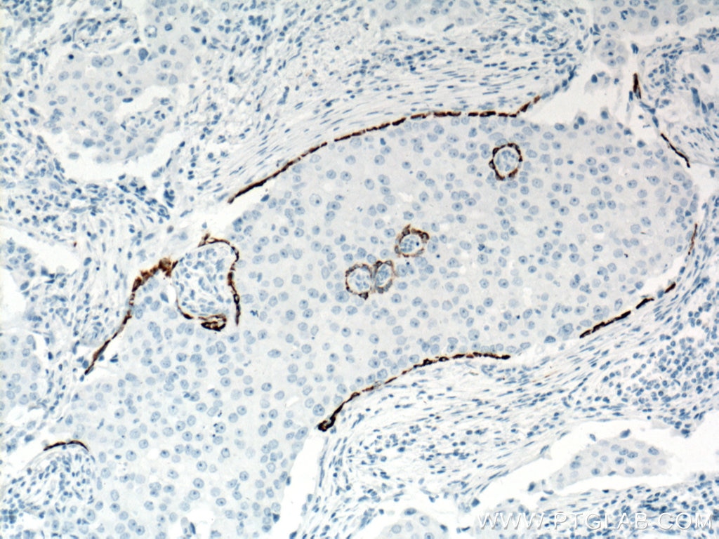
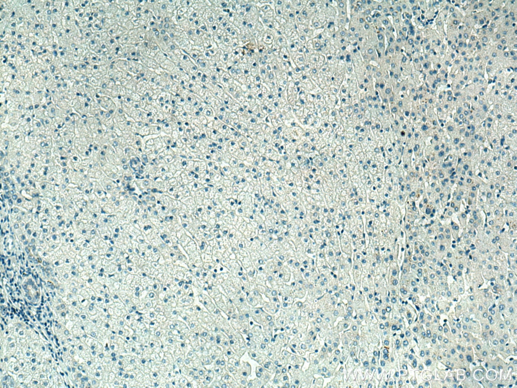
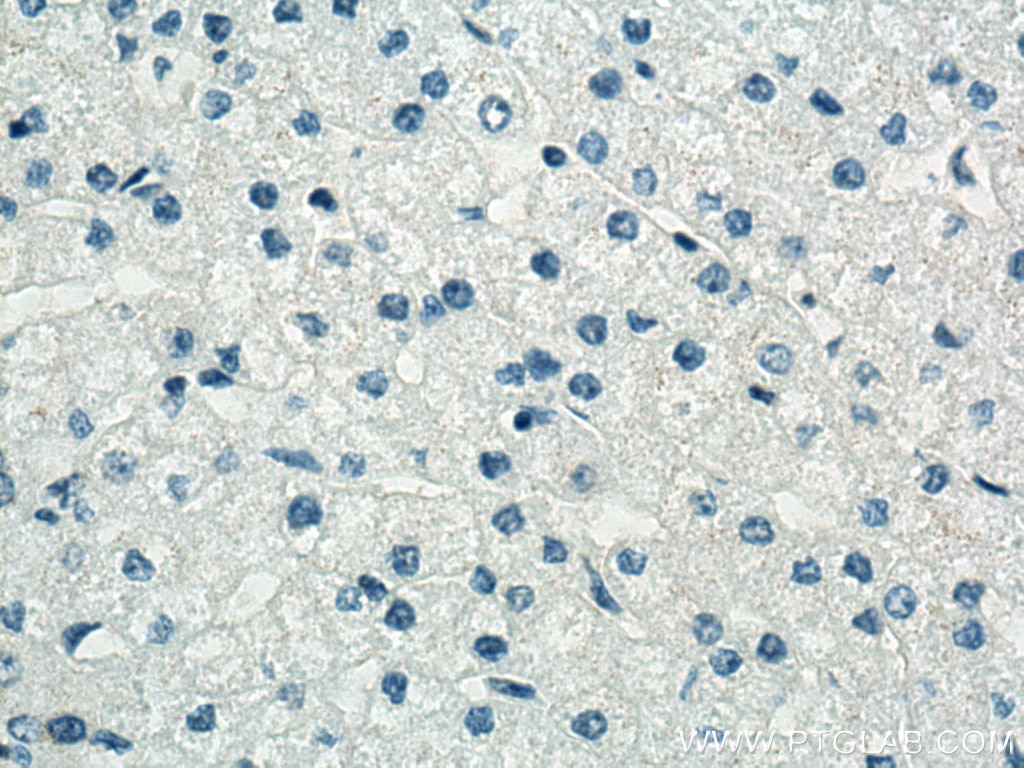
IHC staining of human liver using 66727-1-Ig (same clone as 66727-1-PBS)
Immunohistochemical analysis of paraffin-embedded human liver tissue slide using 66727-1-Ig (Cytokeratin 5 antibody) at dilution of 1:10000 (under 10x lens) showing negative staining. Heat mediated antigen retrieval with Tris-EDTA buffer (pH 9.0). This data was developed using the same antibody clone with 66727-1-PBS in a different storage buffer formulation.
× Immunohistochemical analysis of paraffin-embedded human liver tissue slide using 66727-1-Ig (Cytokeratin 5 antibody) at dilution of 1:10000 (under 10x lens) showing negative staining. Heat mediated antigen retrieval with Tris-EDTA buffer (pH 9.0). This data was developed using the same antibody clone with 66727-1-PBS in a different storage buffer formulation.
")
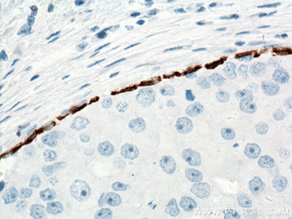
IHC staining of human liver using 66727-1-Ig (same clone as 66727-1-PBS)
Immunohistochemical analysis of paraffin-embedded human liver tissue slide using 66727-1-Ig (Cytokeratin 5 antibody) at dilution of 1:10000 (under 40x lens) showing negative staining. Heat mediated antigen retrieval with Tris-EDTA buffer (pH 9.0). This data was developed using the same antibody clone with 66727-1-PBS in a different storage buffer formulation.
× Immunohistochemical analysis of paraffin-embedded human liver tissue slide using 66727-1-Ig (Cytokeratin 5 antibody) at dilution of 1:10000 (under 40x lens) showing negative staining. Heat mediated antigen retrieval with Tris-EDTA buffer (pH 9.0). This data was developed using the same antibody clone with 66727-1-PBS in a different storage buffer formulation.
")
IF Staining of human oesophagus cancer using 66727-1-Ig (same clone as 66727-1-PBS)
Immunofluorescent analysis of (4% PFA) fixed human oesophagus cancer tissue using Cytokeratin 5 antibody (66727-1-Ig, Clone: 1A1C5 ) at dilution of 1:400 and CoraLite®488-Conjugated AffiniPure Goat Anti-Mouse IgG(H+L). This data was developed using the same antibody clone with 66727-1-PBS in a different storage buffer formulation.
× Immunofluorescent analysis of (4% PFA) fixed human oesophagus cancer tissue using Cytokeratin 5 antibody (66727-1-Ig, Clone: 1A1C5 ) at dilution of 1:400 and CoraLite®488-Conjugated AffiniPure Goat Anti-Mouse IgG(H+L). This data was developed using the same antibody clone with 66727-1-PBS in a different storage buffer formulation.
")
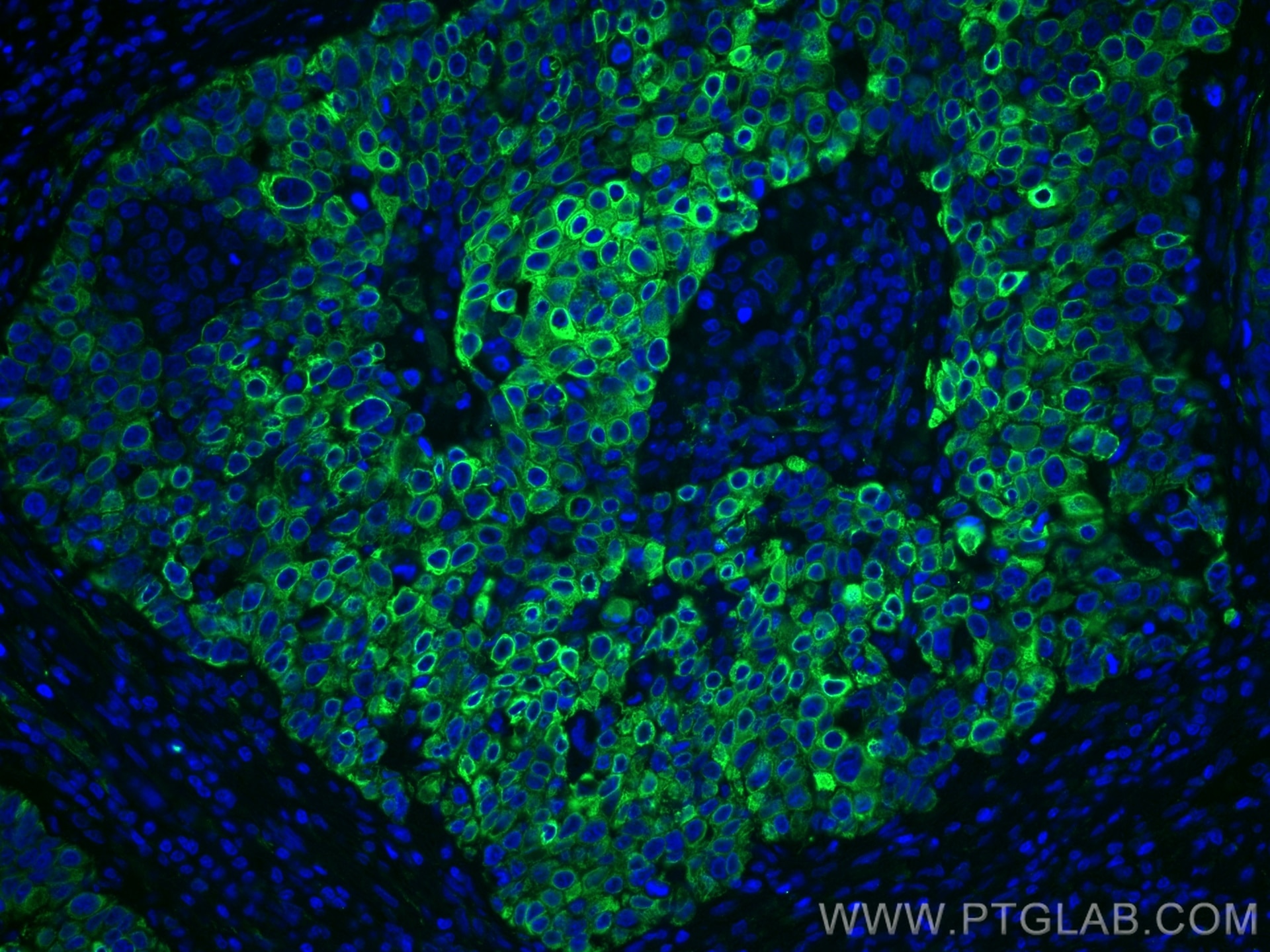
IF Staining of human prostate cancer using 66727-1-Ig (same clone as 66727-1-PBS)
Immunofluorescent analysis of (4% PFA) fixed human prostate cancer tissue using Cytokeratin 5 antibody (66727-1-Ig, Clone: 1A1C5 ) at dilution of 1:400 and CoraLite®488-Conjugated AffiniPure Goat Anti-Mouse IgG(H+L). This data was developed using the same antibody clone with 66727-1-PBS in a different storage buffer formulation.
× Immunofluorescent analysis of (4% PFA) fixed human prostate cancer tissue using Cytokeratin 5 antibody (66727-1-Ig, Clone: 1A1C5 ) at dilution of 1:400 and CoraLite®488-Conjugated AffiniPure Goat Anti-Mouse IgG(H+L). This data was developed using the same antibody clone with 66727-1-PBS in a different storage buffer formulation.
")
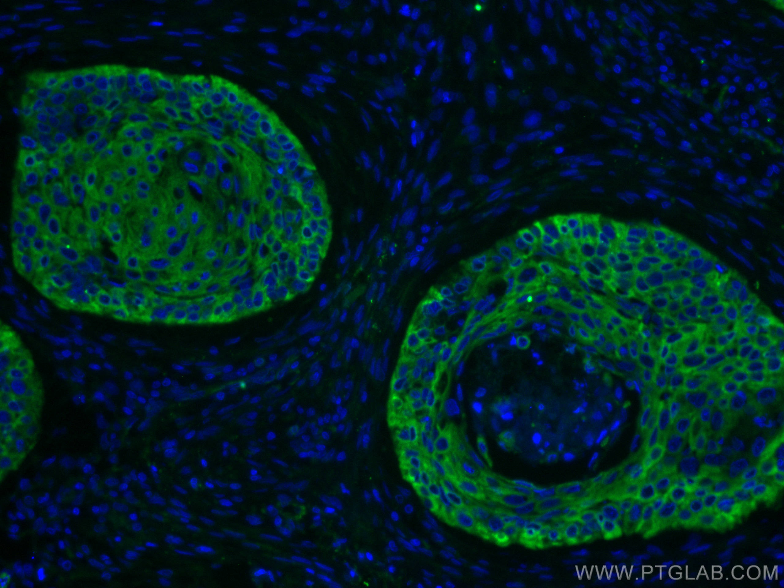
IF Staining of human oesophagus cancer using 66727-1-Ig (same clone as 66727-1-PBS)
Immunofluorescent analysis of (4% PFA) fixed human oesophagus cancer tissue using Cytokeratin 5 antibody (66727-1-Ig, Clone: 1A1C5 ) at dilution of 1:400 and CoraLite®488-Conjugated AffiniPure Goat Anti-Mouse IgG(H+L). This data was developed using the same antibody clone with 66727-1-PBS in a different storage buffer formulation.
× Immunofluorescent analysis of (4% PFA) fixed human oesophagus cancer tissue using Cytokeratin 5 antibody (66727-1-Ig, Clone: 1A1C5 ) at dilution of 1:400 and CoraLite®488-Conjugated AffiniPure Goat Anti-Mouse IgG(H+L). This data was developed using the same antibody clone with 66727-1-PBS in a different storage buffer formulation.
")
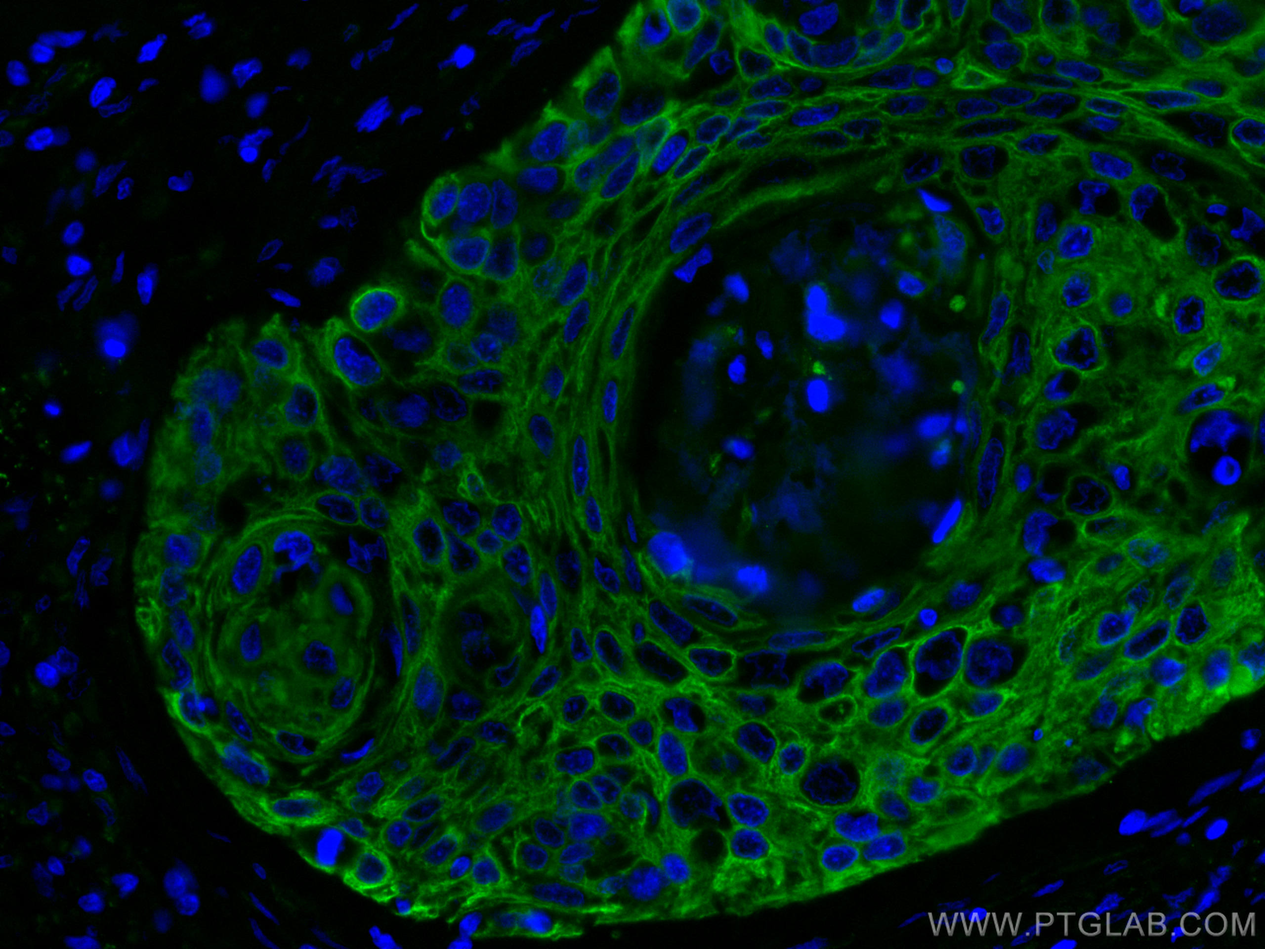
IF Staining of human prostate cancer using 66727-1-Ig (same clone as 66727-1-PBS)
Immunofluorescent analysis of (4% PFA) fixed human prostate cancer tissue using Cytokeratin 5 antibody (66727-1-Ig, Clone: 1A1C5 ) at dilution of 1:400 and CoraLite®488-Conjugated AffiniPure Goat Anti-Mouse IgG(H+L). This data was developed using the same antibody clone with 66727-1-PBS in a different storage buffer formulation.
× Immunofluorescent analysis of (4% PFA) fixed human prostate cancer tissue using Cytokeratin 5 antibody (66727-1-Ig, Clone: 1A1C5 ) at dilution of 1:400 and CoraLite®488-Conjugated AffiniPure Goat Anti-Mouse IgG(H+L). This data was developed using the same antibody clone with 66727-1-PBS in a different storage buffer formulation.
")
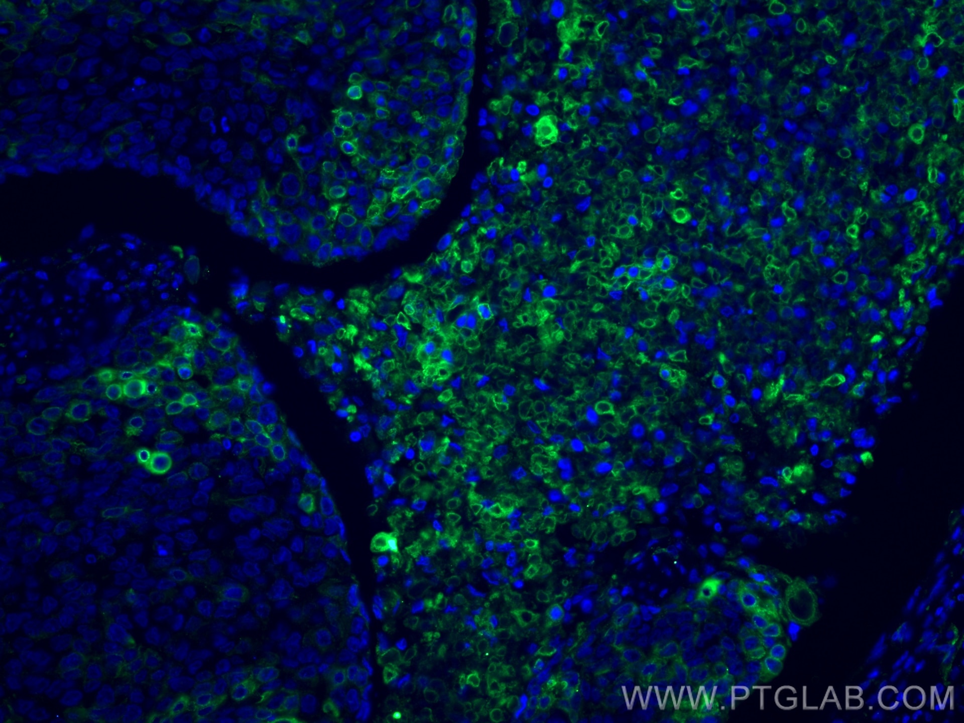
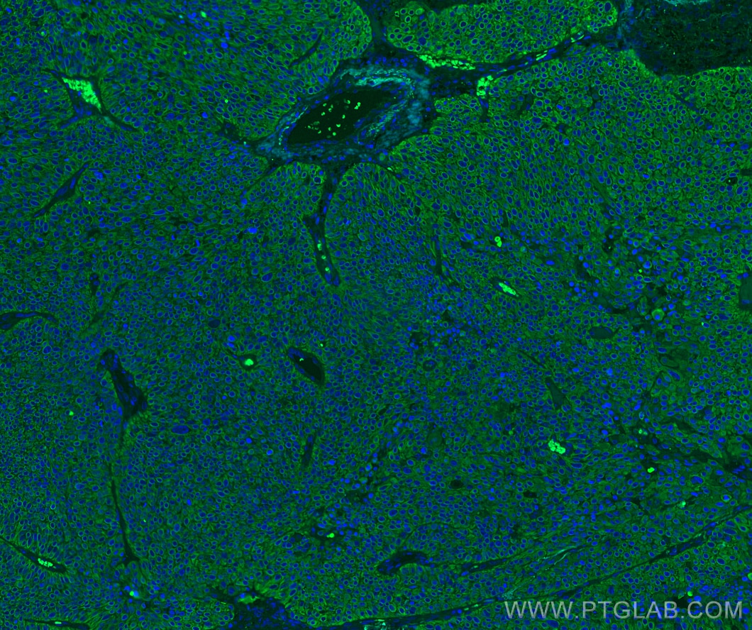
IF Staining of human lung cancer using 66727-1-Ig (same clone as 66727-1-PBS)
Immunofluorescent analysis of (4% PFA) fixed paraffin-embedded human lung cancer tissue using Cytokeratin 5 antibody (66727-1-Ig, Clone: 1A1C5 ) at dilution of 1:400 and CoraLite®488-Conjugated Goat Anti-Mouse IgG(H+L) (SA00013-1). Heat mediated antigen retrieval with Tris-EDTA buffer (pH 9.0). This data was developed using the same antibody clone with 66727-1-PBS in a different storage buffer formulation.
× Immunofluorescent analysis of (4% PFA) fixed paraffin-embedded human lung cancer tissue using Cytokeratin 5 antibody (66727-1-Ig, Clone: 1A1C5 ) at dilution of 1:400 and CoraLite®488-Conjugated Goat Anti-Mouse IgG(H+L) (SA00013-1). Heat mediated antigen retrieval with Tris-EDTA buffer (pH 9.0). This data was developed using the same antibody clone with 66727-1-PBS in a different storage buffer formulation.
")
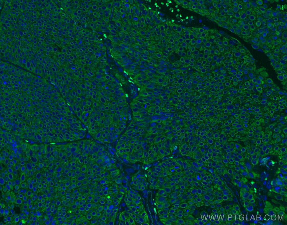
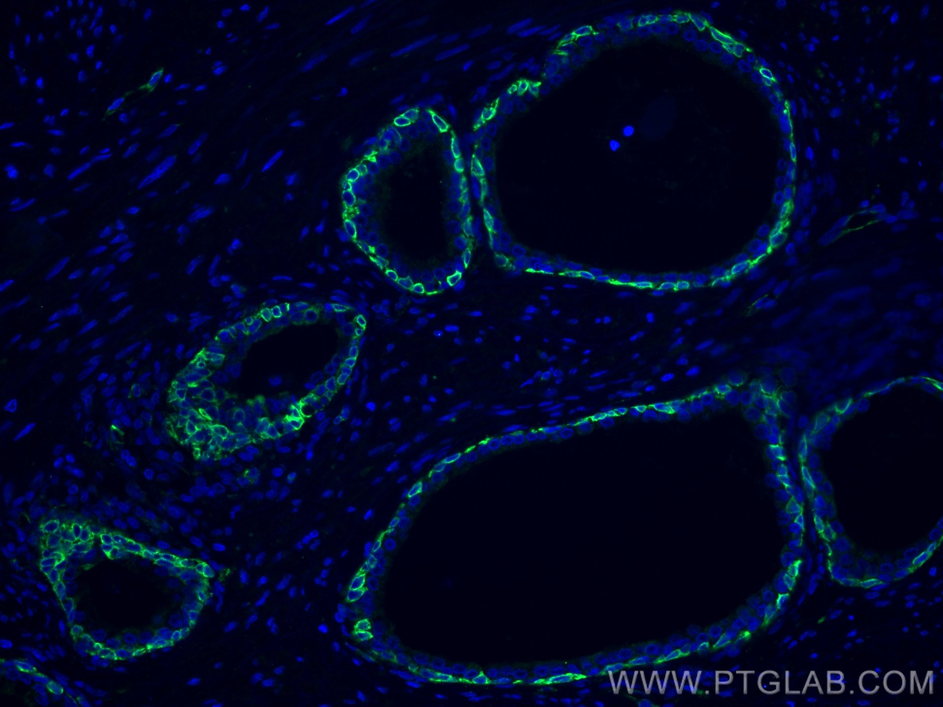
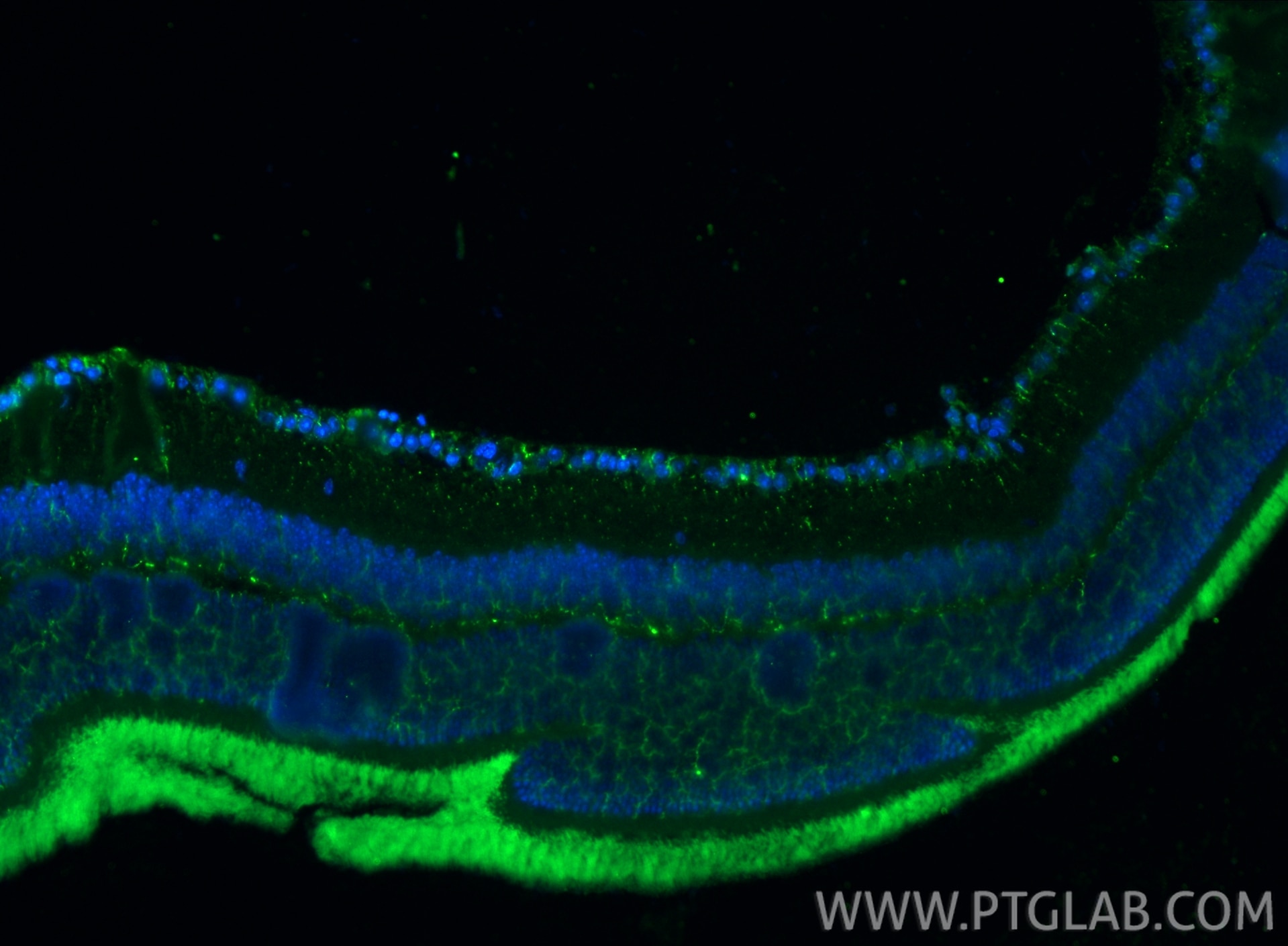
IF Staining of mouse eye using 66727-1-Ig (same clone as 66727-1-PBS)
Immunofluorescent analysis of (4% PFA) fixed paraffin-embedded mouse eye tissue using Cytokeratin 5 antibody (66727-1-Ig, Clone: 1A1C5 ) at dilution of 1:400 and CoraLite®488-Conjugated Goat Anti-Mouse IgG(H+L) (SA00013-1). Heat mediated antigen retrieval with Tris-EDTA buffer (pH 9.0). This data was developed using the same antibody clone with 66727-1-PBS in a different storage buffer formulation.
× Immunofluorescent analysis of (4% PFA) fixed paraffin-embedded mouse eye tissue using Cytokeratin 5 antibody (66727-1-Ig, Clone: 1A1C5 ) at dilution of 1:400 and CoraLite®488-Conjugated Goat Anti-Mouse IgG(H+L) (SA00013-1). Heat mediated antigen retrieval with Tris-EDTA buffer (pH 9.0). This data was developed using the same antibody clone with 66727-1-PBS in a different storage buffer formulation.
")
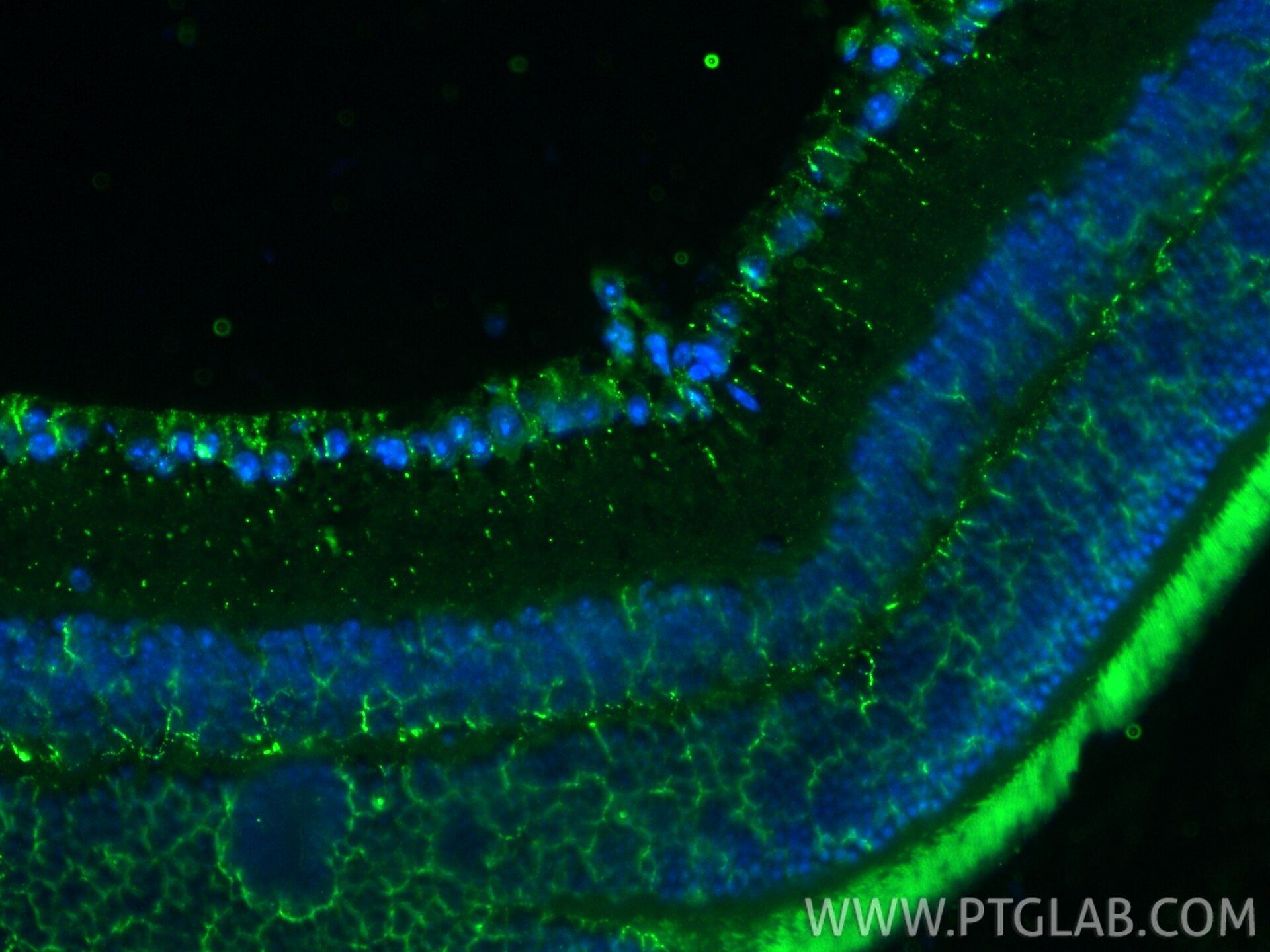
IF Staining of mouse eye using 66727-1-Ig (same clone as 66727-1-PBS)
Immunofluorescent analysis of (4% PFA) fixed paraffin-embedded mouse eye tissue using Cytokeratin 5 antibody (66727-1-Ig, Clone: 1A1C5 ) at dilution of 1:1000 and CoraLite®488-Conjugated Goat Anti-Mouse IgG(H+L) (SA00013-1). Heat mediated antigen retrieval with Tris-EDTA buffer (pH 9.0). This data was developed using the same antibody clone with 66727-1-PBS in a different storage buffer formulation.
× Immunofluorescent analysis of (4% PFA) fixed paraffin-embedded mouse eye tissue using Cytokeratin 5 antibody (66727-1-Ig, Clone: 1A1C5 ) at dilution of 1:1000 and CoraLite®488-Conjugated Goat Anti-Mouse IgG(H+L) (SA00013-1). Heat mediated antigen retrieval with Tris-EDTA buffer (pH 9.0). This data was developed using the same antibody clone with 66727-1-PBS in a different storage buffer formulation.
")
IF Staining of A431 using 66727-1-Ig (same clone as 66727-1-PBS)
Immunofluorescent analysis of (-20°C Methanol) fixed A431 cells using Cytokeratin 5 antibody (66727-1-Ig, Clone: 1A1C5 ) at dilution of 1:400 and CoraLite®488-Conjugated AffiniPure Goat Anti-Mouse IgG(H+L). This data was developed using the same antibody clone with 66727-1-PBS in a different storage buffer formulation.
× Immunofluorescent analysis of (-20°C Methanol) fixed A431 cells using Cytokeratin 5 antibody (66727-1-Ig, Clone: 1A1C5 ) at dilution of 1:400 and CoraLite®488-Conjugated AffiniPure Goat Anti-Mouse IgG(H+L). This data was developed using the same antibody clone with 66727-1-PBS in a different storage buffer formulation.
Proteintech Guarantee
The Proteintech guarantee covers Proteintech antibodies in any species and any application, including those not listed on the datasheet. If the antibody doesn’t perform, you can receive a hassle-free refund or credit note.
Product Information
66727-1-PBS targets Cytokeratin 5 in WB, IHC, IF/ICC, IF-P, ELISA applications and shows reactivity with human, mouse, pig samples.
| Tested Reactivity | human, mouse, pig |
| Host / Isotype | Mouse / IgG1 |
| Class | Monoclonal |
| Type | Antibody |
| Immunogen |
CatNo: Ag24184 Product name: Recombinant human KRT5 protein Source: e coli.-derived, PGEX-4T Tag: GST Domain: 316-491 aa of BC024292 Sequence: THVSDTSVVLSMDNNRNLDLDSIIAEVKAQYEEIANRSRTEAESWYQTKYEELQQTAGRHGDDLRNTKHEISEMNRMIQRLRAEIDNVKKQCANLQNAIADAEQRGELALKDARNKLAELEEALQKAKQDMARLLREYQELMNTKLALDVEIATYRKLLEGEECRLSGEGVGPVNI Predict reactive species |
| Full Name | keratin 5 |
| Calculated Molecular Weight | 590 aa, 62 kDa |
| Observed Molecular Weight | 60 kDa |
| GenBank Accession Number | BC024292 |
| Gene Symbol | Cytokeratin 5 |
| Gene ID (NCBI) | 3852 |
| RRID | AB_2882077 |
| Conjugate | Unconjugated |
| Form | Liquid |
| Purification Method | Protein G purification |
| UNIPROT ID | P13647 |
| Storage Buffer | PBS only, pH 7.3. |
| Storage Conditions | Store at -80°C. |
Background Information
Keratins are a large family of proteins that form the intermediate filament cytoskeleton of epithelial cells. Keratin expression is highly regulated, tissue specific, and varies according to cell-state. Type I keratins consist of acidic, low molecular weight proteins with MW ranging from 40 kDa (KRT19) to 64 kDa (KRT9). Type 2 keratins consist of basic or neutral, high molecular weight proteins with MW from 52 kDa (KRT8) to 67 kDa (KRT18). Keratin 5, one 58-kD type II keratin, is coexpressed with a 50-kD keratin 14 in stratified squamous epithelia. Keratin 5, one 58-kD type II keratin, is coexpressed with a 50-kD tyK14 in stratified squamous epithelia.