

Tested Applications
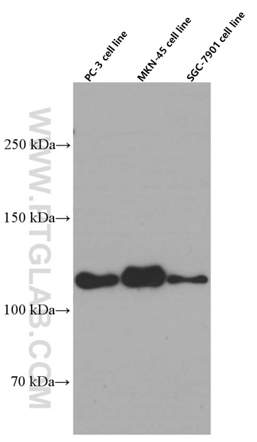
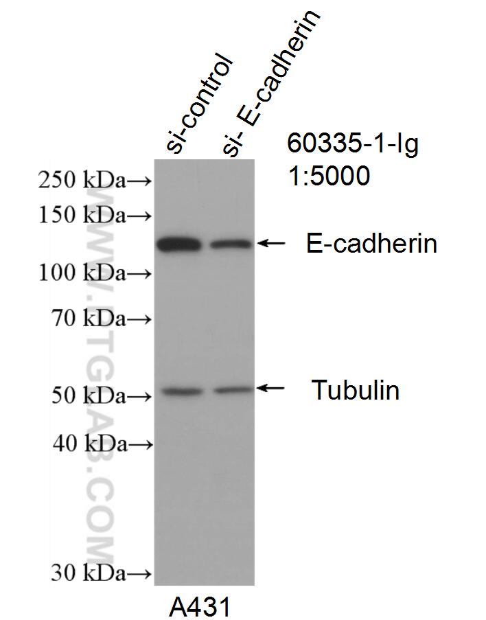
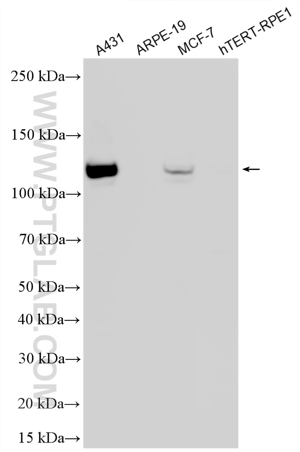
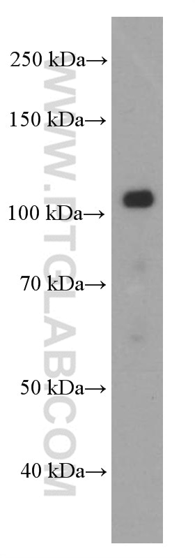
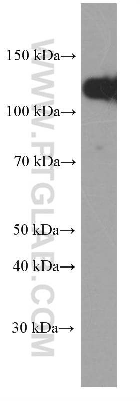
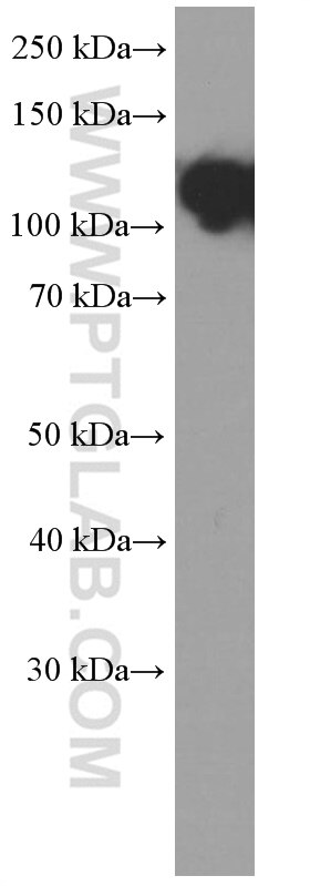
| Positive WB detected in | PC-3 cells, A431 cells, MCF-7 cells, pig brain tissue, MKN-45 cells, SGC-7901 cells |
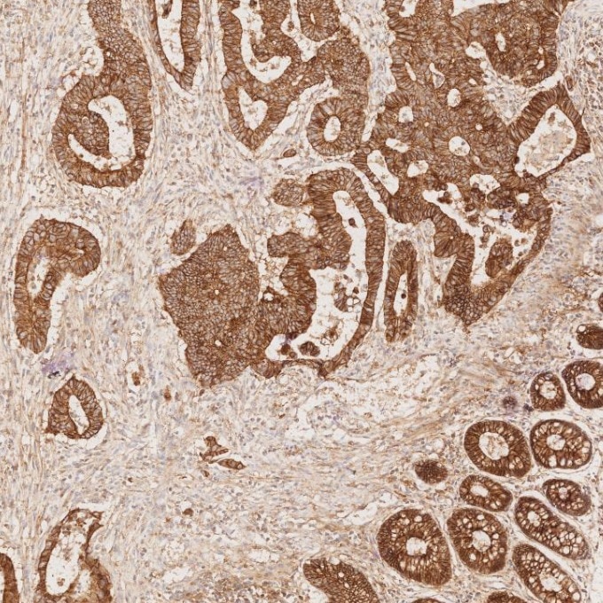
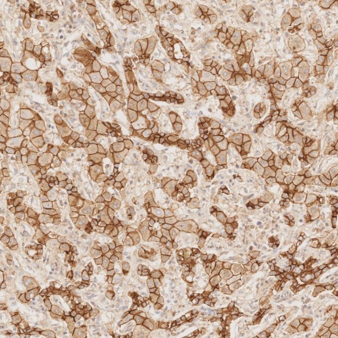
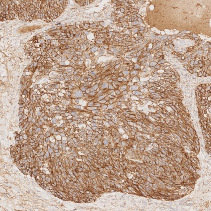
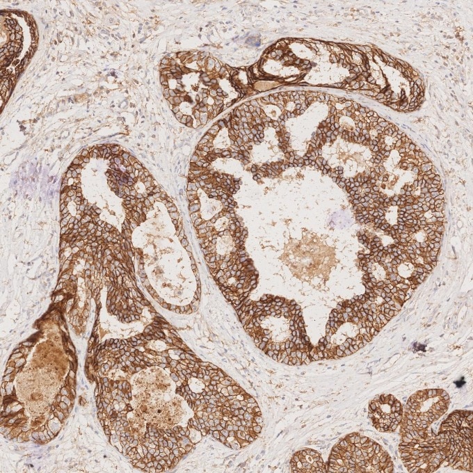
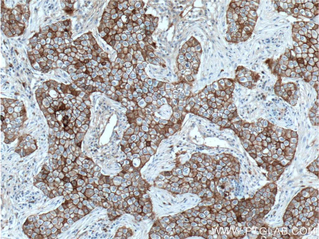
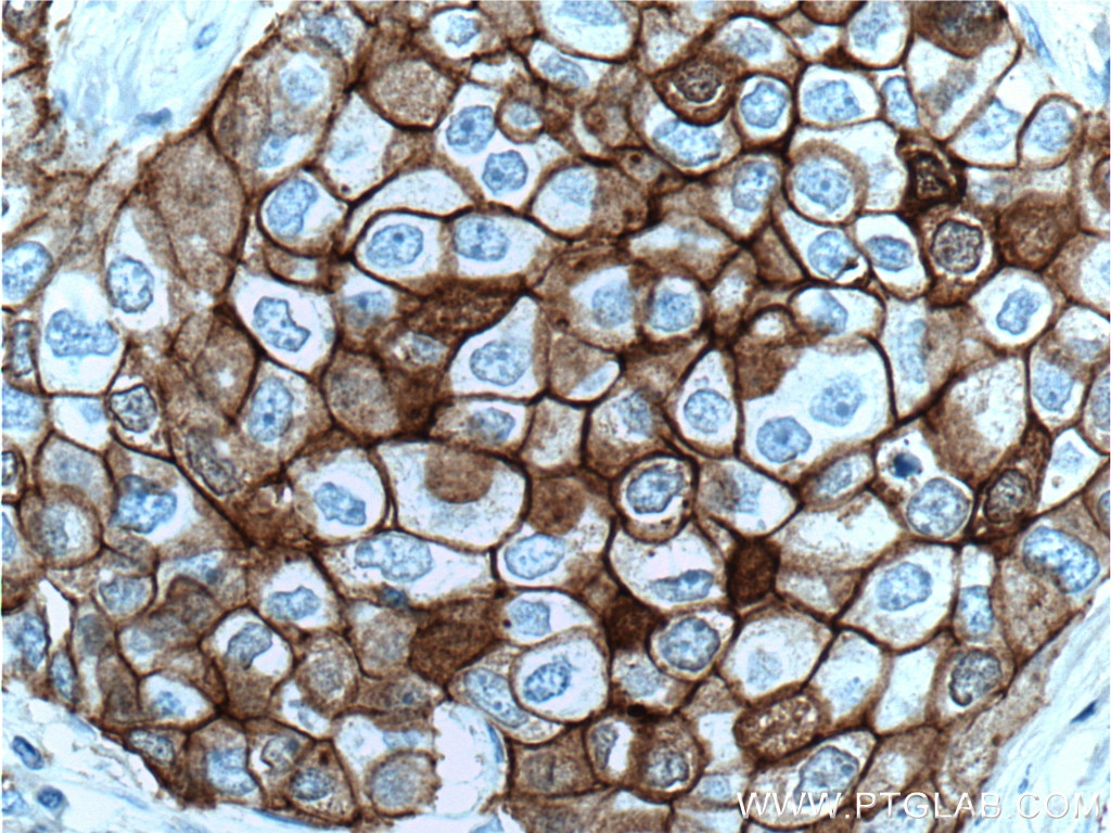
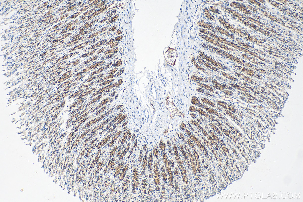
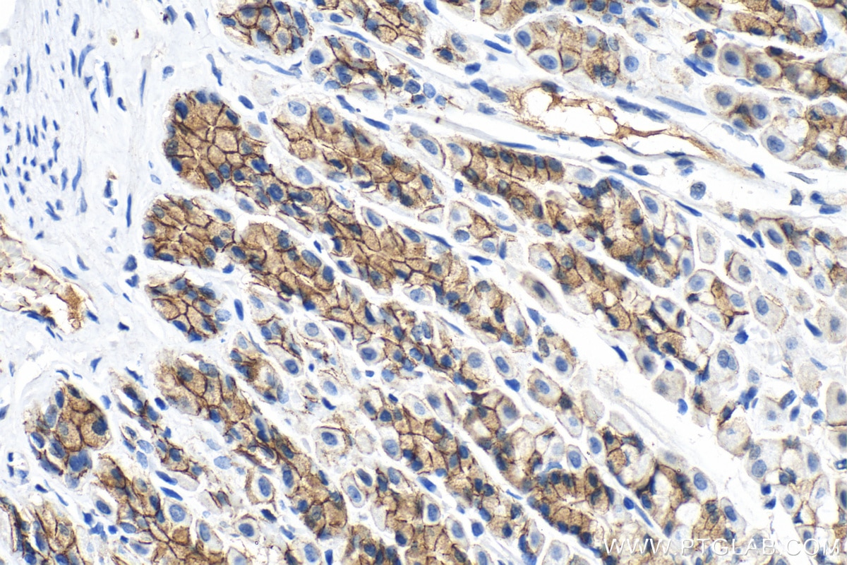
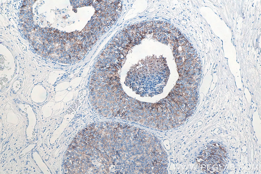
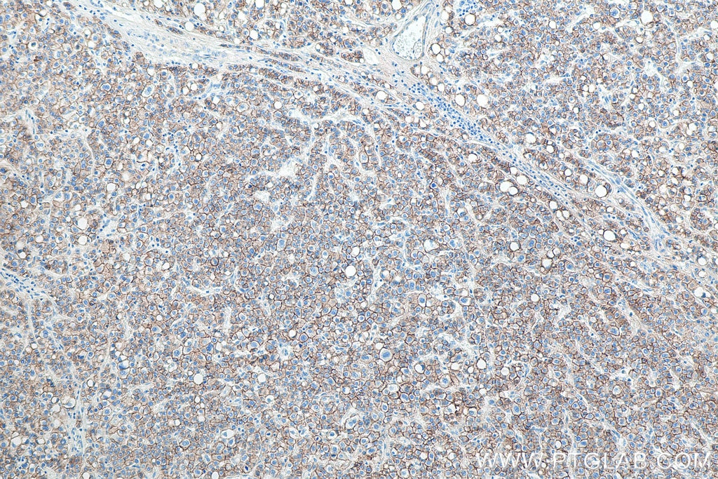
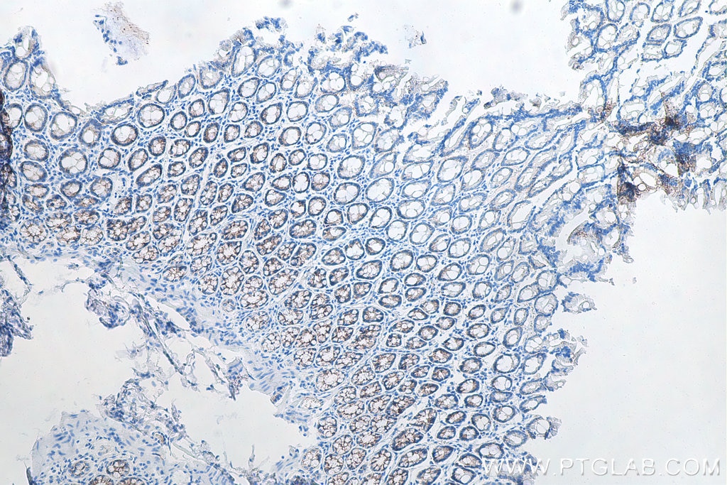
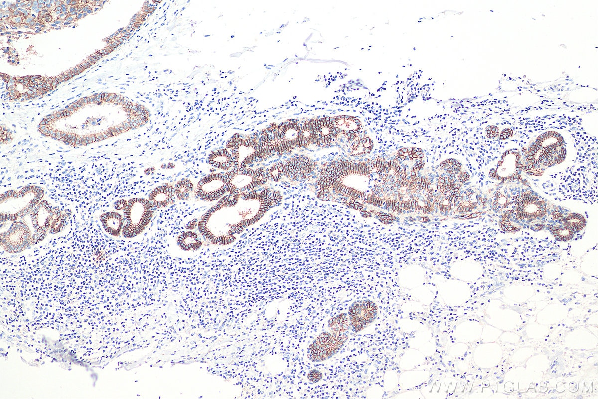
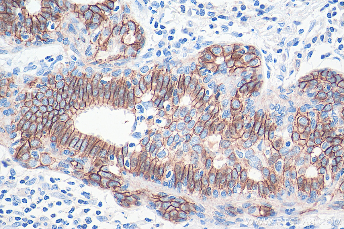
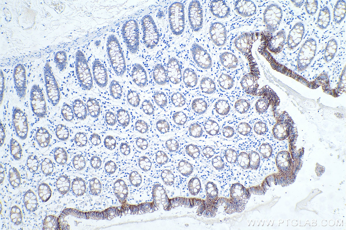
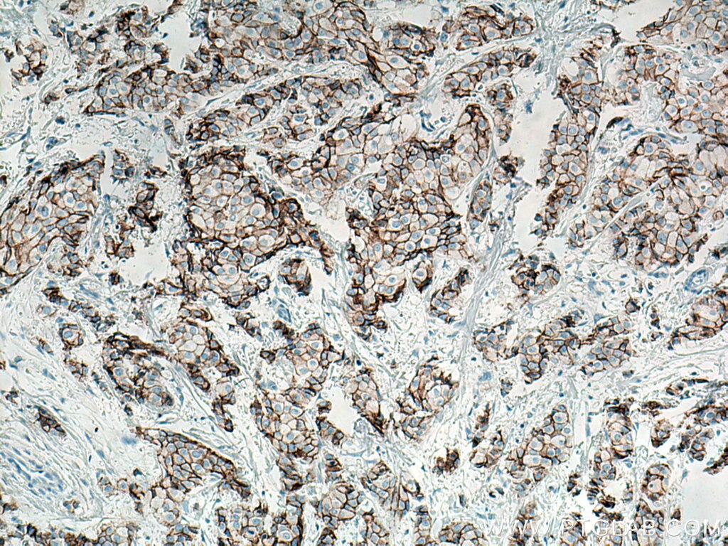
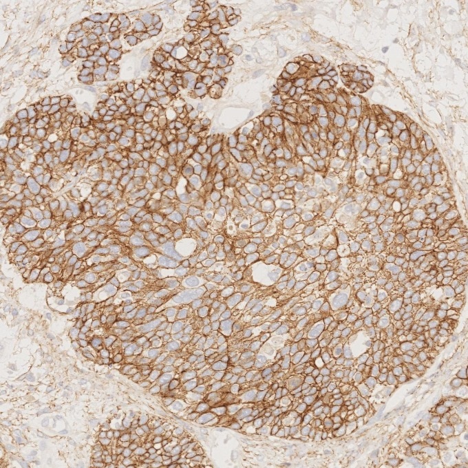
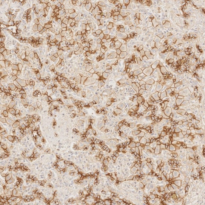
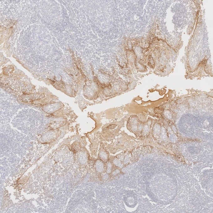
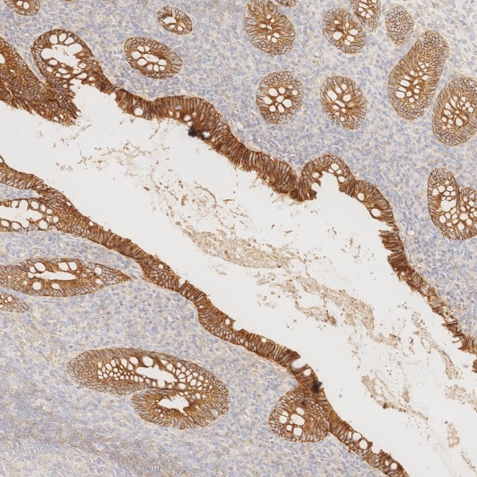
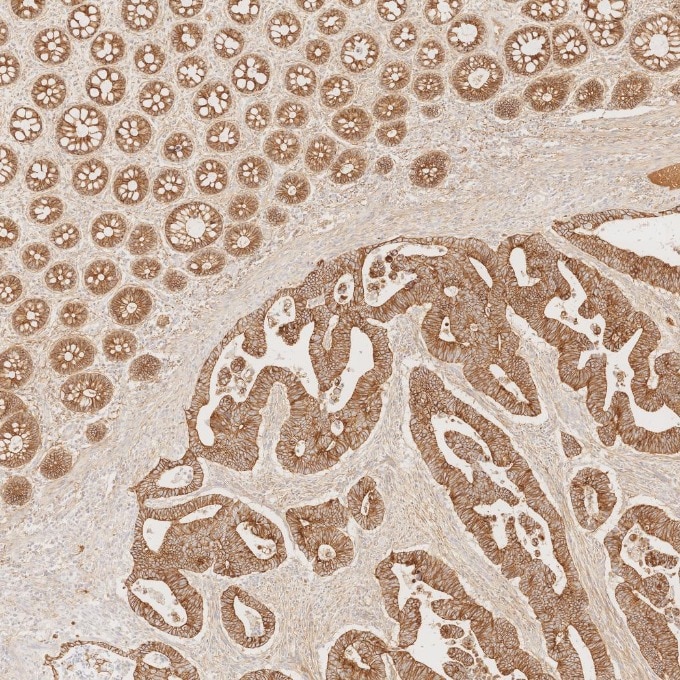
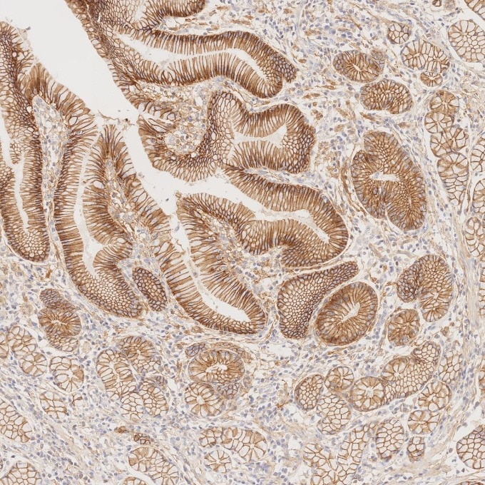
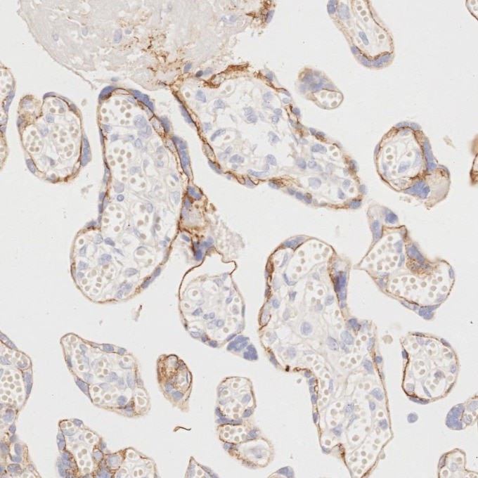
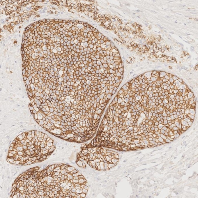
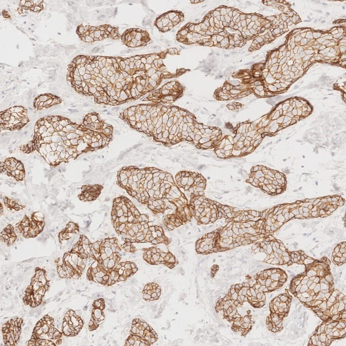
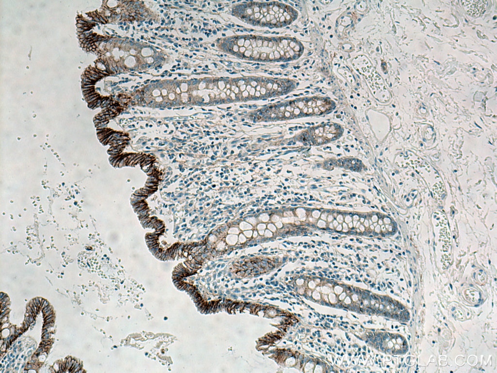
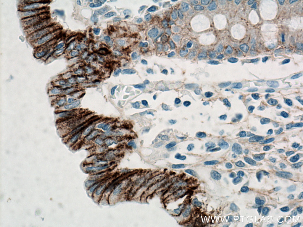
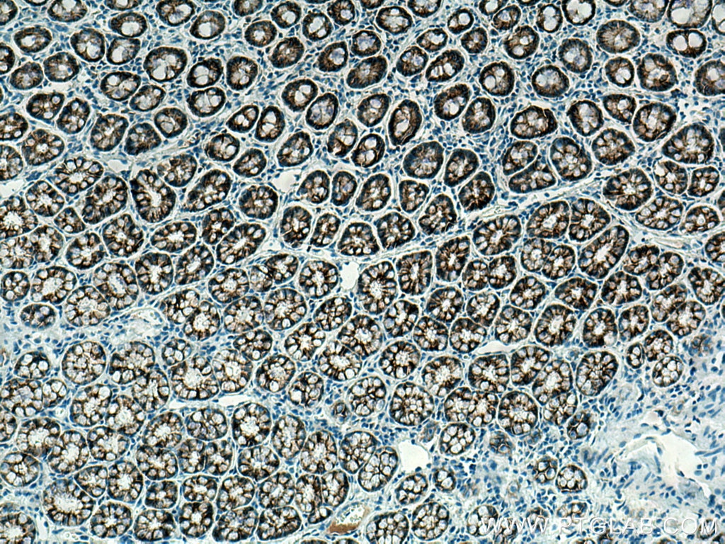
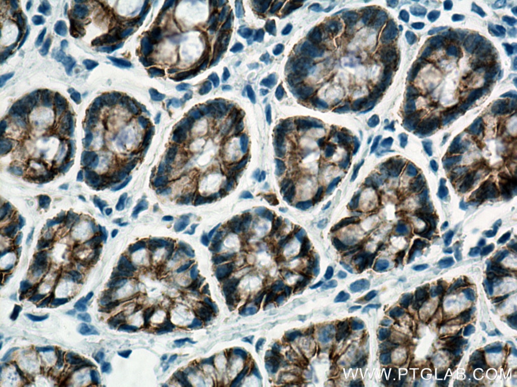
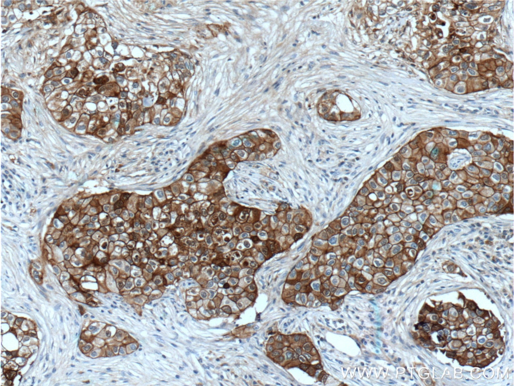
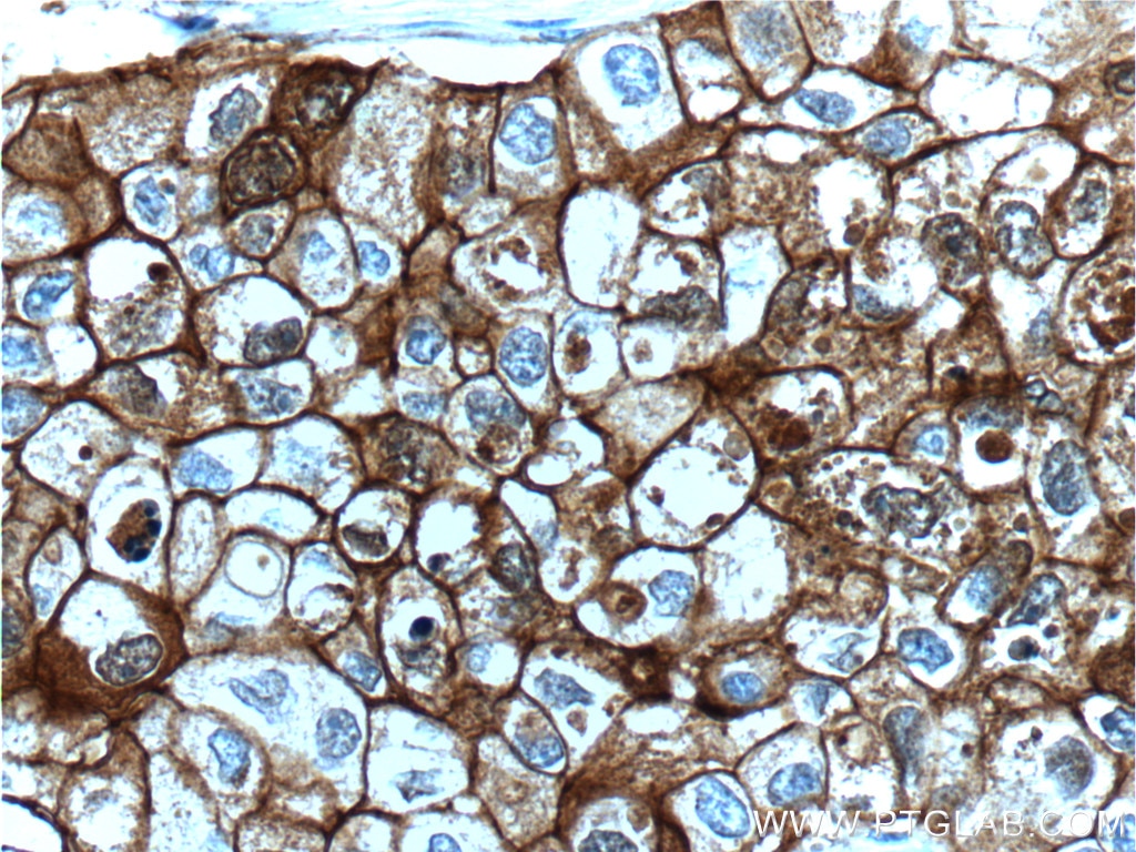
| Positive IHC detected in | human breast cancer tissue, human liver tissue, human colon tissue, human breast cancer tissue, human placenta tissue, human stomach cancer tissue, human colorectal cancer tissue, rat colon tissue, human liver cancer tissue, human appendicitis tissue, human tonsillitis tissue, human ovary cancer tissue, rat stomach tissue Note: suggested antigen retrieval with TE buffer pH 9.0; (*) Alternatively, antigen retrieval may be performed with citrate buffer pH 6.0 |
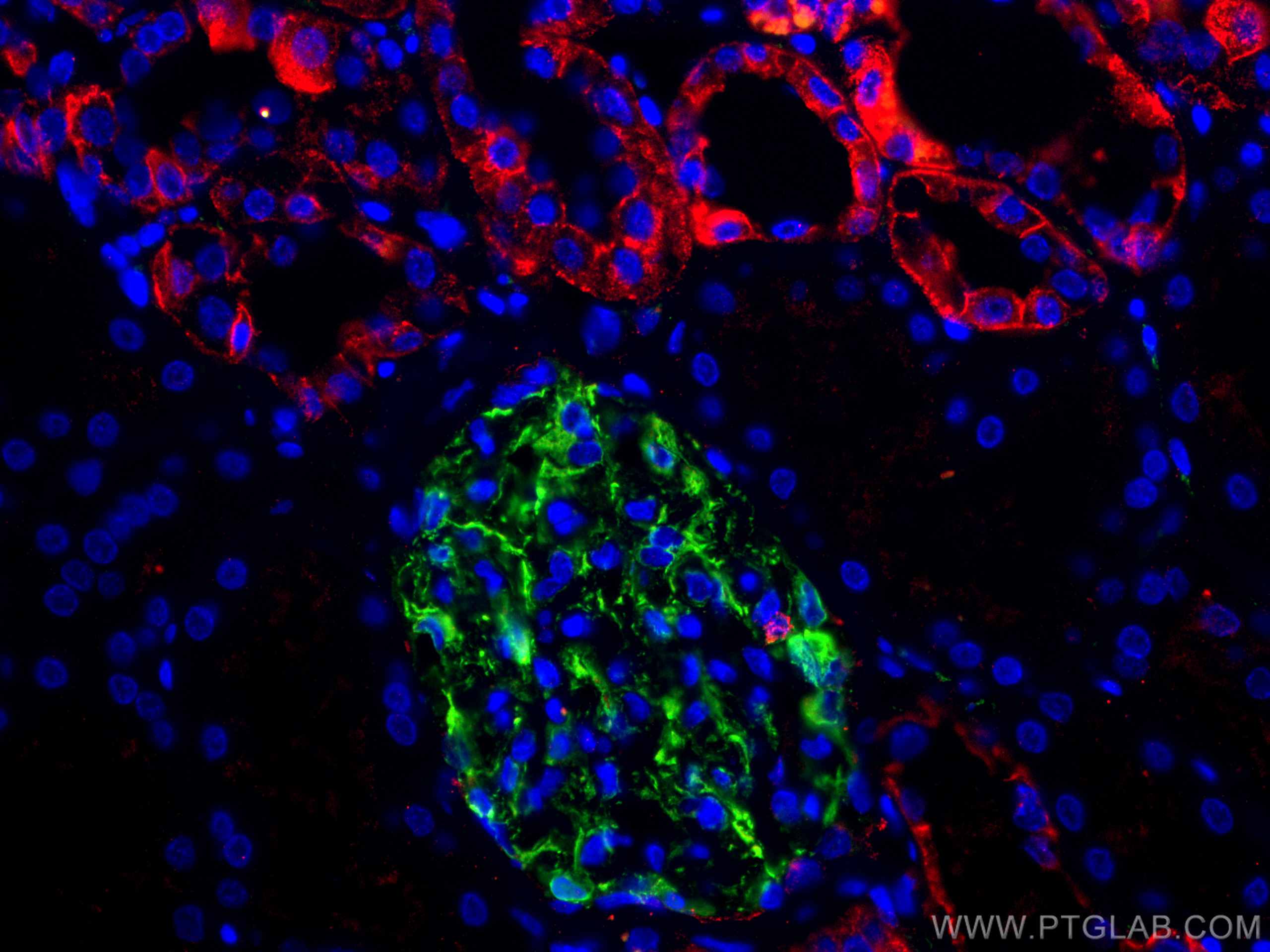
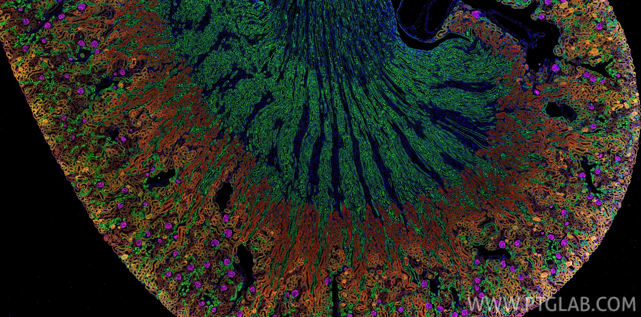
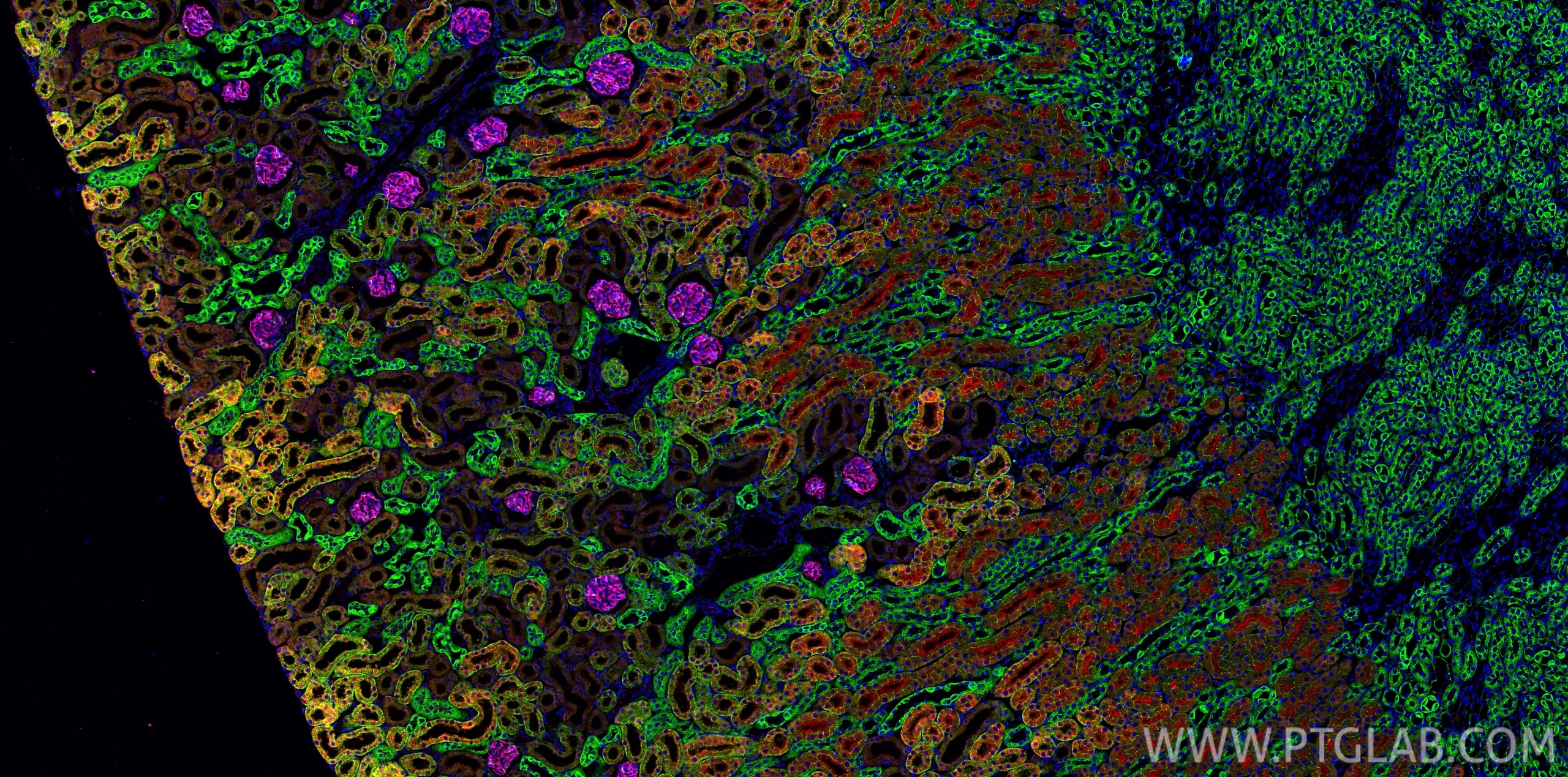
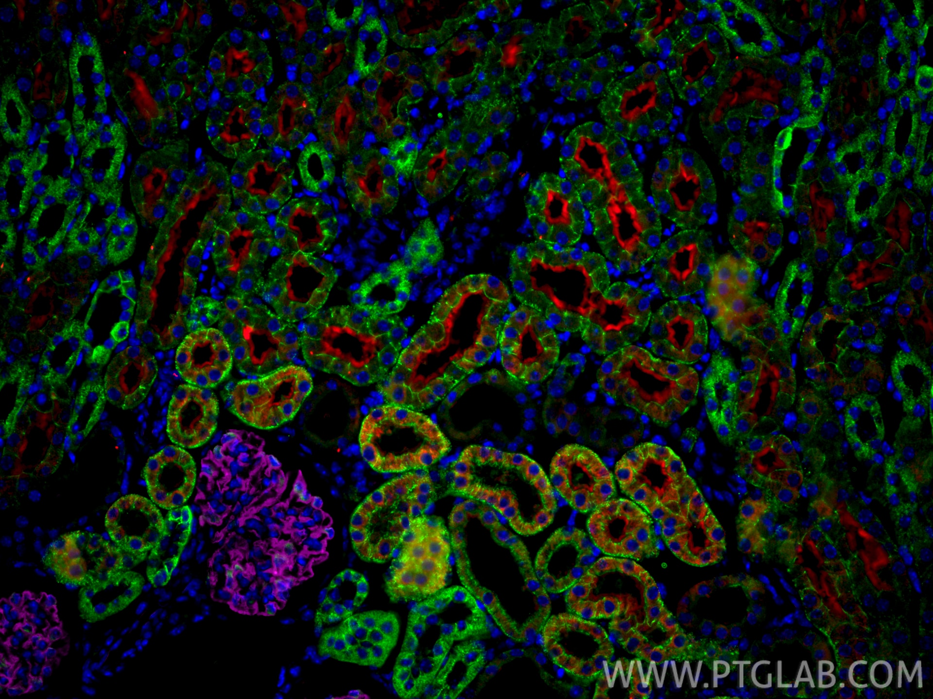
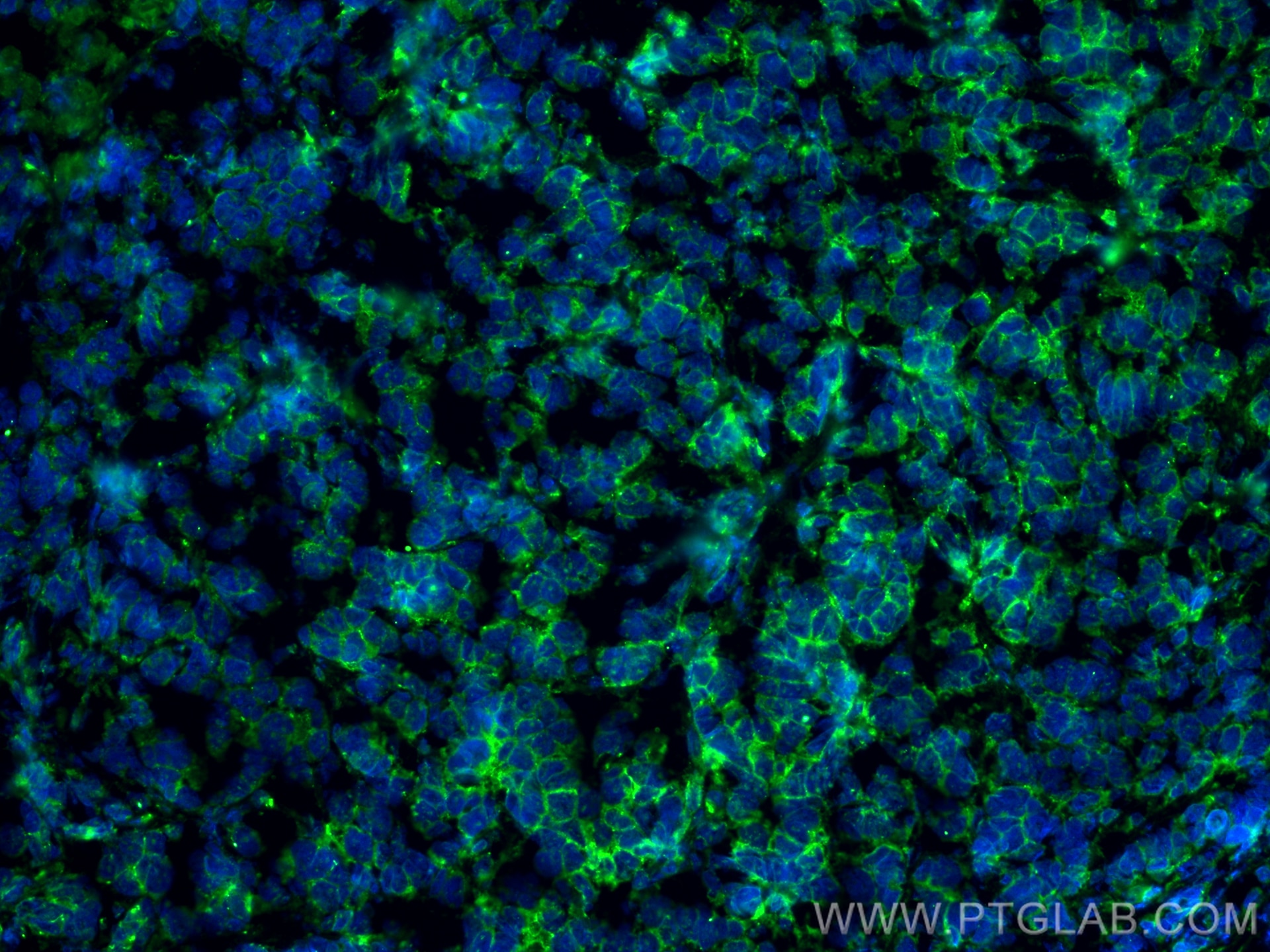
| Positive IF-P detected in | human breast cancer tissue, human kidney tissue, mouse kidney tissue |
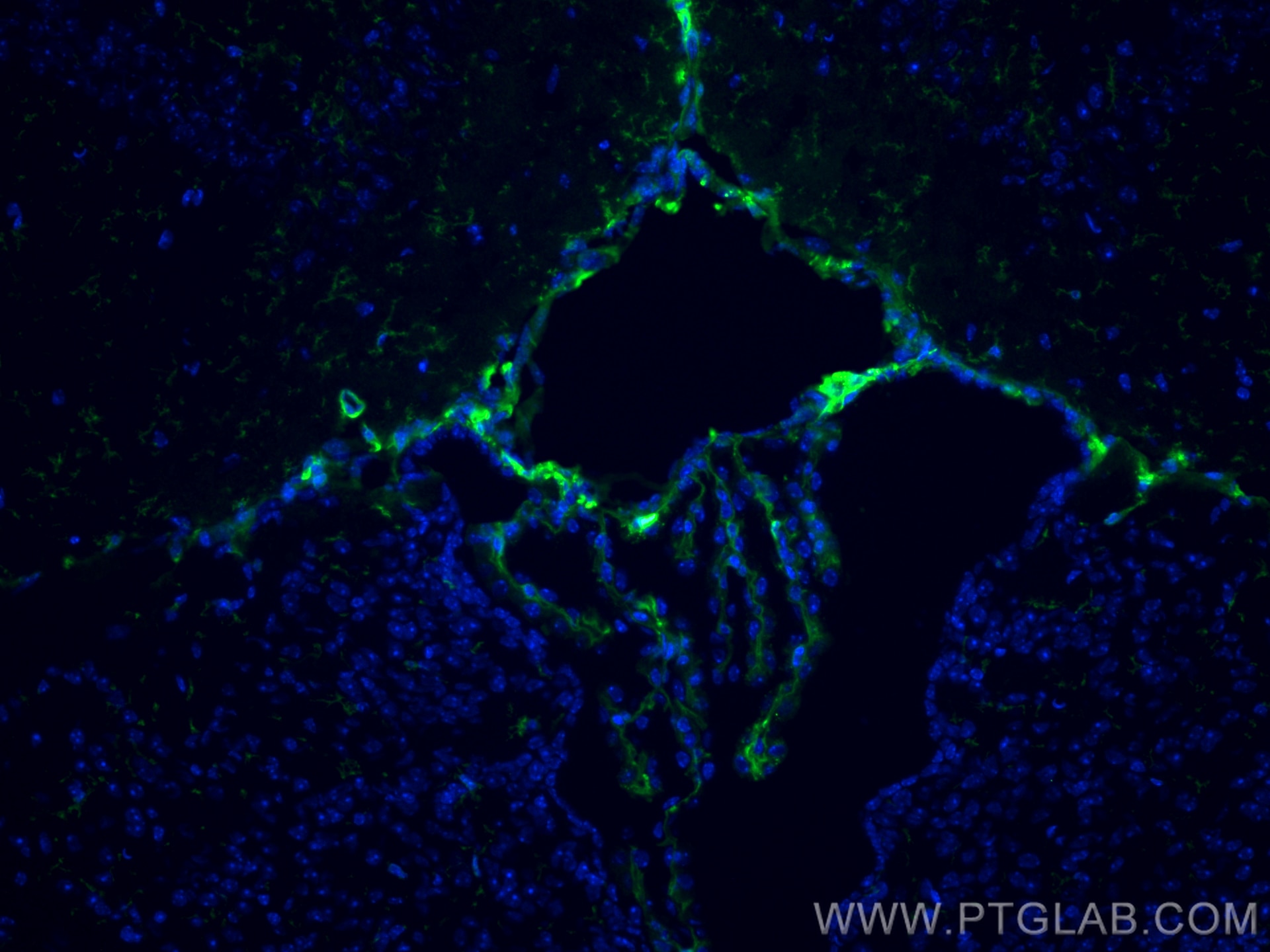
| Positive IF-Fro detected in | mouse brain tissue |
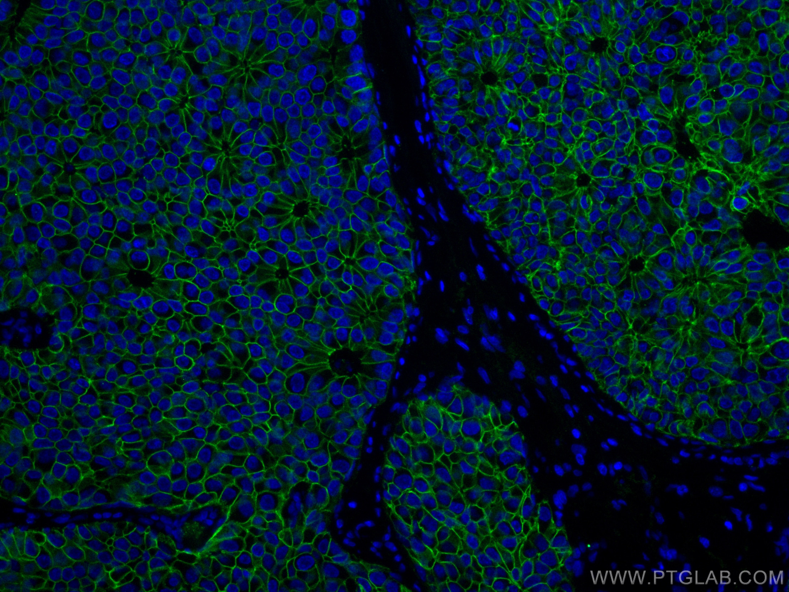
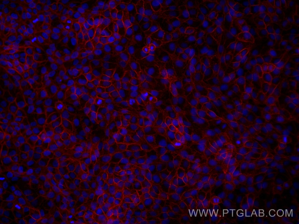
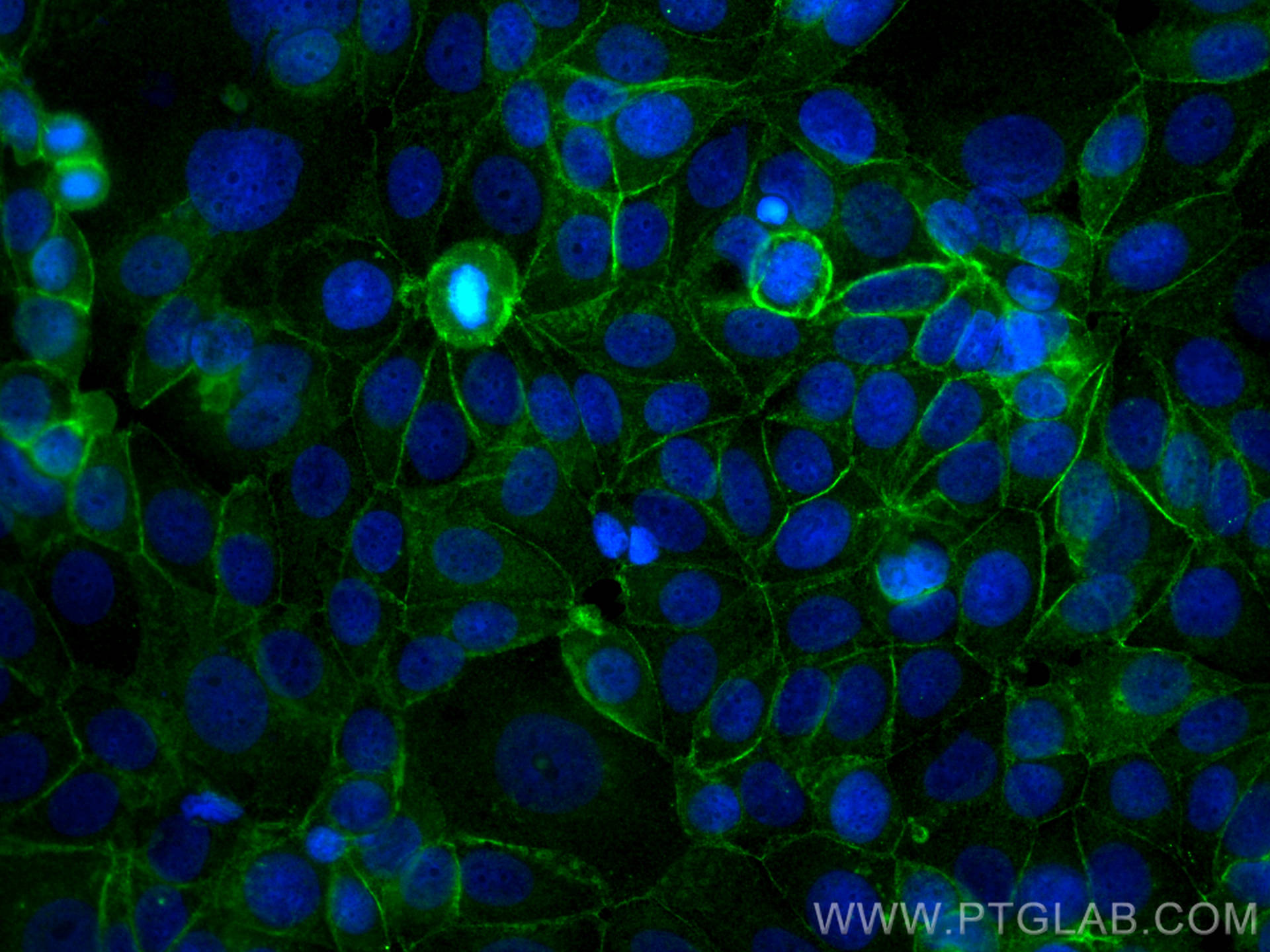
| Positive IF/ICC detected in | MCF-7 cells, mouse breast cancer, HaCaT cells |
Recommended dilution
| Application | Dilution |
|---|---|
| Western Blot (WB) | WB : 1:2000-1:8000 |
| Immunohistochemistry (IHC) | IHC : 1:1000-1:4000 |
| Immunofluorescence (IF)-P | IF-P : 1:200-1:800 |
| Immunofluorescence (IF)-FRO | IF-FRO : 1:200-1:800 |
| Immunofluorescence (IF)/ICC | IF/ICC : 1:200-1:800 |
| It is recommended that this reagent should be titrated in each testing system to obtain optimal results. | |
| Sample-dependent, Check data in validation data gallery. | |
Product Information
60335-1-Ig targets E-cadherin in WB, IHC, IF/ICC, IF-P, IF-Fro, ELISA applications and shows reactivity with human, mouse, rat, pig samples.
| Tested Reactivity | human, mouse, rat, pig |
| Cited Reactivity | human, mouse, rat, pig, canine, monkey |
| Host / Isotype | Mouse / IgG2b |
| Class | Monoclonal |
| Type | Antibody |
| Immunogen |
CatNo: Ag15085 Product name: Recombinant human E-cadherin protein Source: e coli.-derived, PET28a Tag: 6*His Domain: 373-622 aa of BC141838 Sequence: PIFNPTTYKGQVPENEANVVITTLKVTDADAPNTPAWEAVYTILNDDGGQFVVTTNPVNNDGILKTAKGLDFEAKQQYILHVAVTNVVPFEVSLTTSTATVTVDVLDVNEAPIFVPPEKRVEVSEDFGVGQEITSYTAQEPDTFMEQKITYRIWRDTANWLEINPDTGAISTRAELDREDFEHVKNSTYTALIIATDNGSPVATGTGTLLLILSDVNDNAPIPEPRTIFFCERNPKPQVINIIDADLPPI Predict reactive species |
| Full Name | cadherin 1, type 1, E-cadherin (epithelial) |
| Calculated Molecular Weight | 882 aa, 97 kDa |
| Observed Molecular Weight | 120 kDa |
| GenBank Accession Number | BC141838 |
| Gene Symbol | E-cadherin |
| Gene ID (NCBI) | 999 |
| RRID | AB_2881444 |
| Conjugate | Unconjugated |
| Form | Liquid |
| Purification Method | Protein A purification |
| UNIPROT ID | P12830 |
| Storage Buffer | PBS with 0.02% sodium azide and 50% glycerol, pH 7.3. |
| Storage Conditions | Store at -20°C. Stable for one year after shipment. Aliquoting is unnecessary for -20oC storage. 20ul sizes contain 0.1% BSA. |
Background Information
Cadherins are a family of transmembrane glycoproteins that mediate calcium-dependent cell-cell adhesion and play an important role in the maintenance of normal tissue architecture. E-cadherin (epithelial cadherin), also known as CDH1 (cadherin 1) or CAM 120/80, is a classical member of the cadherin superfamily which also include N-, P-, R-, and B-cadherins. E-cadherin is expressed on the cell surface in most epithelial tissues. The extracellular region of E-cadherin establishes calcium-dependent homophilic trans binding, providing specific interaction with adjacent cells, while the cytoplasmic domain is connected to the actin cytoskeleton through the interaction with p120-, α-, β-, and γ-catenin (plakoglobin). E-cadherin is important in the maintenance of the epithelial integrity, and is involved in mechanisms regulating proliferation, differentiation, and survival of epithelial cell. E-cadherin may also play a role in tumorigenesis. It is considered to be an invasion suppressor protein and its loss is an indicator of high tumor aggressiveness.
Protocols
| Product Specific Protocols | |
|---|---|
| IF protocol for E-cadherin antibody 60335-1-Ig | Download protocol |
| IHC protocol for E-cadherin antibody 60335-1-Ig | Download protocol |
| WB protocol for E-cadherin antibody 60335-1-Ig | Download protocol |
| Standard Protocols | |
|---|---|
| Click here to view our Standard Protocols |
Publications
What published studies show
The 60335-1-Ig polyclonal antibody (targeting non-muscle myosin heavy chain II/MYH9) has 318 total citations. These appear across high-impact journals including Nature Communications, Science Advances, EMBO Journal, Cell Reports, Cancer Research, Theranostics, Cell Death & Disease, Oncogene, and Journal of Experimental & Clinical Cancer Research. Research fields center on epithelial-mesenchymal transition (EMT), cancer metastasis, and tumor microenvironment, alongside fibrosis, ferroptosis, and inflammatory diseases, with applications mainly in Western blot, immunohistochemistry, and immunofluorescence.
| Species | Application | Title |
|---|---|---|
Mol Cancer OLFM4 promotes the progression of intestinal metaplasia through activation of the MYH9/GSK3β/β-catenin pathway | ||
Adv Mater Glycated ECM Derived Carbon Dots Inhibit Tumor Vasculogenic Mimicry by Disrupting RAGE Nuclear Translocation and Its Interaction With HMGB1 | ||
Cancer Res Single-Cell and Spatial Transcriptome Profiling Identifies the Transcription Factor BHLHE40 as a Driver of EMT in Metastatic Colorectal Cancer | ||
Cancer Res SRSF9 Forms Phase-separated Condensates to Promote Ovarian Cancer Progression by Inducing RNA Alternative Splicing that is Inhibited by m6A Modification | ||
Nat Commun Self-assembly of hybrid 3D cultures by integrating living and synthetic cells. | ||
Nat Commun Choline metabolism drives metastasis in BRCA1-deficient ovarian cancers by activating FAM3C. |
Reviews
What customers say
Overall, reviewers report positive results with this antibody, particularly for immunofluorescence. Several users noted bright staining with minimal background in Caco-2 and HT-29 cells at dilutions ranging from 1:500 to 1:800. One user confirmed good performance in Western Blot at 1:2000. However, one reviewer experienced no labeling in MCF7 cells using a 1:250 dilution with confocal imaging, suggesting cell-type-specific variability. The majority of feedback is favorable, with most users recommending the product for IF applications.
The reviews below have been submitted by verified Proteintech customers who received an incentive for providing their feedback.
Aqib (Verified Customer) (09-02-2024) | It works quite well. recommended
|
Saba (Verified Customer) (06-14-2022) | The IF staining was very good and satisfying.
|
Silvia (Verified Customer) (02-09-2022) | The antibody worked well on HT-29 cells at 1:800 dilution for IF.
|
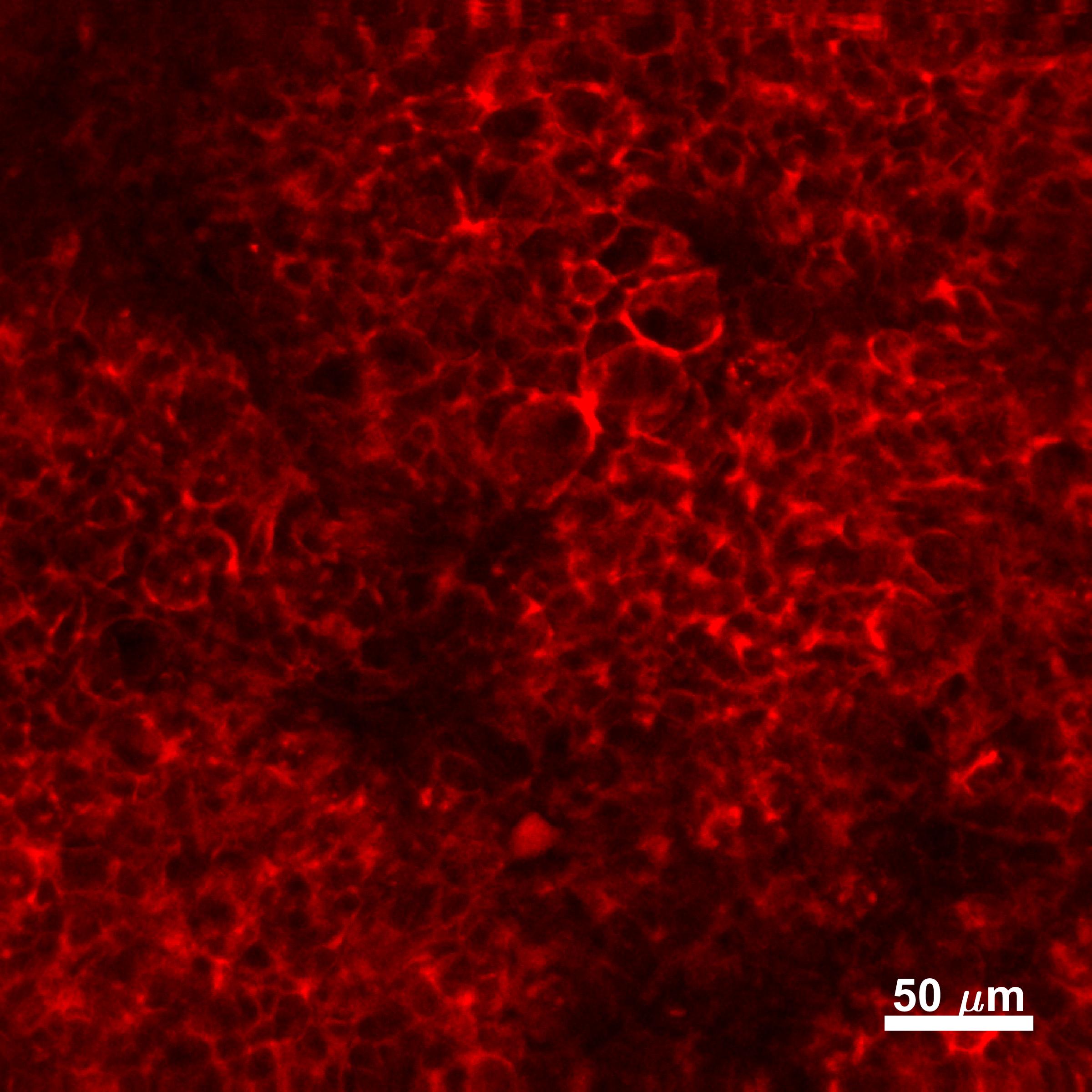
Joshua (Verified Customer) (12-27-2019) | Caco-2 cells fixed in 4% paraformaldehyde. Stained overnight at 4C. Bright stain, minimal background
 |
Louisiane (Verified Customer) (02-06-2019) | Cells were fixed with 4% PFA for 10 min, permeabilized with 0.1% Triton-X100 for 5 min and blocked with 1% FBS/1% BSA in PBS for 3 h. Antibodies were diluted in 1% FBS/1% BSA in PBS. Primary antibody: 2 h. Alexa Fluor anti-mouse secondary antibody (1:250): 1 h.Cells were imaged by confocal microscopy - no labeling was observed.
|














































