The Proteintech guarantee covers Proteintech antibodies in any species and any application, including those not listed on the datasheet. If the antibody doesn’t perform, you can receive a hassle-free refund or credit note.
HeLa cells were subjected to SDS PAGE followed by western blot with 55237-1-AP (ENO1 antibody) at dilution of 1:2000 incubated at room temperature for 1.5 hours.
HeLa cells were subjected to SDS PAGE followed by western blot with 55237-1-AP (ENO1 antibody) at dilution of 1:2000 incubated at room temperature for 1.5 hours.
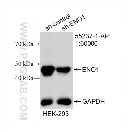
WB analysis of HEK-293 using 55237-1-AP
WB result of ENO1 antibody (55237-1-AP; 1:60000; incubated at room temperature for 1.5 hours) with sh-Control and sh-ENO1 transfected HEK-293 cells.
WB result of ENO1 antibody (55237-1-AP; 1:60000; incubated at room temperature for 1.5 hours) with sh-Control and sh-ENO1 transfected HEK-293 cells.
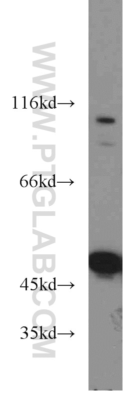
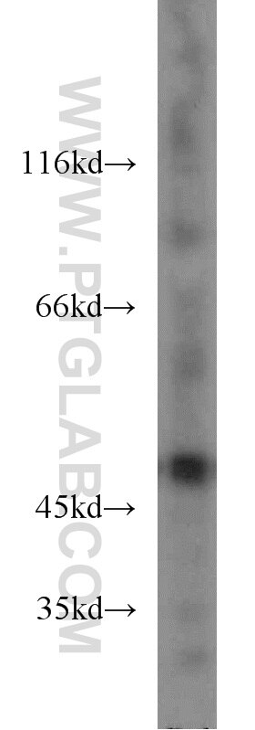
WB analysis of mouse liver using 55237-1-AP
mouse liver tissue were subjected to SDS PAGE followed by western blot with 55237-1-AP (ENO1 antibody) at dilution of 1:2000 incubated at room temperature for 1.5 hours.
mouse liver tissue were subjected to SDS PAGE followed by western blot with 55237-1-AP (ENO1 antibody) at dilution of 1:2000 incubated at room temperature for 1.5 hours.
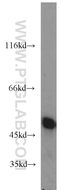
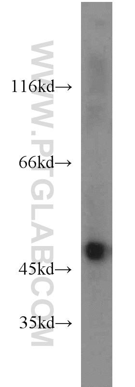
WB analysis of HeLa using 55237-1-AP
HeLa cells were subjected to SDS PAGE followed by western blot with 55237-1-AP (ENO1 antibody) at dilution of 1:2000 incubated at room temperature for 1.5 hours.
HeLa cells were subjected to SDS PAGE followed by western blot with 55237-1-AP (ENO1 antibody) at dilution of 1:2000 incubated at room temperature for 1.5 hours.
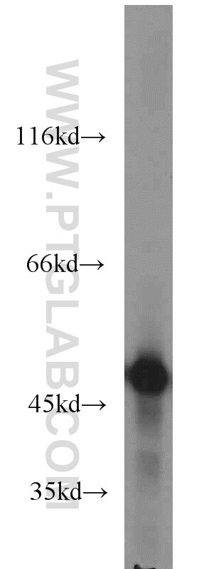
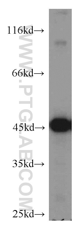
WB analysis of HepG2 using 55237-1-AP
HepG2 cells were subjected to SDS PAGE followed by western blot with 55237-1-AP (ENO1 antibody) at dilution of 1:2000 incubated at room temperature for 1.5 hours.
HepG2 cells were subjected to SDS PAGE followed by western blot with 55237-1-AP (ENO1 antibody) at dilution of 1:2000 incubated at room temperature for 1.5 hours.
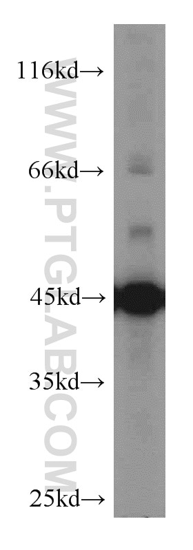
WB analysis of mouse brain using 55237-1-AP
mouse brain tissue were subjected to SDS PAGE followed by western blot with 55237-1-AP (ENO1 antibody) at dilution of 1:500 incubated at room temperature for 1.5 hours.
mouse brain tissue were subjected to SDS PAGE followed by western blot with 55237-1-AP (ENO1 antibody) at dilution of 1:500 incubated at room temperature for 1.5 hours.
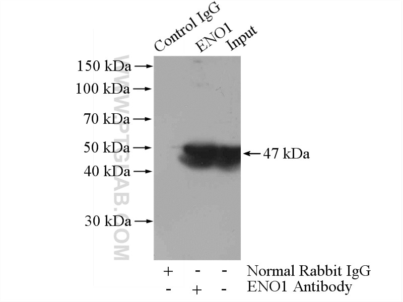
IP experiment of mouse brain using 55237-1-AP
IP result of anti-ENO1 (IP:55237-1-AP, 4ug; Detection:55237-1-AP 1:500) with mouse brain tissue lysate 4000ug.
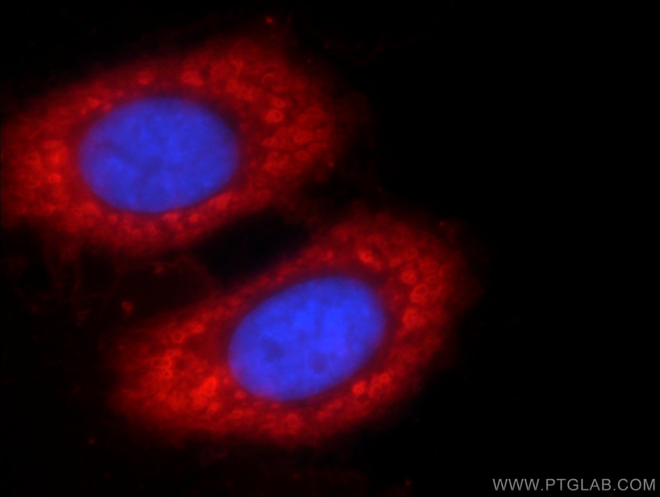
Immunofluorescent analysis of HepG2 cells using 55237-1-AP (ENO1 antibody) at dilution of 1:50 and Rhodamine-Goat anti-Rabbit IgG.
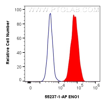
FC experiment of HeLa using 55237-1-AP
1x10^6 HeLa cells were intracellularly stained with 0.25 ug ENO1 Polyclonal antibody (55237-1-AP) and CoraLite®488-Conjugated Goat Anti-Rabbit IgG(H+L) (SA00013-2)(red), or 0.25 ug Isotype Control (blue). Cells were fixed and permeabilized with Intracellular Flow Cytometry Fixation & Permeabilization Buffer Kit (PF00019).
1x10^6 HeLa cells were intracellularly stained with 0.25 ug ENO1 Polyclonal antibody (55237-1-AP) and CoraLite®488-Conjugated Goat Anti-Rabbit IgG(H+L) (SA00013-2)(red), or 0.25 ug Isotype Control (blue). Cells were fixed and permeabilized with Intracellular Flow Cytometry Fixation & Permeabilization Buffer Kit (PF00019).
human brain tissue, human pancreas tissue, human skeletal muscle tissue Note: suggested antigen retrieval with TE buffer pH 9.0; (*) Alternatively, antigen retrieval may be performed with citrate buffer pH 6.0
Positive IF/ICC detected in
HepG2 cells
Positive FC (Intra) detected in
HeLa cells
Recommended dilution
Application
Dilution
Western Blot (WB)
WB : 1:1000-1:4000
Immunoprecipitation (IP)
IP : 0.5-4.0 ug for 1.0-3.0 mg of total protein lysate
Immunohistochemistry (IHC)
IHC : 1:20-1:200
Immunofluorescence (IF)/ICC
IF/ICC : 1:20-1:200
Flow Cytometry (FC) (INTRA)
FC (INTRA) : 0.25 ug per 10^6 cells in a 100 µl suspension
It is recommended that this reagent should be titrated in each testing system to obtain optimal results.
Sample-dependent, Check data in validation data gallery.
PBS with 0.02% sodium azide and 50% glycerol, pH 7.3.
Storage Conditions
Store at -20°C. Stable for one year after shipment. Aliquoting is unnecessary for -20oC storage. 20ul sizes contain 0.1% BSA.
Background Information
ENO1, also named as NNE, ENO1L1, MBPB1, MPB1 and MBP1, belongs to the enolase family. ENO1 is a metabolic enzyme involved in the synthesis of pyruvate. It also acts as a plasminogen receptor and mediates the activation of plasmin and extracellular matrix degradation. In tumor cells, ENO1 is up-regulated and supports the Warburg effect; it is expressed at the cell surface, where it promotes cancer invasion, and is subjected to a specific array of post-translational modifications, namely acetylation, methylation and phosphorylation. ENO1 overexpression and post-translational modifications could be of diagnostic and prognostic value in many cancer types. (PMID: 27814656). This antibody is specific to ENO1 and has no cross reaction with ENO2 and ENO3.
Integrating multi-omics and machine learning to decipher the molecular pathways of bisphenol a-associated lactylation-related genes driving bladder cancer.
HeLa cells were subjected to SDS PAGE followed by western blot with 55237-1-AP (ENO1 antibody) at dilution of 1:2000 incubated at room temperature for 1.5 hours.
WB analysis of HEK-293 using 55237-1-AP
WB result of ENO1 antibody (55237-1-AP; 1:60000; incubated at room temperature for 1.5 hours) with sh-Control and sh-ENO1 transfected HEK-293 cells.
WB analysis of mouse liver using 55237-1-AP
mouse liver tissue were subjected to SDS PAGE followed by western blot with 55237-1-AP (ENO1 antibody) at dilution of 1:2000 incubated at room temperature for 1.5 hours.
WB analysis of HeLa using 55237-1-AP
HeLa cells were subjected to SDS PAGE followed by western blot with 55237-1-AP (ENO1 antibody) at dilution of 1:2000 incubated at room temperature for 1.5 hours.
WB analysis of HepG2 using 55237-1-AP
HepG2 cells were subjected to SDS PAGE followed by western blot with 55237-1-AP (ENO1 antibody) at dilution of 1:2000 incubated at room temperature for 1.5 hours.
WB analysis of HepG2 using 55237-1-AP
HepG2 cells were subjected to SDS PAGE followed by western blot with 55237-1-AP (ENO1 antibody) at dilution of 1:2000 incubated at room temperature for 1.5 hours.
WB analysis of mouse brain using 55237-1-AP
mouse brain tissue were subjected to SDS PAGE followed by western blot with 55237-1-AP (ENO1 antibody) at dilution of 1:500 incubated at room temperature for 1.5 hours.
WB analysis of mouse brain using 55237-1-AP
mouse brain tissue were subjected to SDS PAGE followed by western blot with 55237-1-AP (ENO1 antibody) at dilution of 1:500 incubated at room temperature for 1.5 hours.
IHC Figures
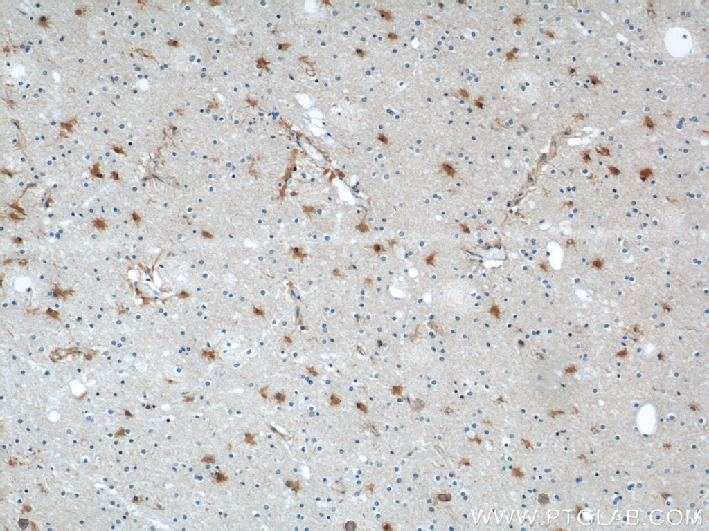
IHC staining of human brain using 55237-1-AP
Immunohistochemical analysis of paraffin-embedded human brain using 55237-1-AP (ENO1 antibody) at dilution of 1:100 (under 10x lens).
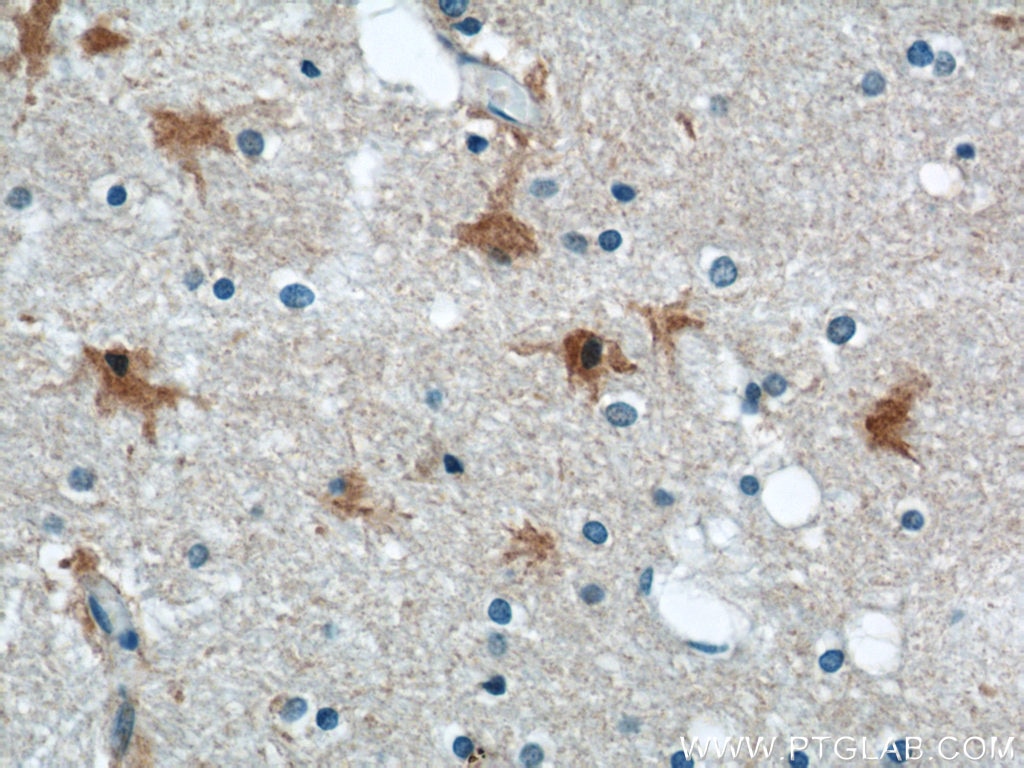
IHC staining of human brain using 55237-1-AP
Immunohistochemical analysis of paraffin-embedded human brain using 55237-1-AP (ENO1 antibody) at dilution of 1:100 (under 40x lens).
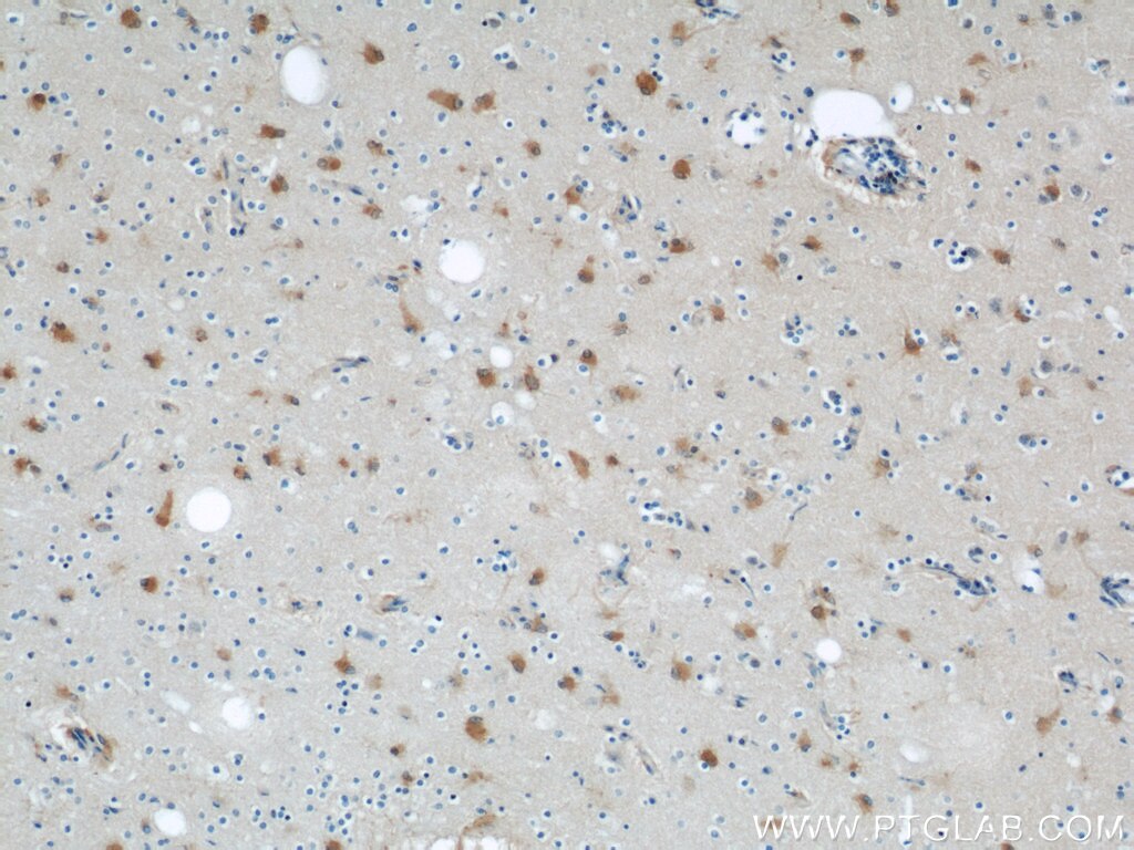
IHC staining of human brain using 55237-1-AP
Immunohistochemical analysis of paraffin-embedded human brain using 55237-1-AP (ENO1 antibody) at dilution of 1:100 (under 10x lens).
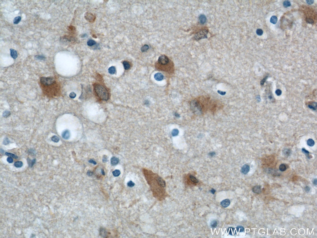
IHC staining of human brain using 55237-1-AP
Immunohistochemical analysis of paraffin-embedded human brain using 55237-1-AP (ENO1 antibody) at dilution of 1:100 (under 40x lens).
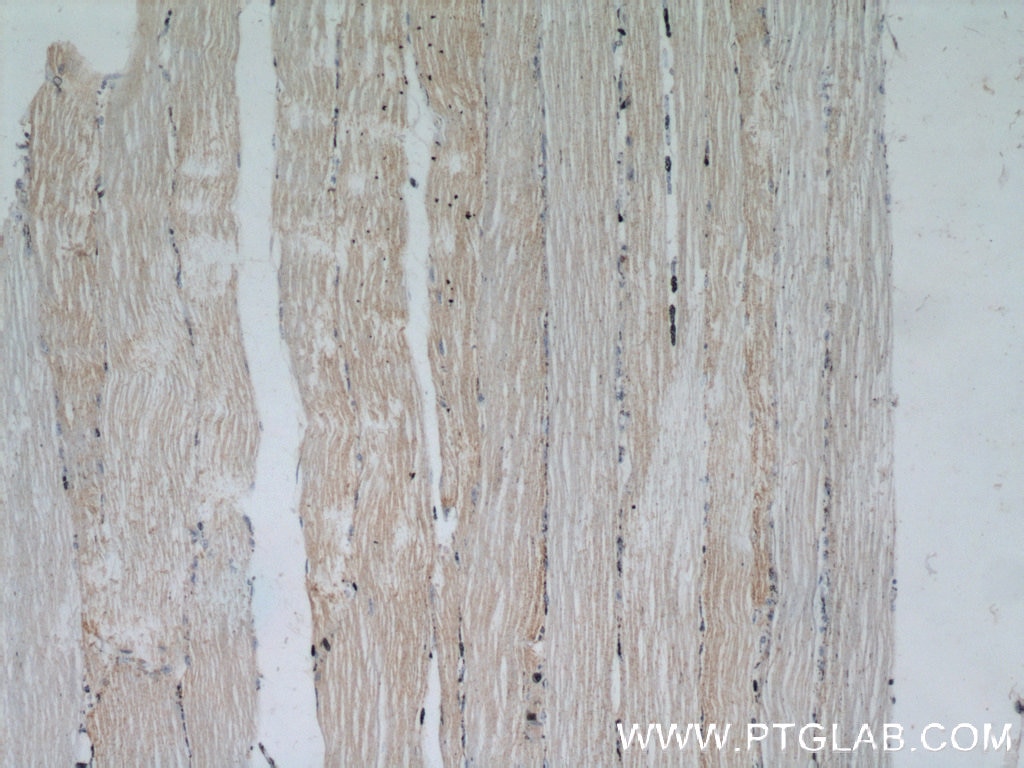
IHC staining of human skeletal muscle using 55237-1-AP
Immunohistochemical analysis of paraffin-embedded human skeletal muscle using 55237-1-AP (ENO1 antibody) at dilution of 1:100 (under 10x lens).
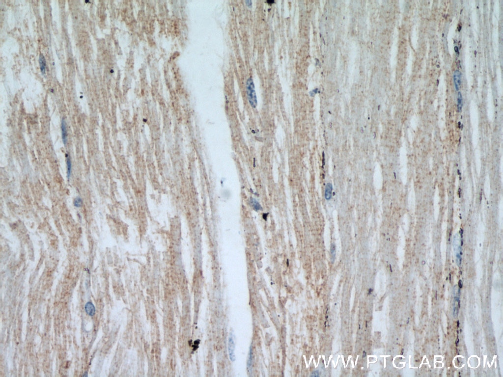
IHC staining of human skeletal muscle using 55237-1-AP
Immunohistochemical analysis of paraffin-embedded human skeletal muscle using 55237-1-AP (ENO1 antibody) at dilution of 1:100 (under 40x lens).
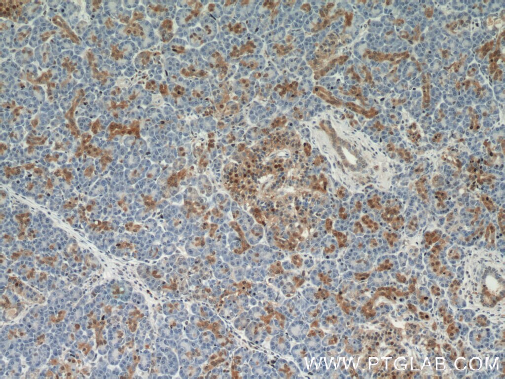
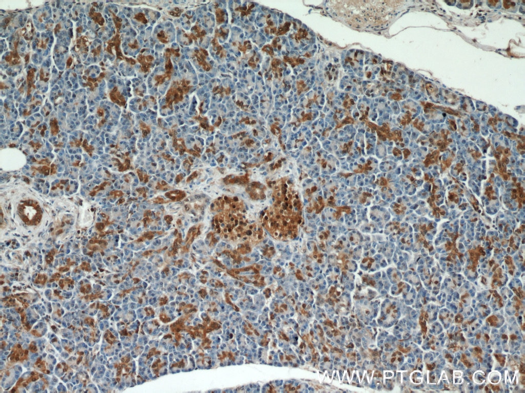
IHC staining of human pancreas using 55237-1-AP
Immunohistochemical analysis of paraffin-embedded human pancreas using 55237-1-AP (ENO1 antibody) at dilution of 1:100 (under 10x lens).
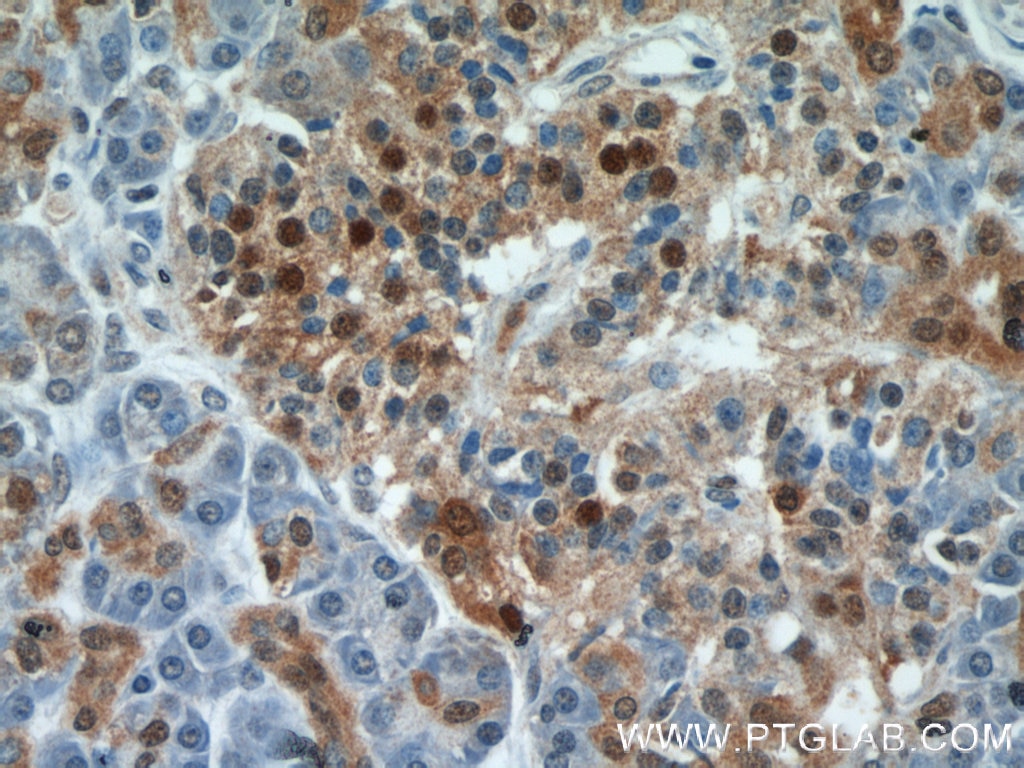
IHC staining of human pancreas using 55237-1-AP
Immunohistochemical analysis of paraffin-embedded human pancreas using 55237-1-AP (ENO1 antibody) at dilution of 1:100 (under 40x lens).
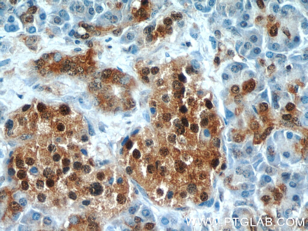
IHC staining of human pancreas using 55237-1-AP
Immunohistochemical analysis of paraffin-embedded human pancreas using 55237-1-AP (ENO1 antibody) at dilution of 1:100 (under 10x lens).
IHC staining of human pancreas using 55237-1-AP
Immunohistochemical analysis of paraffin-embedded human pancreas using 55237-1-AP (ENO1 antibody) at dilution of 1:100 (under 40x lens).
IP Figures
IP experiment of mouse brain using 55237-1-AP
IP result of anti-ENO1 (IP:55237-1-AP, 4ug; Detection:55237-1-AP 1:500) with mouse brain tissue lysate 4000ug.
IF/ICC Figures
IF Staining of HepG2 using 55237-1-AP
Immunofluorescent analysis of HepG2 cells using 55237-1-AP (ENO1 antibody) at dilution of 1:50 and Rhodamine-Goat anti-Rabbit IgG.
FC (INTRA) Figures
FC experiment of HeLa using 55237-1-AP
1x10^6 HeLa cells were intracellularly stained with 0.25 ug ENO1 Polyclonal antibody (55237-1-AP) and CoraLite®488-Conjugated Goat Anti-Rabbit IgG(H+L) (SA00013-2)(red), or 0.25 ug Isotype Control (blue). Cells were fixed and permeabilized with Intracellular Flow Cytometry Fixation & Permeabilization Buffer Kit (PF00019).
The species listed in Tested Reactivity are in-house verified and applicable species. For unlisted species, please refer to the homology analysis of the immunogen sequence and related species. For rabbit polyclonal antibodies, homology >70% is recommended. For mouse monoclonal antibodies and rabbit recombinant antibodies, homology >90% is recommended. Generally, the higher the homology, the greater the applicability. However, there will be certain differences in protein expression in different species, tissues or cells. Therefore, the homology analysis results are for reference only and do not serve as a guarantee.
At Proteintech, we pride ourselves on our antibody quality, customer service and transparency. As such, we are comparing our antibodies with other vendors, enabling easy identification and comparisons of key data to help you choose the suitable antibody for your needs.
We have selected the top cited antibodies from these vendors for you to compare.
Proteintech
KD/KO VALIDATED
ENO1 Polyclonal antibody
Catalog Number
55237-1-AP
Citations
3
Dilutions
WB : 1:1000-1:4000 IP : 0.5-4.0 ug for IP and 0.5-4.0 ug for 1.0-3.0 mg of total protein lysate for WB IHC : 1:20-1:200 IF/ICC : 1:20-1:200 FC (INTRA) : 0.25 ug per 10^6 cells in a 100 µl suspension
Applications
WB, IHC, IF/ICC, FC (Intra), IP, ELISA
Reactivity
human, mouse
Product Guarantee
Covers any species including not listed on datasheet
Covers any applications including not listed on datasheet