Tested Applications
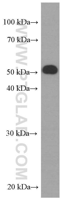
| Positive WB detected in | HepG2 cells |
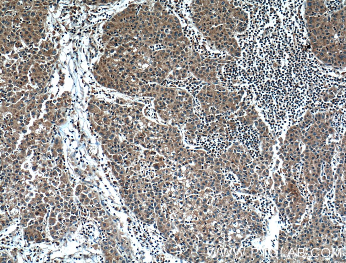
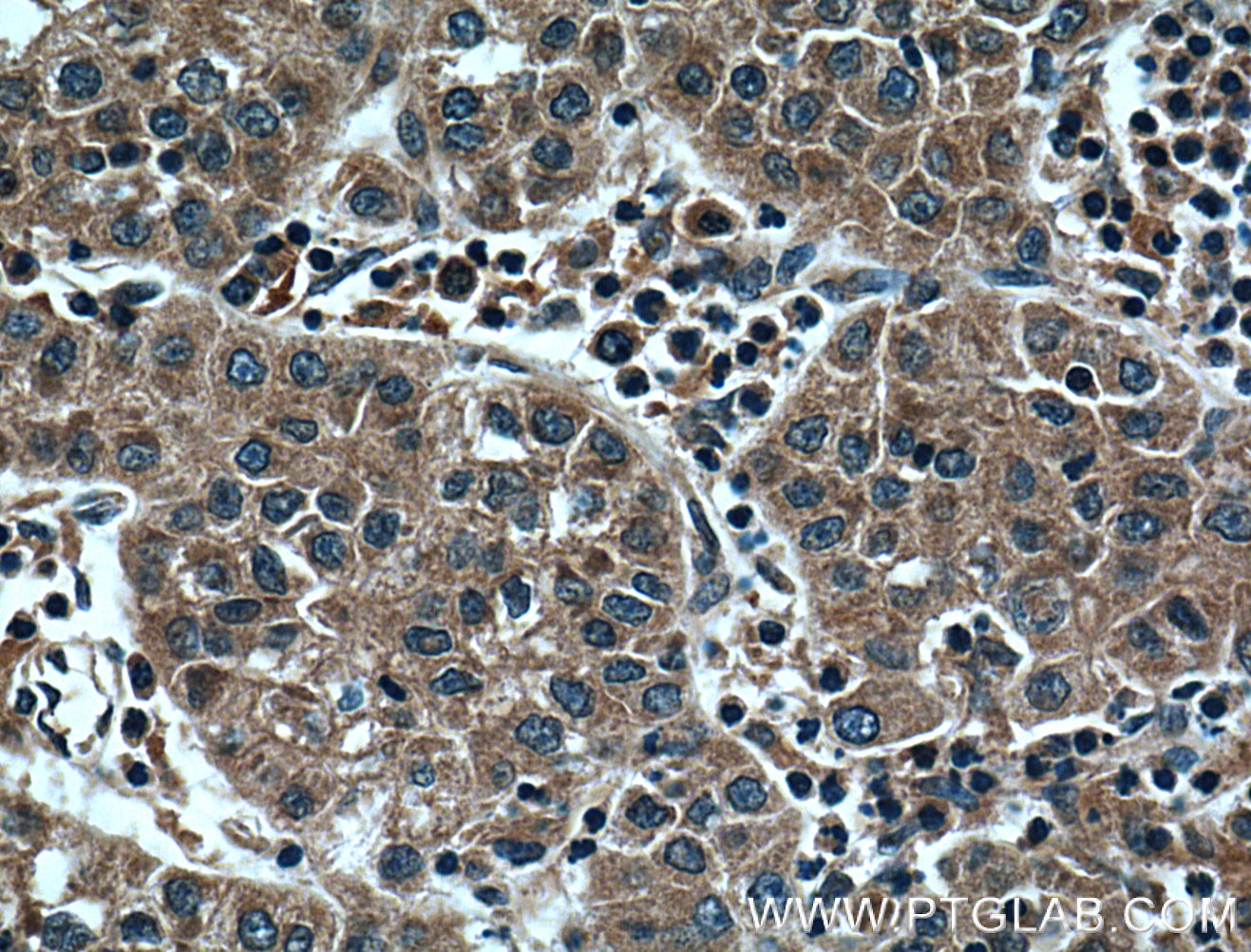
| Positive IHC detected in | human liver cancer tissue Note: suggested antigen retrieval with TE buffer pH 9.0; (*) Alternatively, antigen retrieval may be performed with citrate buffer pH 6.0 |
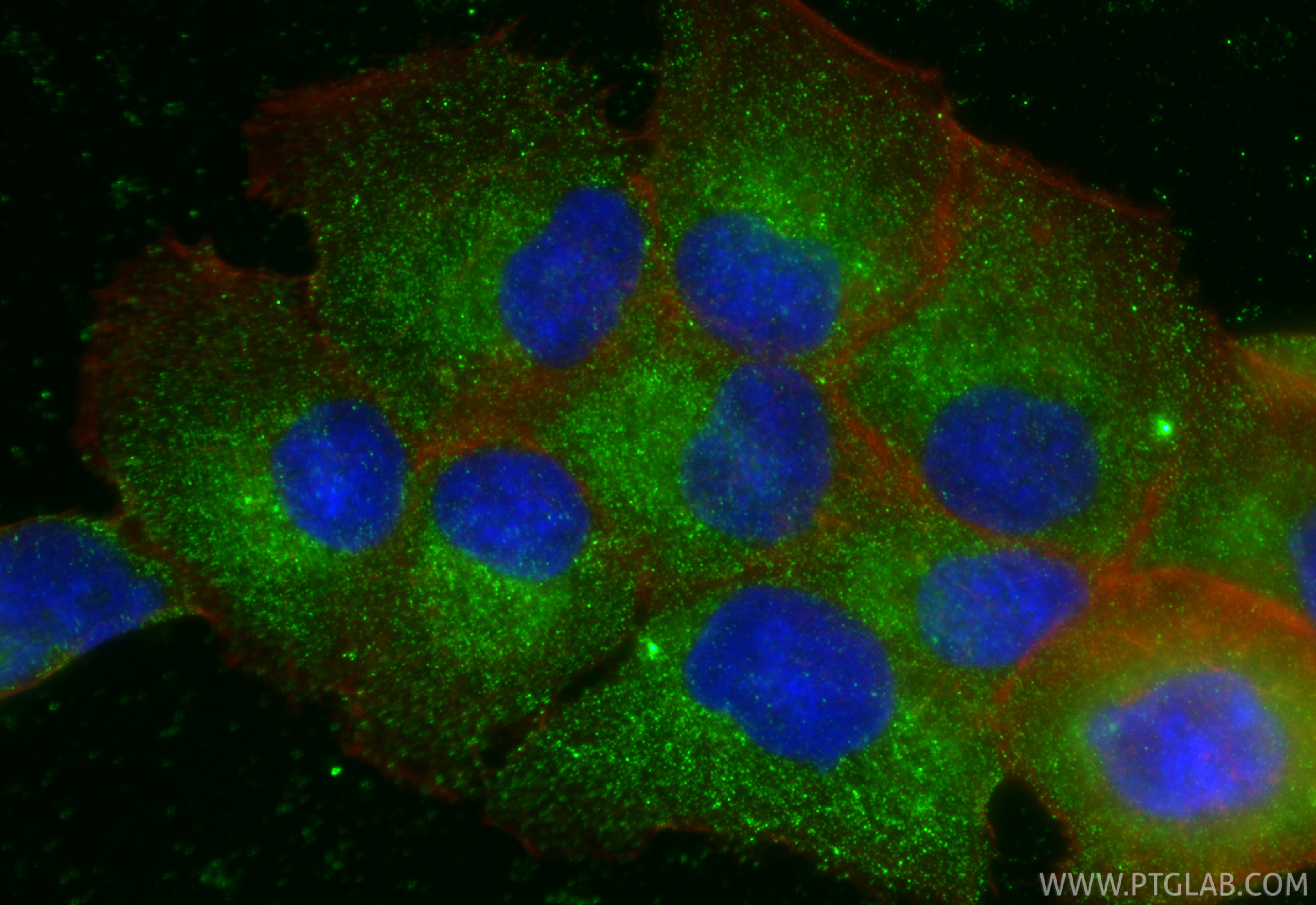
| Positive IF/ICC detected in | DU 145 cells |
Recommended dilution
| Application | Dilution |
|---|---|
| Western Blot (WB) | WB : 1:500-1:2000 |
| Immunohistochemistry (IHC) | IHC : 1:200-1:1000 |
| Immunofluorescence (IF)/ICC | IF/ICC : 1:500-1:2000 |
| It is recommended that this reagent should be titrated in each testing system to obtain optimal results. | |
| Sample-dependent, Check data in validation data gallery. | |
Published Applications
| KD/KO | See 1 publications below |
| WB | See 3 publications below |
| IHC | See 1 publications below |
| IF | See 1 publications below |
Product Information
66226-1-Ig targets HPSE in WB, IHC, IF/ICC, ELISA applications and shows reactivity with human samples.
| Tested Reactivity | human |
| Cited Reactivity | human, mouse |
| Host / Isotype | Mouse / IgG1 |
| Class | Monoclonal |
| Type | Antibody |
| Immunogen |
CatNo: Ag10067 Product name: Recombinant human HPSE protein Source: e coli.-derived, PET28a Tag: 6*His Domain: 159-543 aa of BC051321 Sequence: KFKNSTYSRSSVDVLYTFANCSGLDLIFGLNALLRTADLQWNSSNAQLLLDYCSSKGYNISWELGNEPNSFLKKADIFINGSQLGEDFIQLHKLLRKSTFKNAKLYGPDVGQPRRKTAKMLKSFLKAGGEVIDSVTWHHYYLNGRTATREDFLNPDVLDIFISSVQKVFQVVESTRPGKKVWLGETSSAYGGGAPLLSDTFAAGFMWLDKLGLSARMGIEVVMRQVFFGAGNYHLVDENFDPLPDYWLSLLFKKLVGTKVLMASVQGSKRRKLRVYLHCTNTDNPRYKEGDLTLYAINLHNVTKYLRLPYPFSNKQVDKYLLRPLGPHGLLSKSVQLNGLTLKMVDDQTLPPLMEKPLRPGSSLGLPAFSYSFFVIRNAKVAACI Predict reactive species |
| Full Name | heparanase |
| Calculated Molecular Weight | 543 aa, 61 kDa |
| Observed Molecular Weight | 50 kDa |
| GenBank Accession Number | BC051321 |
| Gene Symbol | HPSE |
| Gene ID (NCBI) | 10855 |
| RRID | AB_2881617 |
| Conjugate | Unconjugated |
| Form | Liquid |
| Purification Method | Protein G purification |
| UNIPROT ID | Q9Y251 |
| Storage Buffer | PBS with 0.02% sodium azide and 50% glycerol, pH 7.3. |
| Storage Conditions | Store at -20°C. Stable for one year after shipment. Aliquoting is unnecessary for -20oC storage. 20ul sizes contain 0.1% BSA. |
Background Information
HPSE(Heparanase) is also named as HEP, HPA, HPA1, HPR1, HPSE1, HSE1 and belongs to the glycosyl hydrolase 79 family. It is a endoglycosidase that cleaves heparan sulfate proteoglycans (HSPGs) into heparan sulfate side chains and core proteoglycans. HPSE is essential in the disassembly of the extracellular matrix (ECM) by invading cells. It has 3 isoforms produced by alternative splicing with the molecular weight of 61 kDa, 55 kDa and 53 kDa. The full length protein has six glycosylation sites. The cleavage of the 65 kDa form leads to the generation of a linker peptide, and 8 kDa and 50 kDa products. The active form, the 8/50 kDa heterodimer, is resistant to degradation and glycosylation of the 50 kDa subunit appears to be essential for its solubility.
Protocols
| Product Specific Protocols | |
|---|---|
| IF protocol for HPSE antibody 66226-1-Ig | Download protocol |
| IHC protocol for HPSE antibody 66226-1-Ig | Download protocol |
| WB protocol for HPSE antibody 66226-1-Ig | Download protocol |
| Standard Protocols | |
|---|---|
| Click here to view our Standard Protocols |
Publications
| Species | Application | Title |
|---|---|---|
Cells A New Synthesized Dicarboxylated Oxy-Heparin Efficiently Attenuates Tumor Growth and Metastasis | ||
Mar Drugs A Marine λ-Oligocarrageenan Inhibits Migratory and Invasive Ability of MDA-MB-231 Human Breast Cancer Cells through Actions on Heparanase Metabolism and MMP-14/MMP-2 Axis. | ||
Front Oncol Heparanase Promotes Tumor Growth and Liver Metastasis of Colorectal Cancer Cells by Activating the p38/MMP1 Axis.
|
Reviews
The reviews below have been submitted by verified Proteintech customers who received an incentive for providing their feedback.
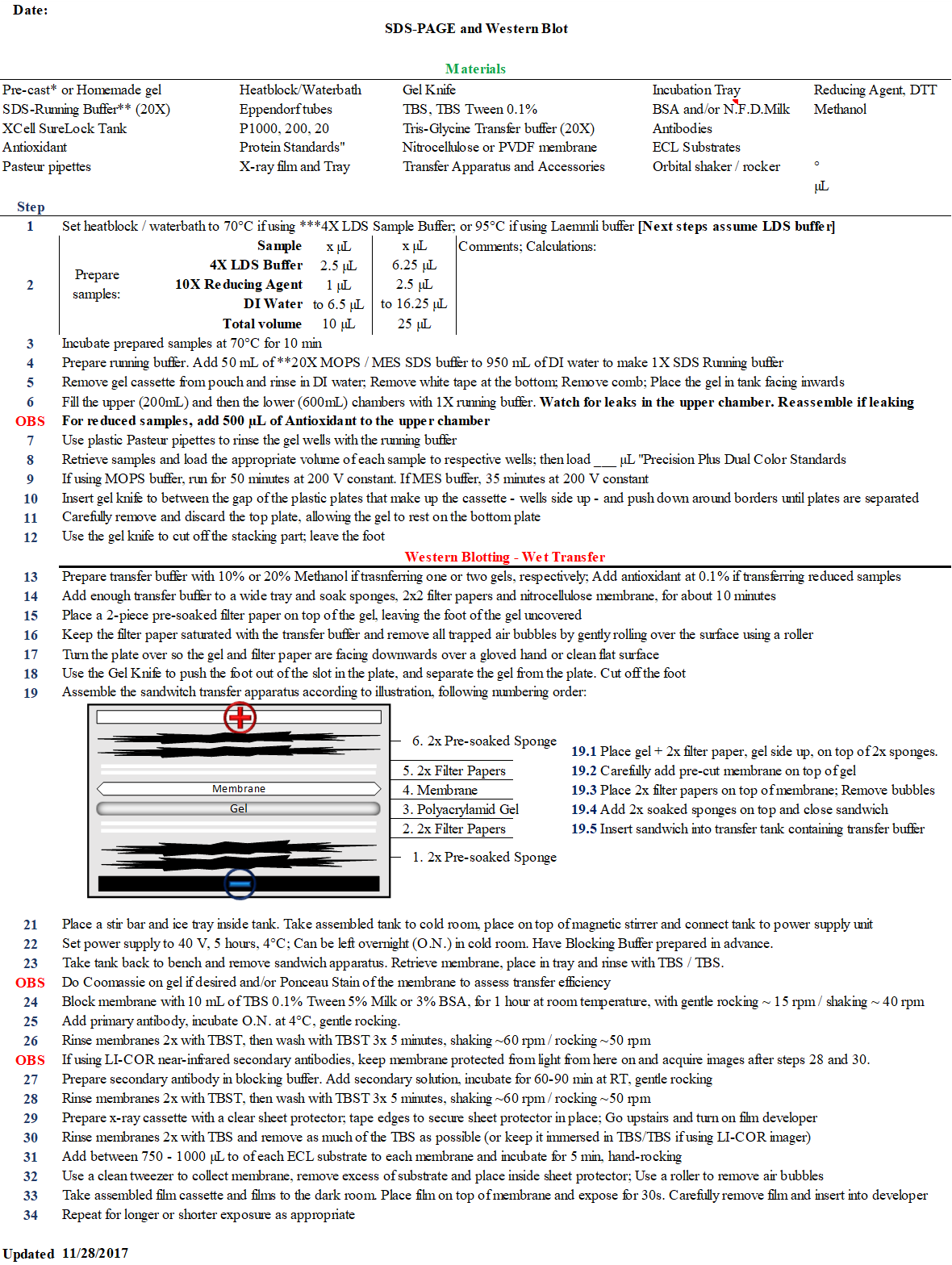
FH Fabio Henrique (Verified Customer) (01-17-2019) | Tested in dot blot and western blot formats.Dot blot performance: (Figure 1)The antibody performed according to expectations - the cell lines tested (HS-27A, HS-5, C4-2B, MCF-7 and PC-3) were probed for HPSE via qPCR and the dot blot data mostly agreed with the transcript expression. Brief methods: Cells were cultured until 70% confluent and medium was replaced with serum-free Opti-MEM. After 48hrs, conditioned medium (CM) was collected and concentrated 50X using a 10 kDa molecular weight cut-off spin column. 50 uL of 50X CM was blotted in duplicate onto each well of dot blot apparatus for 1 hr. The membrane (nitrocellulose) was blocked for 1 hr @ RT in PBS + 0.1%Tween20 + 5% Non-fat dry milk. Anti-HPSE was diluted in blocking buffer at 1 ug/mL (1:2000) and incubated with membrane overnight at 4*C. Sheep anti-mouse HRP-conjugated secondary was added after washes at dilution 1:50000 and incubated at RT for 1 hr. ECL substrate used was SuperSignal West Dura. Western blot performance:Poor. I initially tested this product against cell lysates (C4-2B and MCF-7), but due to weak/lack of signal, I also tested in the positive control HEPG2 lysate. Figure 2: For C4-2B and MCF-7, different concentrations of lysate were tested and 2 weak bands were detected after 5-minute exposure, one around 50 kDa and another around 65 kDa. That may be HPSE, but I expected stronger signal considering loading amount of 40 ug of lysate. Figure 3: When testing HEPG2 (30 ug lsyate per lane), the results were not comparable to the data available on the website. Two bands were detected, a strong one at around 65 kDa and another at around 75 kDa. Brief methods:Lysates were collected using RIPA buffer and Halt Protease inhibitor, then sonicated and quantified using BCA assay. Pre-cast 10% Bis-tris gels were used for PAGE, running at 200 V for 50 minutes in MOPS SDS buffer. Transfer was done using Tris-glycine buffer with 10% methanol - 40 V for 5 hours inside cold room. Efficiency of transfer was measured using Ponceau Stain and Coomassie blue. Blocking of membrane was in 5% milk in PBST0.1%. Anti-HPSE at 1:1000 and 1:2000 was incubated overnight at 4*C. Developed was described above. Exposure time: 1 second. Detailed protocol can be found in excel sheet provided.
 |

