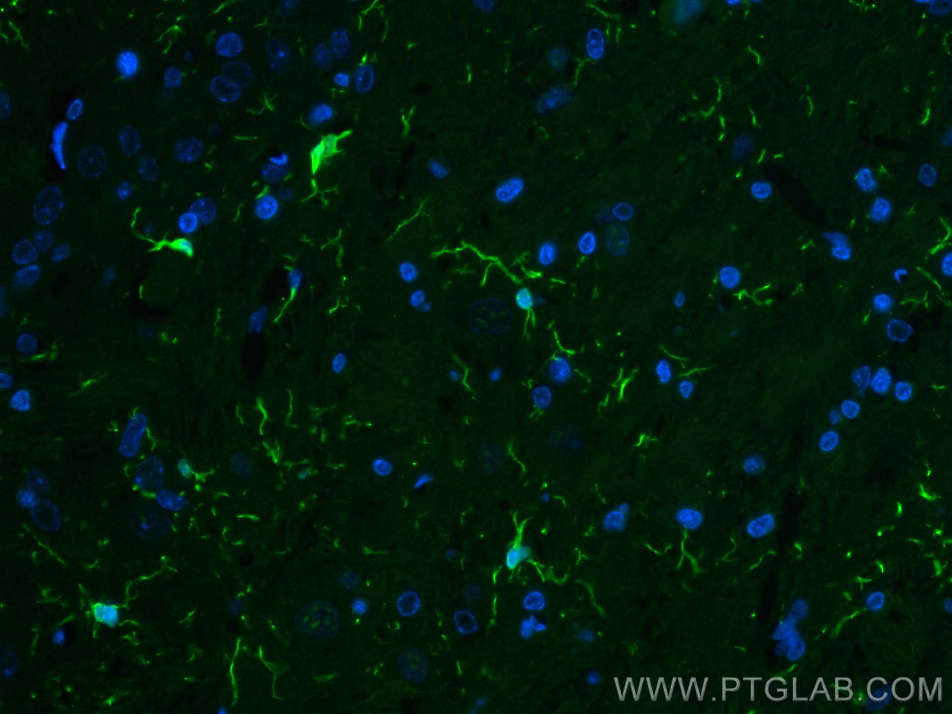
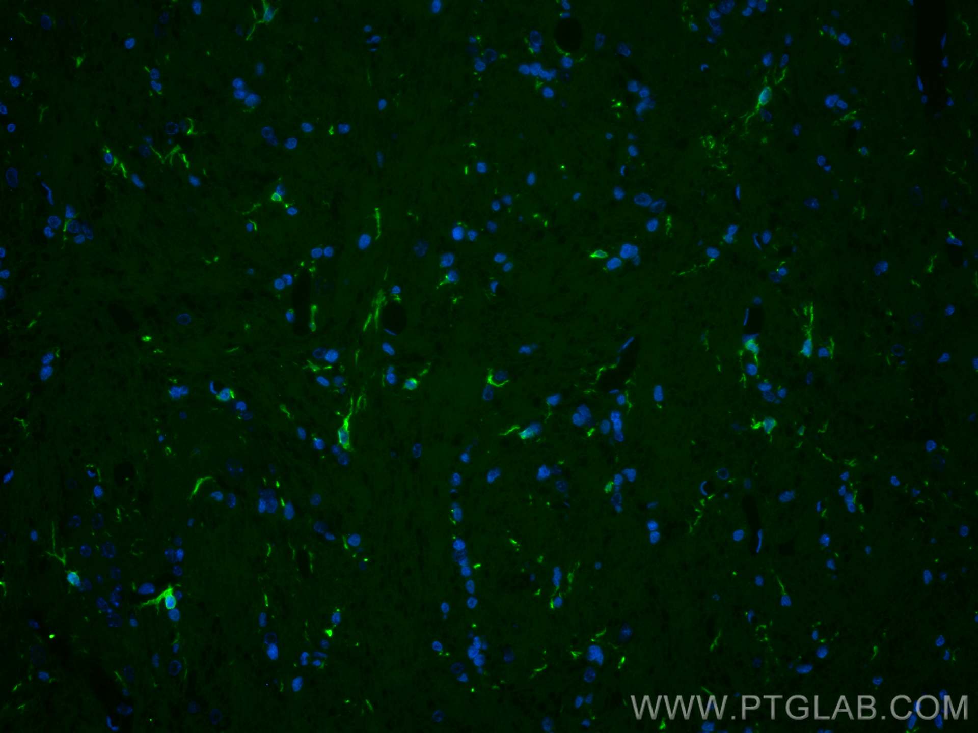
Tested Applications
| Positive IF-P detected in | rat brain tissue |
Recommended dilution
| Application | Dilution |
|---|---|
| Immunofluorescence (IF)-P | IF-P : 1:50-1:500 |
| It is recommended that this reagent should be titrated in each testing system to obtain optimal results. | |
| Sample-dependent, Check data in validation data gallery. | |
Published Applications
| WB | See 1 publications below |
Product Information
CL488-10904 targets IBA1 in WB, IF-P applications and shows reactivity with human, mouse, rat samples.
| Tested Reactivity | human, mouse, rat |
| Cited Reactivity | mouse |
| Host / Isotype | Rabbit / IgG |
| Class | Polyclonal |
| Type | Antibody |
| Immunogen |
CatNo: Ag1363 Product name: Recombinant human IBA1 protein Source: e coli.-derived, PGEX-4T Tag: GST Domain: 1-147 aa of BC009474 Sequence: MSQTRDLQGGKAFGLLKAQQEERLDEINKQFLDDPKYSSDEDLPSKLEGFKEKYMEFDLNGNGDIDIMSLKRMLEKLGVPKTHLELKKLIGEVSSGSGETFSYPDFLRMMLGKRSAILKMILMYEEKAREKEKPTGPPAKKAISELP Predict reactive species |
| Full Name | allograft inflammatory factor 1 |
| Calculated Molecular Weight | 17 kDa |
| GenBank Accession Number | BC009474 |
| Gene Symbol | IBA1 |
| Gene ID (NCBI) | 199 |
| ENSEMBL Gene ID | ENSG00000204472 |
| RRID | AB_2919009 |
| Conjugate | CoraLite® Plus 488 Fluorescent Dye |
| Excitation/Emission Maxima Wavelengths | 493 nm / 522 nm |
| Excitation Laser | Blue laser (488 nm) |
| Form | Liquid |
| Purification Method | Antigen affinity purification |
| UNIPROT ID | P55008 |
| Storage Buffer | PBS with 50% glycerol, 0.05% Proclin300, 0.5% BSA, pH 7.3. |
| Storage Conditions | Store at -20°C. Avoid exposure to light. Stable for one year after shipment. Aliquoting is unnecessary for -20oC storage. |
Background Information
What is the molecular weight of IBA1?
The molecular weight of IBA1 is 16.7 kD.
What is IBA1?
Ionized calcium-binding adaptor molecule 1 (IBA1), also known as Allograft inflammatory factor-1 (AIF-1), is an inflammation-responsive scaffold protein expressed and secreted from macrophages. Microglia response factor (MRF-1) and daintain are also similar, and likely identical, proteins (PMIDs: 29749461, 9630473, 23792284).
What is the function of IBA1?
IBA1 is necessary for macrophage survival, and it is also a key molecule in proinflammatory activity (PMID: 29749461).
Where is IBA1 localized?
IBA1 is a cytoplasmic protein, often expressed in immunocytes, macrophages, and microglia. It can be used as a marker for normal (not 'dark') microglia in brain tissue, as IBA1 is expressed by all microglial cell subpopulations (PMIDs: 29749461, 11943136, 26847266, 9630473).
Is IBA1 upregulated in active immunophages?
Yes; its expression is associated with inflammatory activity (PMID: 29749461).
How do IBA1-positive microglia differ from microglia that express less or no IBA1?
'Dark' microglia is a recently described phenotype associated with Alzheimer's disease pathology and chronic stress; these dark microglia express decreased IBA1 (often punctiform), and show distinct ultrastructural differences from 'normal' microglia as well as condensed and electron-dense cytoplasm and nucleoplasm. Normal microglia generally display strong, diffuse expression of IBA1. IBA1-positive microglial processes in normal conditions also have a strong tendency to exclusively contact synaptic elements, such as axon terminals, dendritic spines, and synaptic clefts (PMIDs: 29992181, 26847266).
How is IBA1 related to Alzheimer's disease and pain?
Exacerbated immunoactivity of IBA1, particularly in proximity to amyloid plaques, is prominently featured in AD pathology. IBA1, along with other microglial markers, are also associated with pain, and robust microglial reactions often follow spinal cord injury (PMIDs: 29992181, 23792284).
Protocols
| Product Specific Protocols | |
|---|---|
| IF protocol for CL Plus 488 IBA1 antibody CL488-10904 | Download protocol |
| Standard Protocols | |
|---|---|
| Click here to view our Standard Protocols |