

Tested Applications
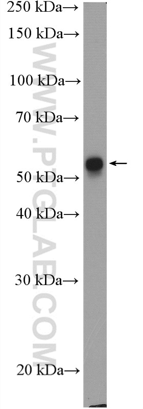
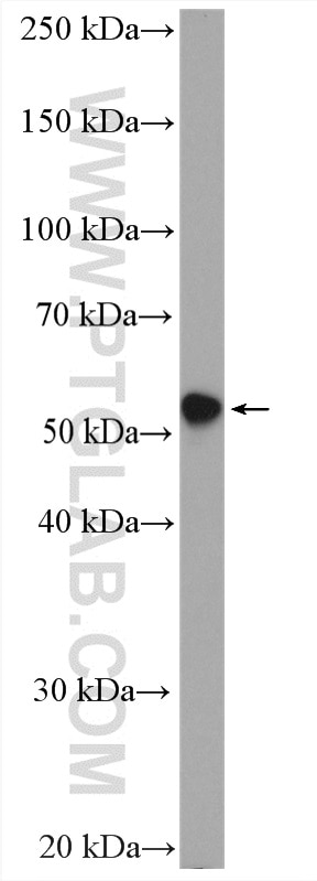
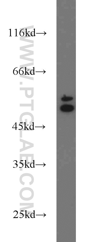
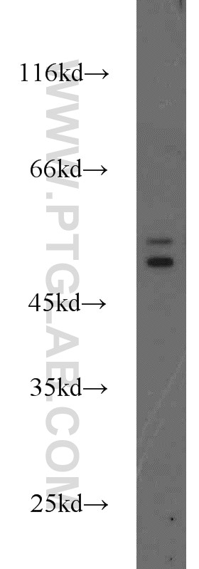
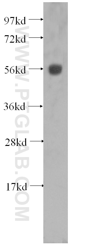
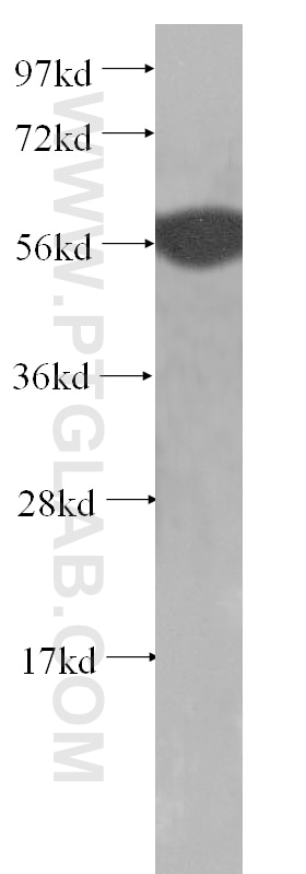
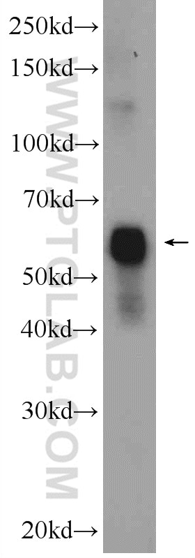
| Positive WB detected in | A431 cells, MCF-7 cells, HeLa cells, A375 cells, rat skin tissue, U-251 cells |
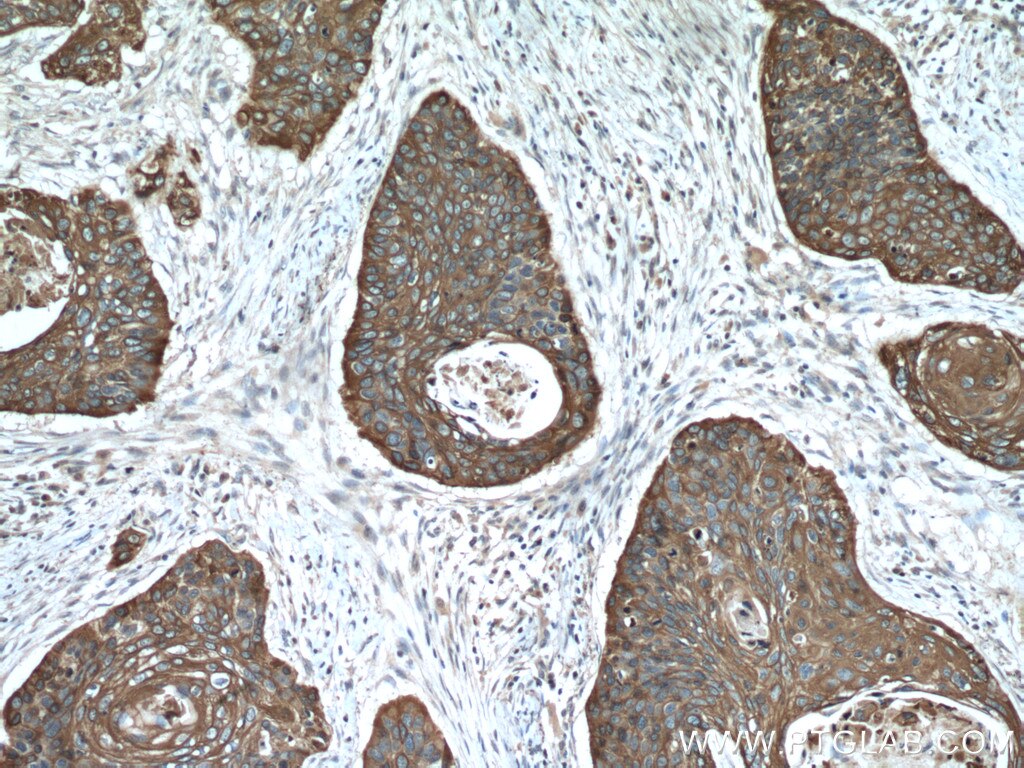
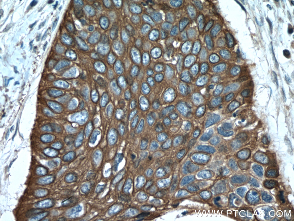
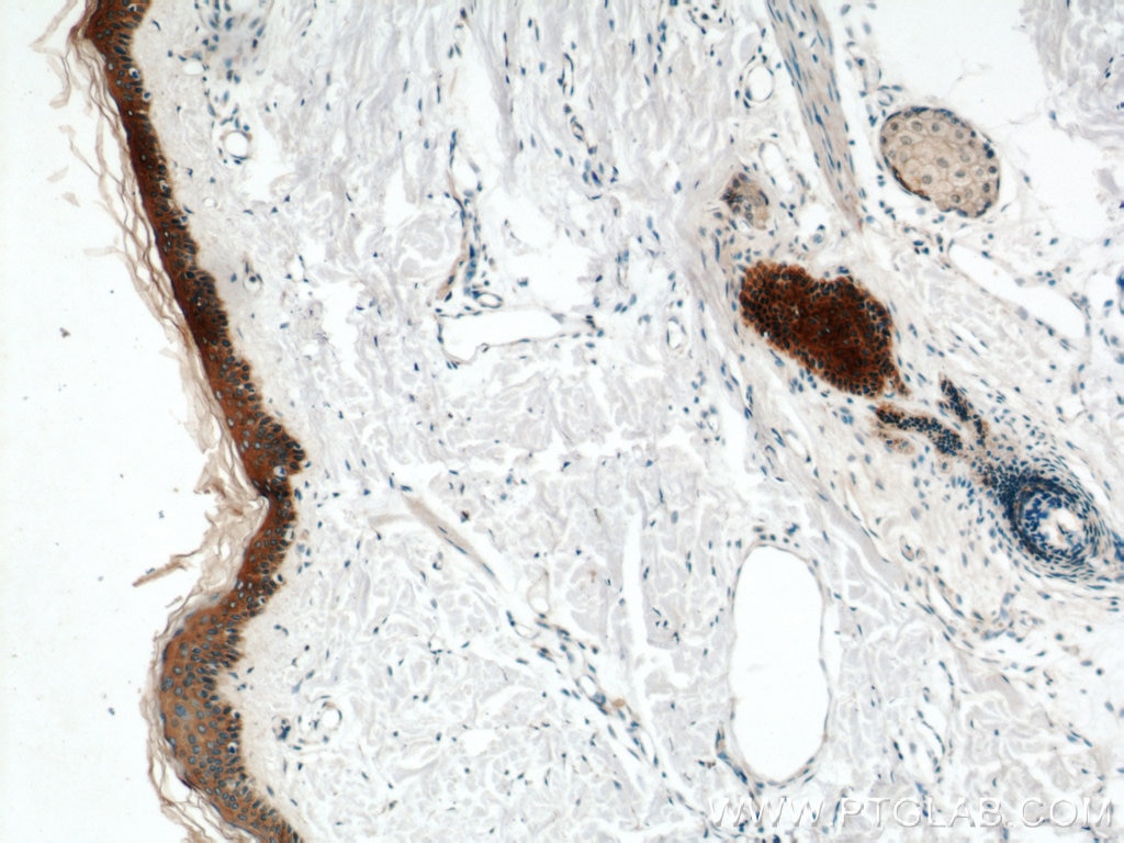
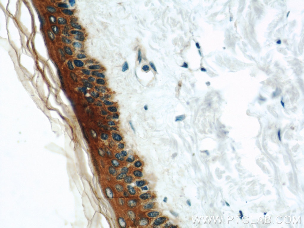
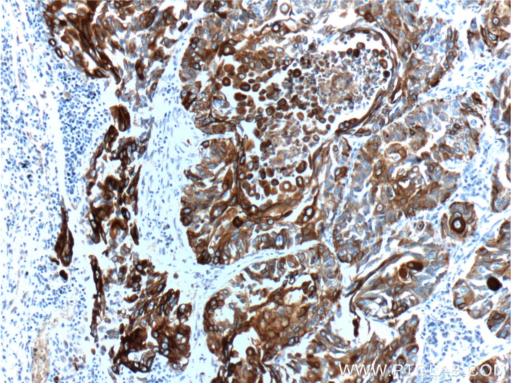
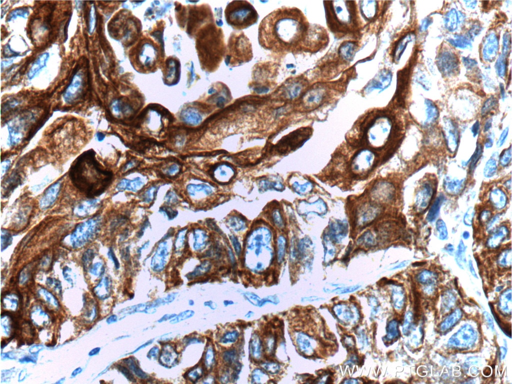
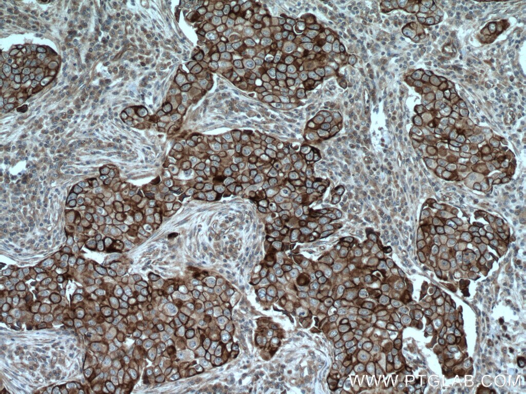
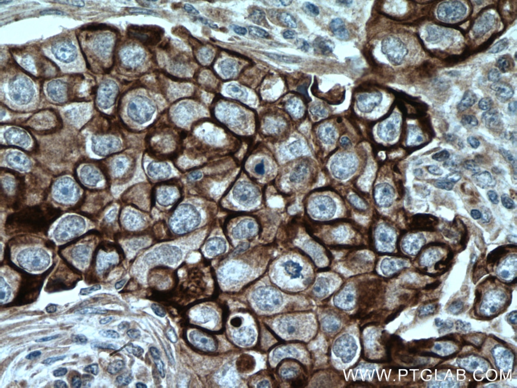
| Positive IHC detected in | human oesophagus cancer tissue, human breast cancer tissue, human skin tissue, human lung cancer tissue Note: suggested antigen retrieval with TE buffer pH 9.0; (*) Alternatively, antigen retrieval may be performed with citrate buffer pH 6.0 |
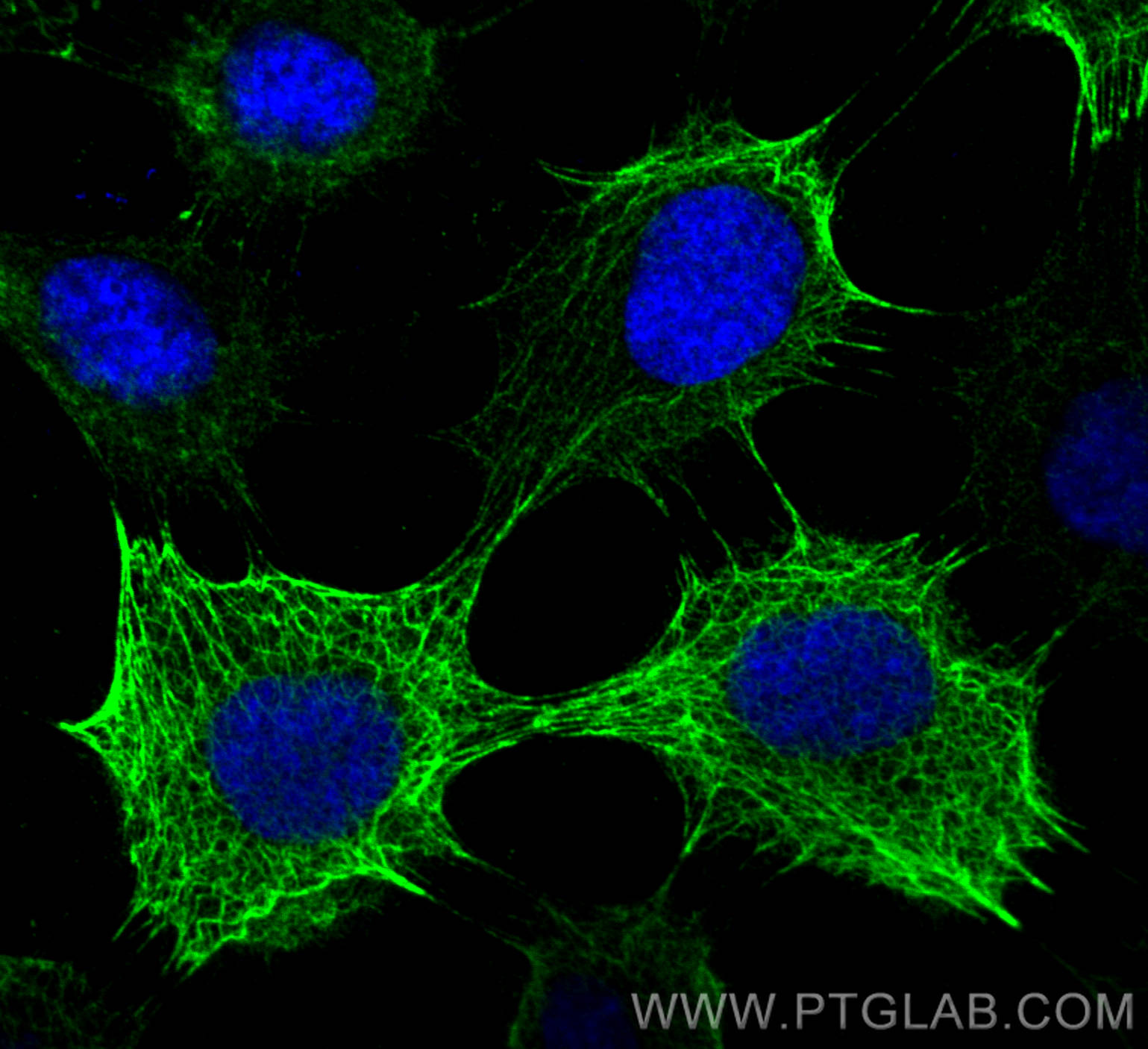
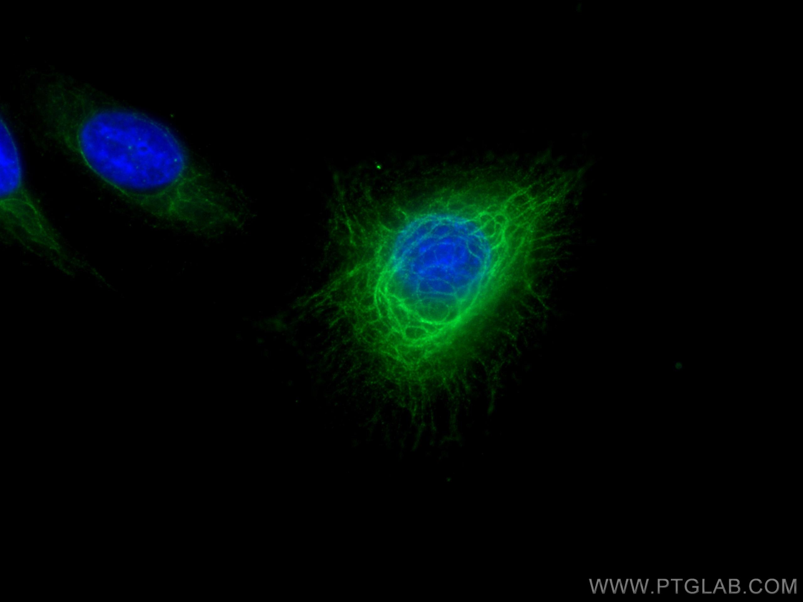
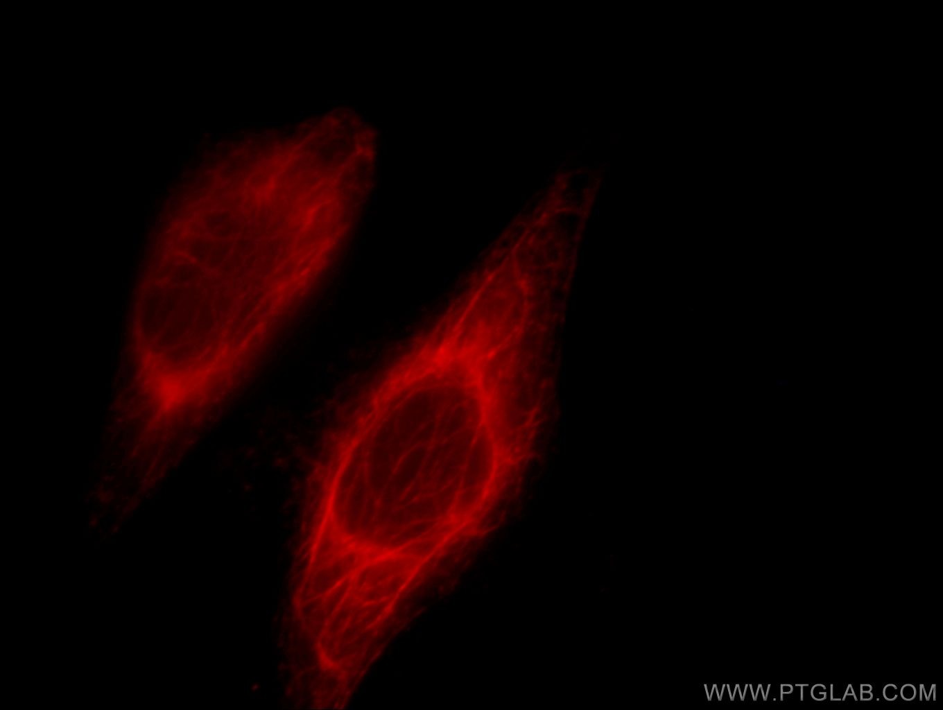
| Positive IF/ICC detected in | A431 cells, HeLa cells |
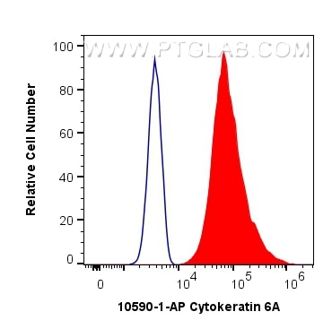
| Positive FC (Intra) detected in | A431 cells |
Recommended dilution
| Application | Dilution |
|---|---|
| Western Blot (WB) | WB : 1:10000-1:50000 |
| Immunohistochemistry (IHC) | IHC : 1:200-1:800 |
| Immunofluorescence (IF)/ICC | IF/ICC : 1:50-1:500 |
| Flow Cytometry (FC) (INTRA) | FC (INTRA) : 0.40 ug per 10^6 cells in a 100 µl suspension |
| It is recommended that this reagent should be titrated in each testing system to obtain optimal results. | |
| Sample-dependent, Check data in validation data gallery. | |
Product Information
10590-1-AP targets Cytokeratin 6A in WB, IHC, IF/ICC, FC (Intra), CoIP, ChIP, ELISA applications and shows reactivity with human, rat samples.
| Tested Reactivity | human, rat |
| Cited Reactivity | human, mouse, rat |
| Host / Isotype | Rabbit / IgG |
| Class | Polyclonal |
| Type | Antibody |
| Immunogen |
CatNo: Ag0882 Product name: Recombinant human KRT6A protein Source: e coli.-derived, PGEX-4T Tag: GST Domain: 364-564 aa of BC008807 Sequence: QVTAGRHGDDLRNTKQEIAEINRMIQRLRSEIDHVKKQCANLQAAIADAEQRGEMALKDAKNKLEGLEDALQKAKQDLARLLKEYQELMNVKLALDVEIATYRKLLEGEECRLNGEGVGQVNISVVQSTVSSGYGGASGVGSGLGLGGGSSYSYGSGLGVGGGFSSSSGRAIGGGLSSVGGGSSTIKYTTTSSSSRKSYKH Predict reactive species |
| Full Name | keratin 6A |
| Calculated Molecular Weight | 60 kDa |
| Observed Molecular Weight | 56 kDa |
| GenBank Accession Number | BC008807 |
| Gene Symbol | Cytokeratin 6A |
| Gene ID (NCBI) | 3853 |
| RRID | AB_2134306 |
| Conjugate | Unconjugated |
| Form | Liquid |
| Purification Method | Antigen affinity purification |
| UNIPROT ID | P02538 |
| Storage Buffer | PBS with 0.02% sodium azide and 50% glycerol, pH 7.3. |
| Storage Conditions | Store at -20°C. Stable for one year after shipment. Aliquoting is unnecessary for -20oC storage. 20ul sizes contain 0.1% BSA. |
Background Information
Keratins are a large family of proteins that form the intermediate filament cytoskeleton of epithelial cells, which are classified into two major sequence types. Type I keratins are a group of acidic intermediate filament proteins, including K9-K23, and the hair keratins Ha1-Ha8. Type II keratins are the basic or neutral courterparts to the acidic type I keratins, including K1-K8, and the hair keratins, Hb1-Hb6. Keratin 6 is a type II keratin. It is used as marker for epidermal hyperproliferation and differentiation. Three are three keratin 6 isoforms: ketatin 6A,6B and 6C. They share more than 99% identical DNA sequence.
Protocols
| Product Specific Protocols | |
|---|---|
| FC protocol for Cytokeratin 6A antibody 10590-1-AP | Download protocol |
| IF protocol for Cytokeratin 6A antibody 10590-1-AP | Download protocol |
| IHC protocol for Cytokeratin 6A antibody 10590-1-AP | Download protocol |
| WB protocol for Cytokeratin 6A antibody 10590-1-AP | Download protocol |
| Standard Protocols | |
|---|---|
| Click here to view our Standard Protocols |
Publications
What published studies show
The KRT6A (Keratin 6A) antibody, catalog number 10590-1-AP, has 52 citations. These appear in journals including Nature Communications, Cancer Cell, Immunity, Cell Reports, Molecular Cancer, Journal of Allergy and Clinical Immunology, and STAR Protocols. Research fields span dermatology (psoriasis, skin inflammation, wound healing), oncology (esophageal, colorectal, pancreatic, breast, and lung cancers), keratinocyte biology, and immunology. Applications include WB, IHC, IF, and CoIP.
| Species | Application | Title |
|---|---|---|
Signal Transduct Target Ther Krüppel-like factor 4 downregulation during esophageal cancer formation facilitates immune evasion and impairs immunotherapy | ||
Cancer Cell KMT2C deficiency drives transdifferentiation of double-negative prostate cancer and confer resistance to AR-targeted therapy | ||
Mol Cancer Integrated single-cell and spatial transcriptomic profiling decodes lineage plasticity and immune microenvironment remodeling in prostate cancer progression. | ||
Immunity Excessive Polyamine Generation in Keratinocytes Promotes Self-RNA Sensing by Dendritic Cells in Psoriasis. | ||
Cancer Res A Genome-Wide Synthetic Lethal Screen Identifies Spermidine Synthase as a Target to Enhance Erdafitinib Efficacy in FGFR-Mutant Bladder Cancer | ||
Drug Resist Updat Targeting keratin 6 A overcomes gemcitabine resistance by restoring equilibrative nucleoside transporter 1 and TAM-mediated metabolic compensation in pancreatic cancer.
|
Reviews
What customers say
The antibody performed excellently in IHC on human NSCLC patient-derived tissue microarrays. Used at a 1:100 dilution with pH 9 TE buffer for DAB staining, it produced perfect results. The reviewer highly recommends this antibody for immunohistochemistry applications.
The reviews below have been submitted by verified Proteintech customers who received an incentive for providing their feedback.
Nikolett (Verified Customer) (03-31-2026) | Antibody was used on human NSCLC patient derived TMA and was working perfectly at pH9 TE, as 1:100 for DAB IHC stainings. I highly recommend this antibody.
|




















