Tested Applications
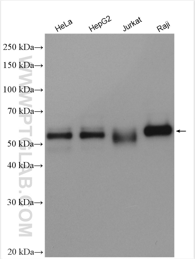
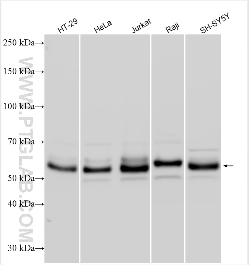
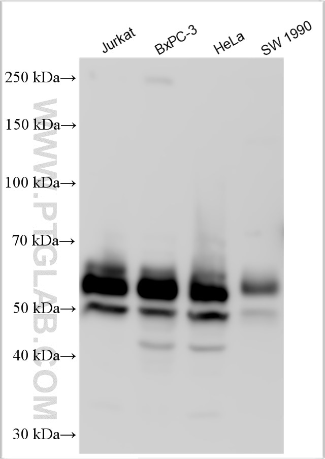
| Positive WB detected in | HT-29 cells, Jurkat cells, HeLa cells, A549 cells, HL-60 cells, human placenta tissue, Raji cells, SH-SY5Y cells, HepG2 cells, BxPC-3 cells, SW 1990 cells, Jurkat cells and Raji cells. |
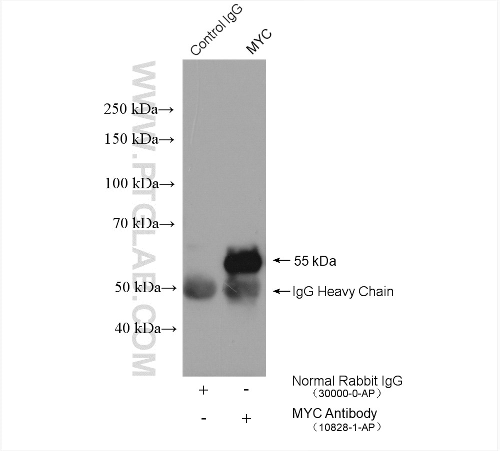
| Positive IP detected in | MCF-7 cells |
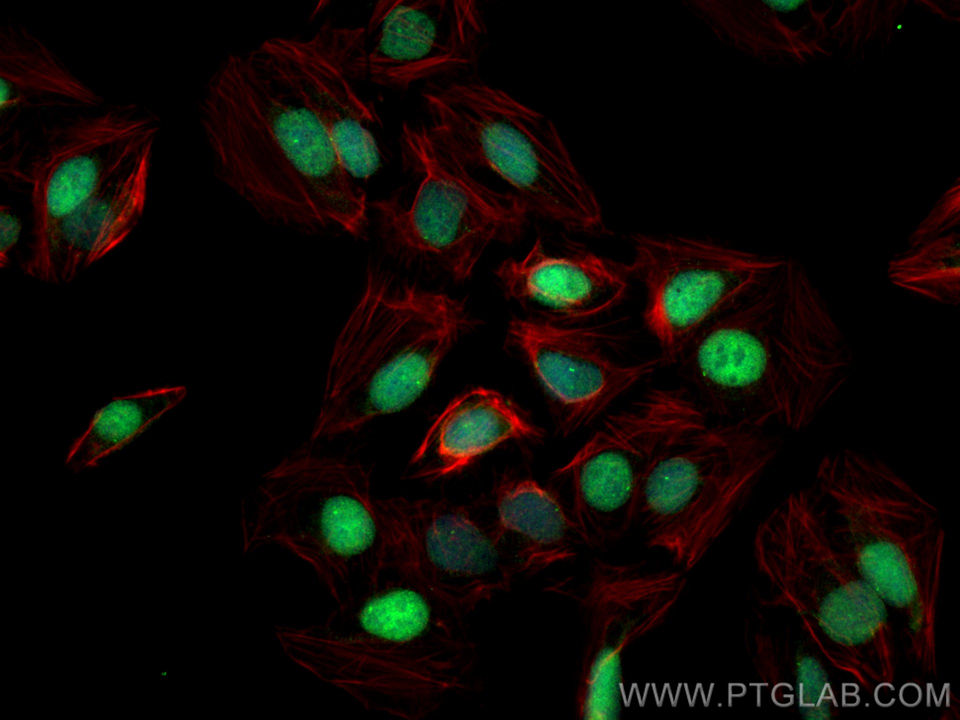
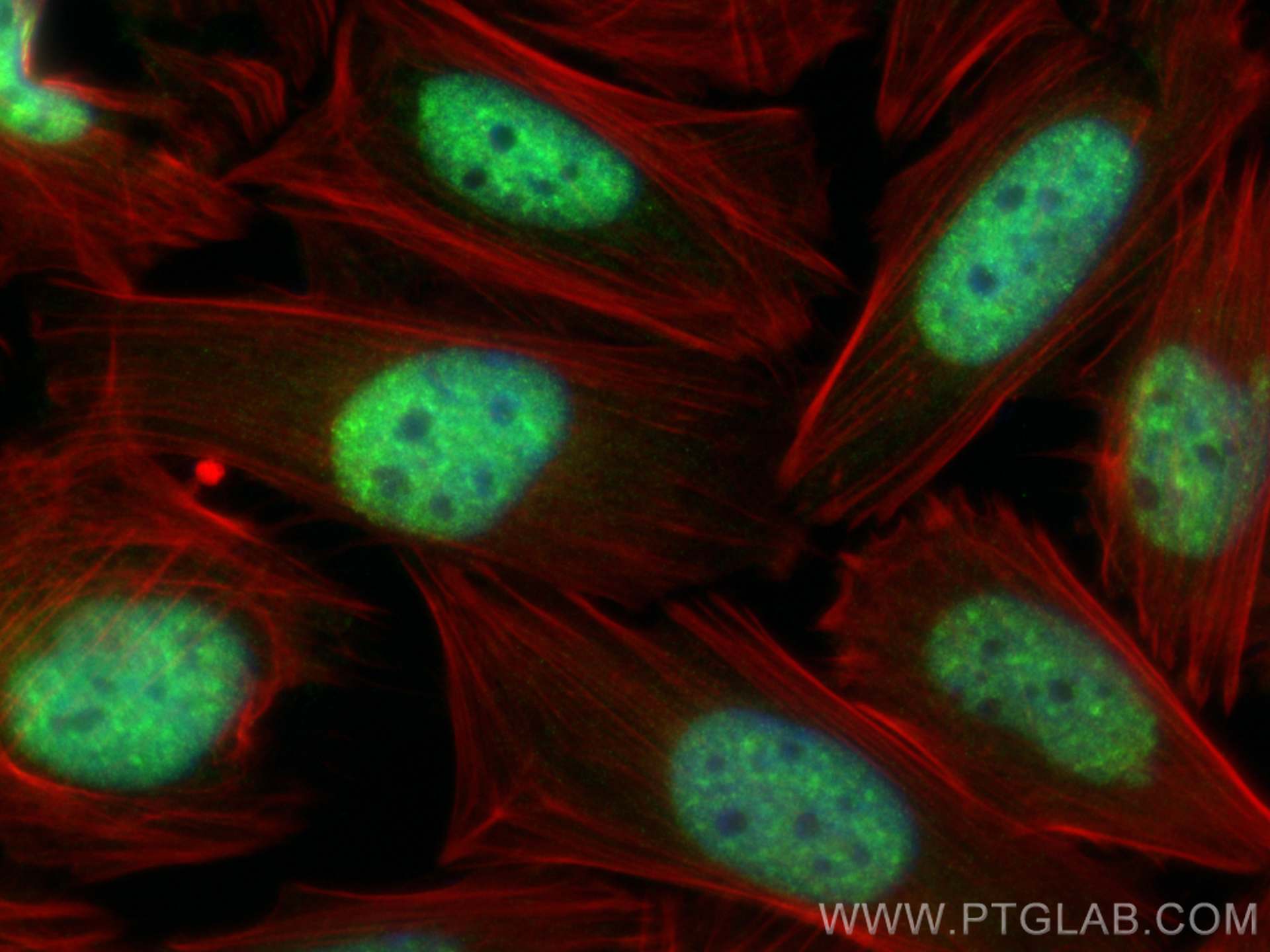
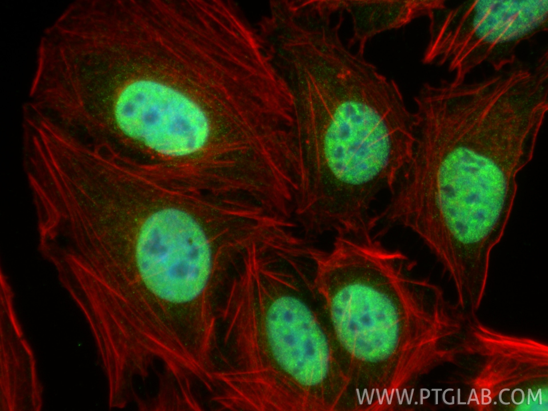
| Positive IF/ICC detected in | U2OS cells, HEK-293 cells, Saos-2 cells |
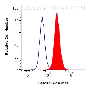
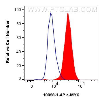
| Positive FC (Intra) detected in | HeLa cells, NCCIT cells |
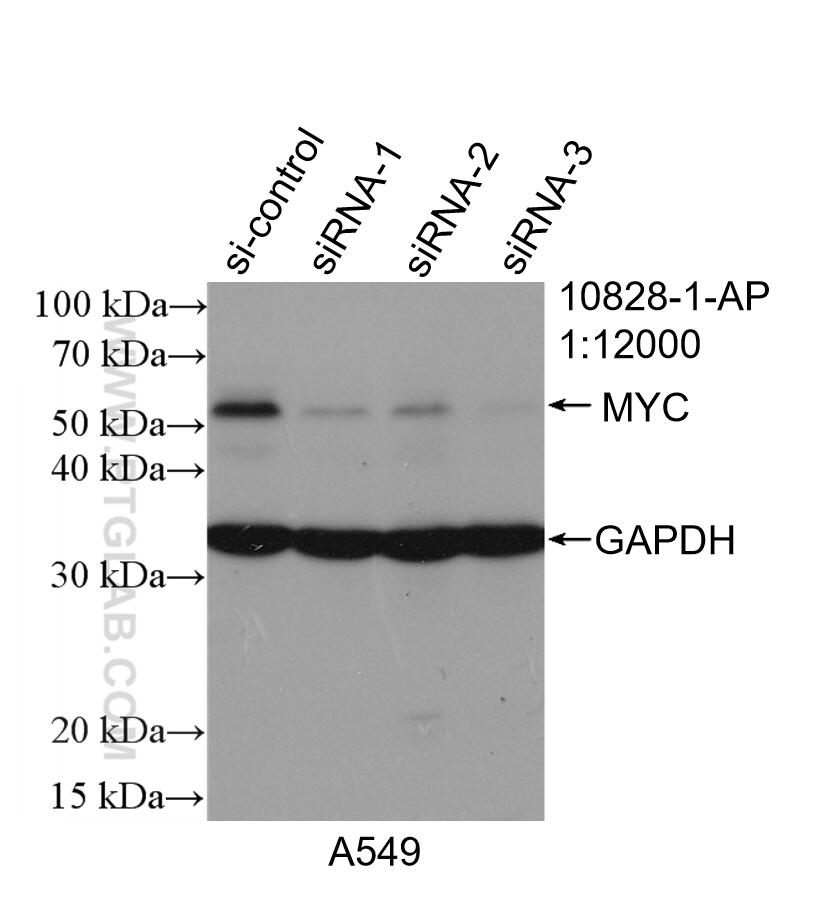
This antibody is not for the detetion of MYC-tag.
Recommended dilution
| Application | Dilution |
|---|---|
| Western Blot (WB) | WB : 1:2000-1:12000 |
| Immunoprecipitation (IP) | IP : 0.5-4.0 ug for 1.0-3.0 mg of total protein lysate |
| Immunofluorescence (IF)/ICC | IF/ICC : 1:200-1:800 |
| Flow Cytometry (FC) (INTRA) | FC (INTRA) : 0.40 ug per 10^6 cells in a 100 µl suspension |
| It is recommended that this reagent should be titrated in each testing system to obtain optimal results. | |
| Sample-dependent, Check data in validation data gallery. | |
Product Information
10828-1-AP targets c-MYC in WB, IF/ICC, FC (Intra), IP, CoIP, ChIP, RIP, ELISA, EMSA applications and shows reactivity with human samples.
| Tested Reactivity | human |
| Cited Reactivity | human, mouse, rat, pig, chicken, sheep, silkworm, p. lilacinum |
| Host / Isotype | Rabbit / IgG |
| Class | Polyclonal |
| Type | Antibody |
| Immunogen |
CatNo: Ag1263 Product name: Recombinant human MYC protein Source: e coli.-derived, PGEX-4T Tag: GST Domain: 16-454 aa of BC000141 Sequence: MPLNVSFTNRNYDLDYDSVQPYFYCDEEENFYQQQQQSELQPPAPSEDIWKKFELLPTPPLSPSRRSGLCSPSYVAVTPFSLRGDNDGGGGSFSTADQLEMVTELLGGDMVNQSFICDPDDETFIKNIIIQDCMWSGFSAAAKLVSEKLASYQAARKDSGSPNPARGHSVCSTSSLYLQDLSAAASECIDPSVVFPYPLNDSSSPKSCASQDSSAFSPSSDSLLSSTESSPQGSPEPLVLHEETPPTTSSDSEEEQEDEEEIDVVSVEKRQAPGKRSESGSPSAGGHSKPPHSPLVLKRCHVSTHQHNYAAPPSTRKDYPAAKRVKLDSVRVLRQISNNRKCTSPRSSDTEENVKRRTHNVLERQRRNELKRSFFALRDQIPELENNEKAPKVVILKKATAYILSVQAEEQKLISEEDLLRKRREQLKHKLEQLRNSCA Predict reactive species |
| Full Name | v-myc myelocytomatosis viral oncogene homolog (avian) |
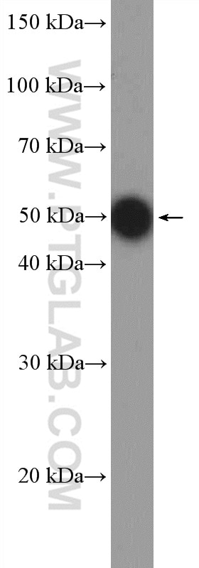
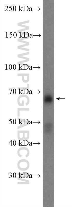
| Calculated Molecular Weight | 49 kDa |
| Observed Molecular Weight | 62-65 kDa, 50 kDa |
| GenBank Accession Number | BC000141 |
| Gene Symbol | MYC |
| Gene ID (NCBI) | 4609 |
| RRID | AB_2148585 |
| Conjugate | Unconjugated |
| Form | Liquid |
| Purification Method | Antigen affinity purification |
| UNIPROT ID | P01106 |
| Storage Buffer | PBS with 0.02% sodium azide and 50% glycerol, pH 7.3. |
| Storage Conditions | Store at -20°C. Stable for one year after shipment. Aliquoting is unnecessary for -20oC storage. 20ul sizes contain 0.1% BSA. |
Background Information
Function
c-Myc (also known as Myc), together with l-Myc and n-Myc, belongs to the Myc family of transcription factors. c-Myc has a basic helix-loop-helix leucine zipper domain and through heterodimerization can bind and regulate the transcriptional activity of genes, either by repression or activation. It is a key player in the regulation of cell growth and cell cycle progression and acts as a proto-oncogene. The latest research reveals a new key mechanism of MYC protein, which is independent of its classical transcriptional regulation function. It was found that under the stress of transcription, MYC was polymerized through its specific RNA binding domain (RBRIII), and exocrine was targeted at the nuclear R loop and double-stranded RNA. This process can effectively eliminate the immunogenic RNA-DNA hybrid from R-loop, thus preventing it from activating TLR3-TBK1 natural immune signaling pathway. This mechanism is very important for MYC to maintain tumor growth and achieve immune escape in vivo. (PMID: 41576951)
Tissue specificity
c-Myc is ubiquitously expressed in almost all cell types and its expression positively correlates with tissue proliferative capacity. c-Myc is also expressed during embryogenesis and is upregulated in many cancer types.
Involvement in disease
· Upregulated in many cancer types, especially in aggressive, poorly differentiated tumors.
· Mutations in the MYC gene and breakpoint translocations within the MYC gene cause Burkitt lymphoma.
Isoforms
There are 3 different isoforms of c-Myc: c-Myc1, c-Myc2, and c-MycS (PMID: 16260605). They differ in molecular size, can be preferentially expressed during cell growth, and are reported to be functionally distinct. The 50kDa band recognized by antibody is the native form of MYC, while the other bands, between 60-70kDa, are the phosphorylated form of MYC (PMID: 12189186).
Post-translational modifications
c-Myc is subject to various post-translational modifications, including phosphorylation, acetylation, and ubiquitinylation (PMID: 16987807), which regulate its activity.
Cellular localization
c-Myc localizes to the nucleus but can also be present in the cytoplasm of certain cancer types.
Protocols
| Product Specific Protocols | |
|---|---|
| FC protocol for c-MYC antibody 10828-1-AP | Download protocol |
| IF protocol for c-MYC antibody 10828-1-AP | Download protocol |
| IP protocol for c-MYC antibody 10828-1-AP | Download protocol |
| WB protocol for c-MYC antibody 10828-1-AP | Download protocol |
| Standard Protocols | |
|---|---|
| Click here to view our Standard Protocols |
Publications
| Species | Application | Title |
|---|---|---|
Signal Transduct Target Ther MDIG-mediated H3K9me3 demethylation upregulates Myc by activating OTX2 and facilitates liver regeneration
| ||
Signal Transduct Target Ther Overexpression of CIP2A is associated with poor prognosis in multiple myeloma. | ||
Gastroenterology Nucleolar HEATR1 upregulated by mTORC1 signaling promotes hepatocellular carcinoma growth by dominating ribosome biogenesis and proteome homeostasis | ||
Cancer Cell p53 Is a Master Regulator of Proteostasis in SMARCB1-Deficient Malignant Rhabdoid Tumors.
| ||
Cell Metab Efferocytosis induces macrophage proliferation to help resolve tissue injury.
|
Reviews
The reviews below have been submitted by verified Proteintech customers who received an incentive for providing their feedback.
FH Katie (Verified Customer) (11-27-2025) | Other antibodies c-MYC antibodies I had tried from other companies had failed to work but this one worked perfectly first use, with very clean bands. Would highly recommend.
|
FH Ying (Verified Customer) (10-30-2025) | It’s good work on cell
|
FH Takeshi (Verified Customer) (02-03-2025) | Comparing the expression abundance of c-Myc between iPS Cells and Primary Human Hepatocytes, this antibody detects both isoforms with very high signal to noise ratio.
|
FH Molly (Verified Customer) (05-02-2023) | The antibody worked well for western blotting following the recommended protocol.
|
FH Anna-Maria (Verified Customer) (08-13-2019) | This antibody worked as expected for western blotting
|
FH Christine (Verified Customer) (11-13-2018) | The antibody worked better than the Abcam c-Myc antibody. However, I did not get clear nuclear staining but rather a mix of nuclear staining with cytoplasmic staining. This could be due non-optimized conditions.
|
FH Claudio (Verified Customer) (12-14-2017) | We tested the antibody in IHC/IFL on mouse brain tumors that showed c-myc upregulation at the mRNA level.
|