Tested Applications
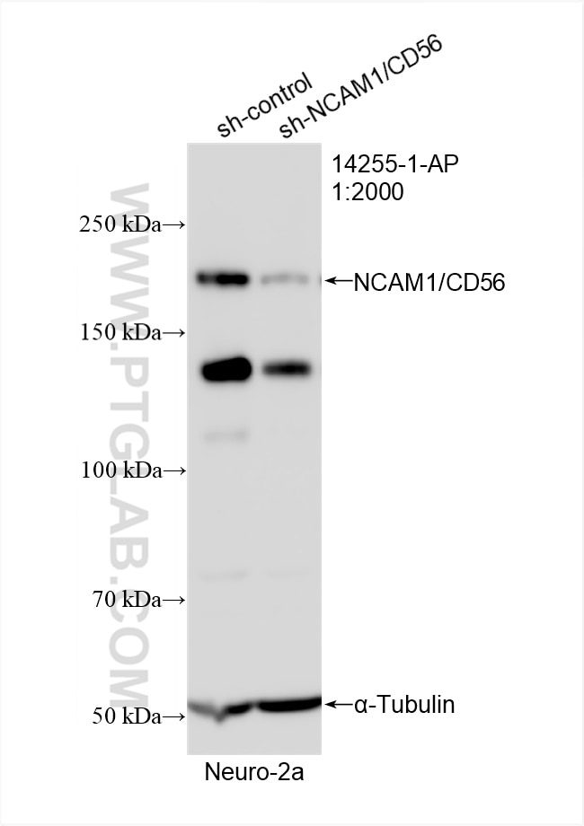
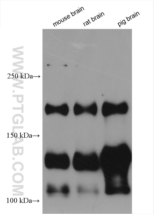
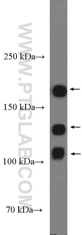
| Positive WB detected in | mouse brain tissue, Neuro-2a cells, rat brain tissue, pig brain tissue |
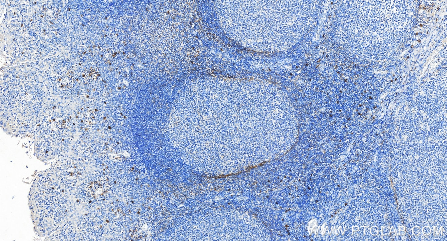
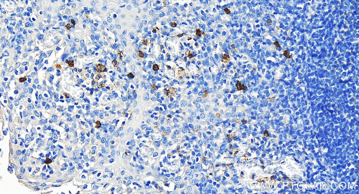
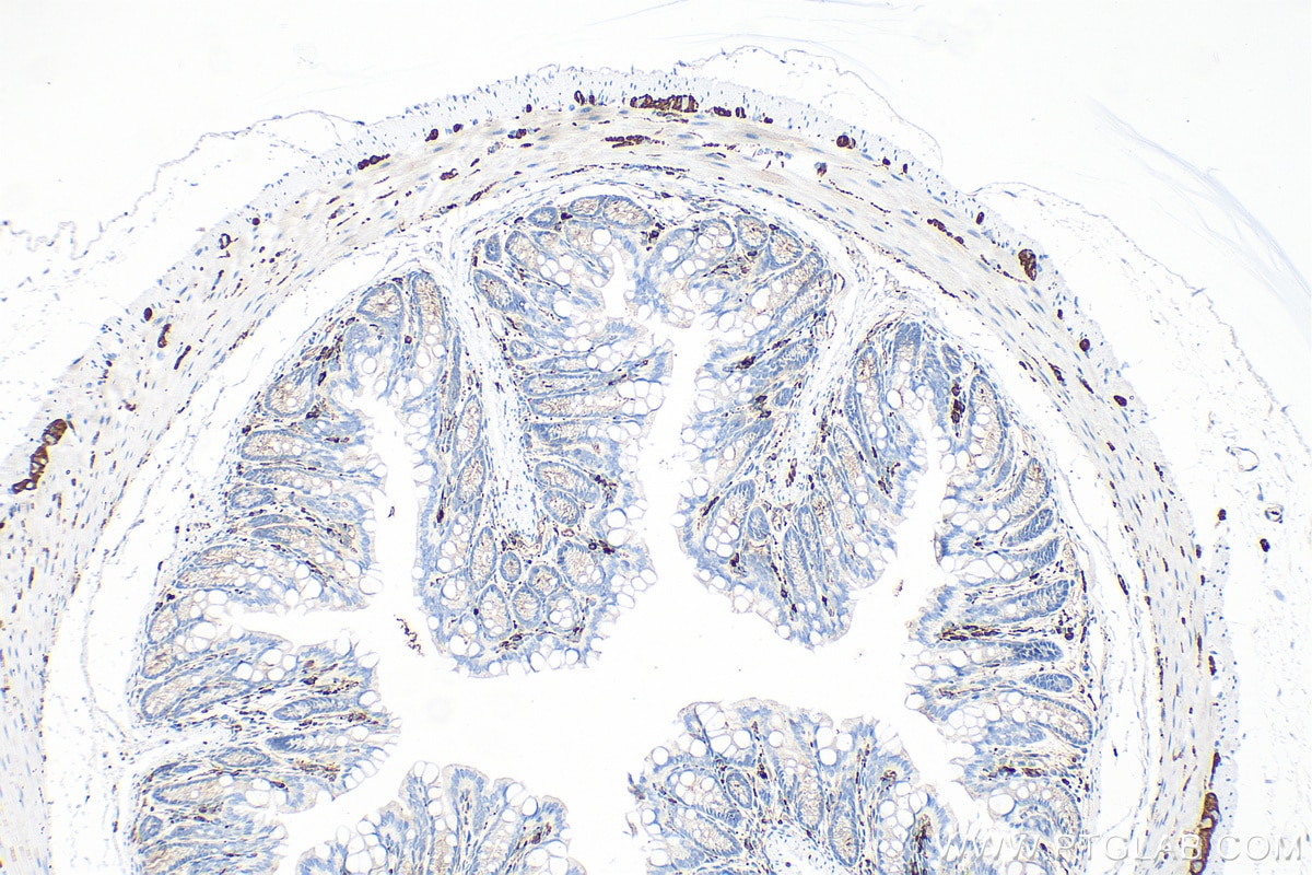
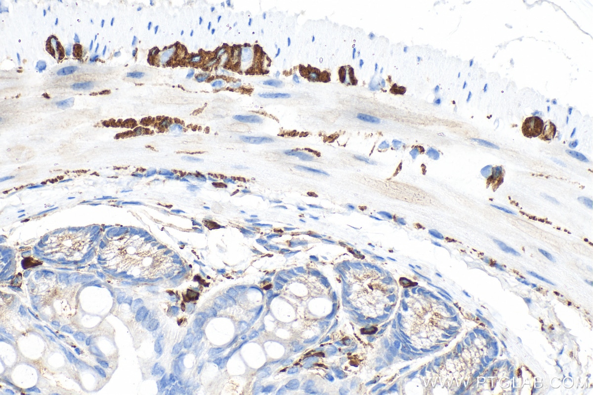
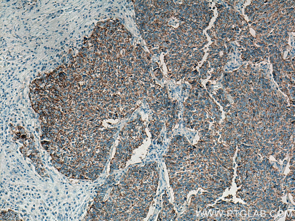
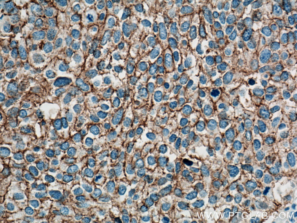
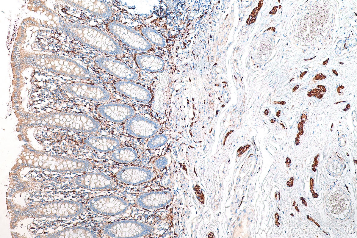
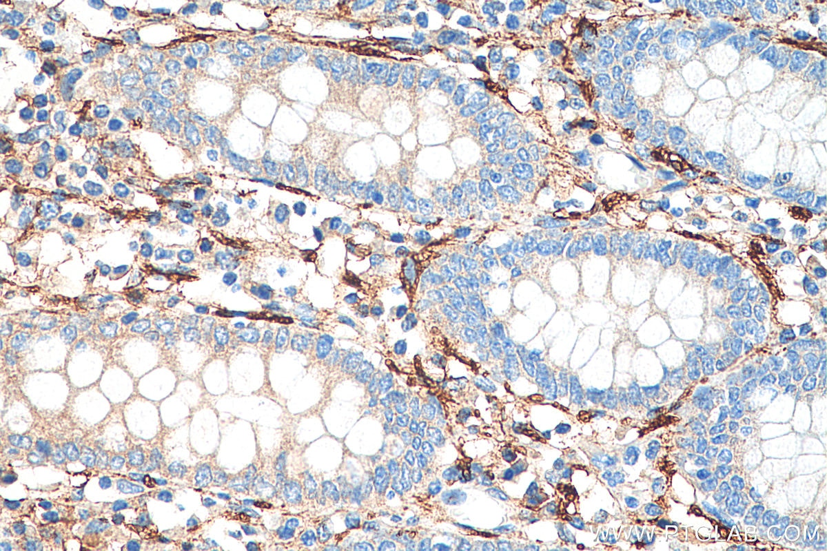
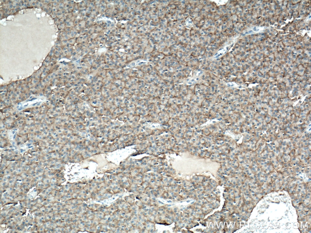
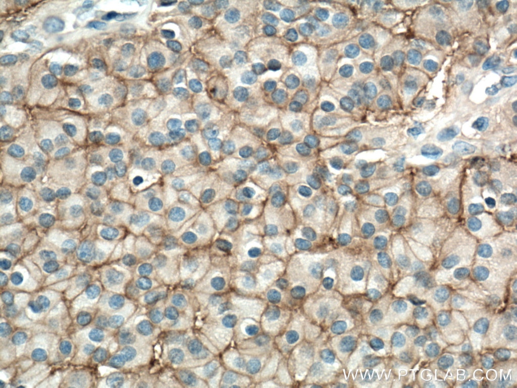
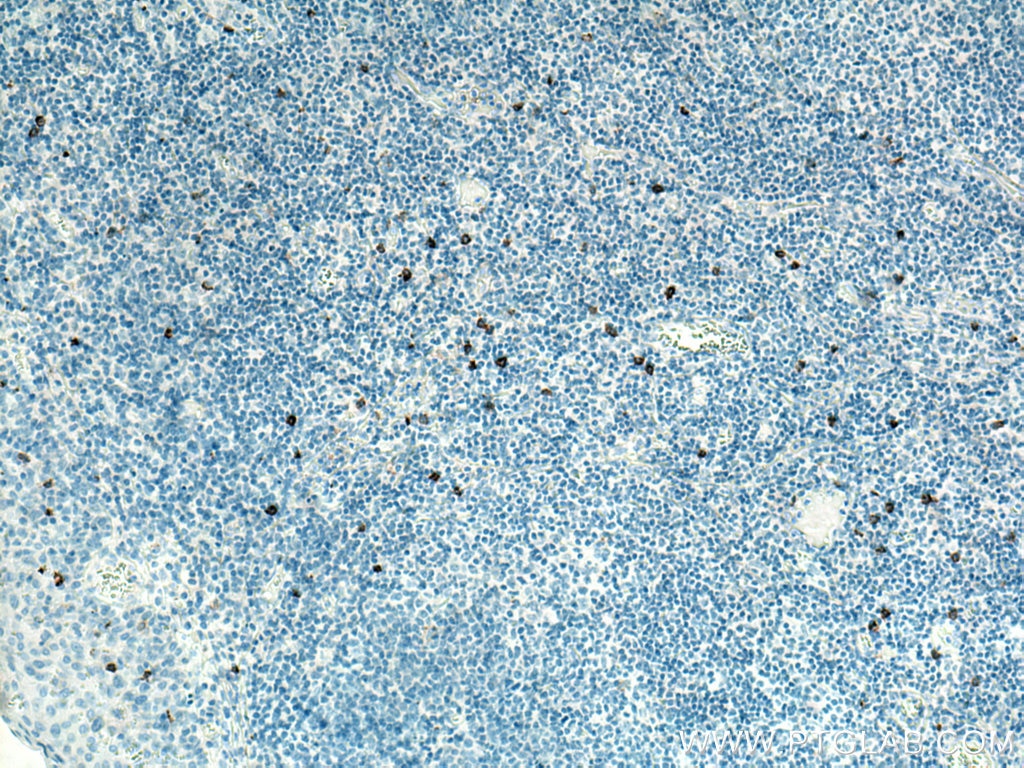
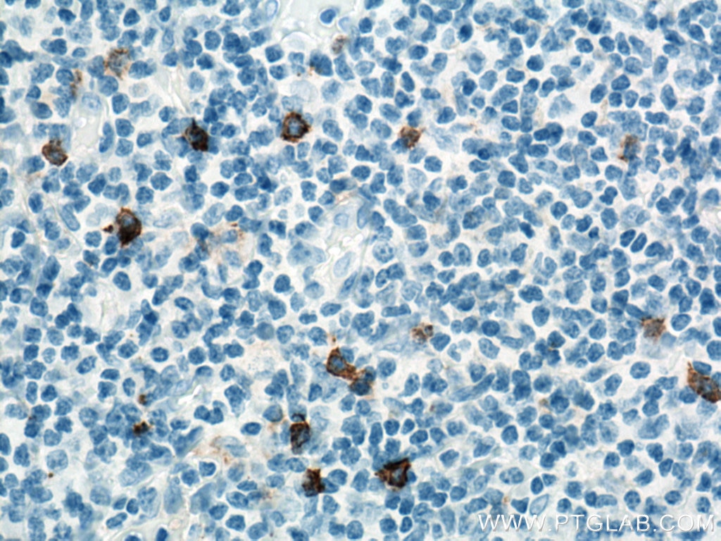
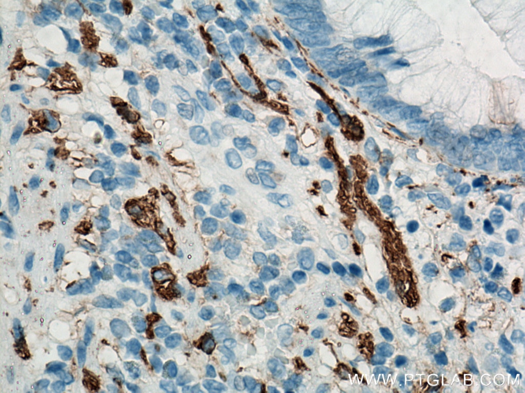
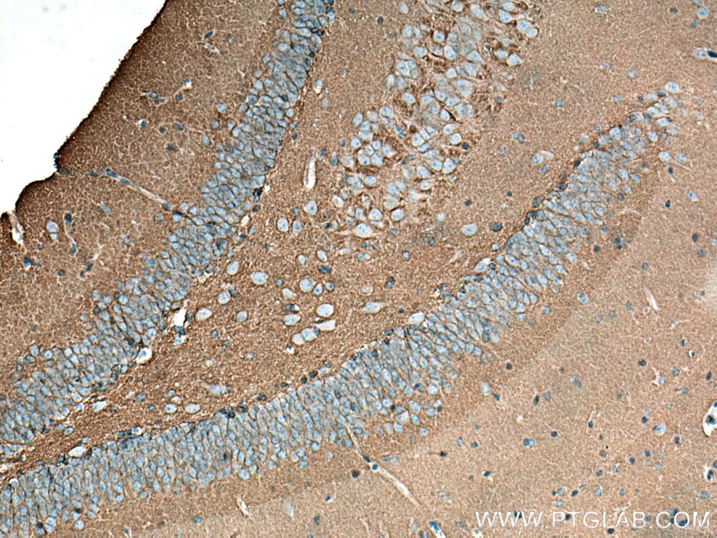
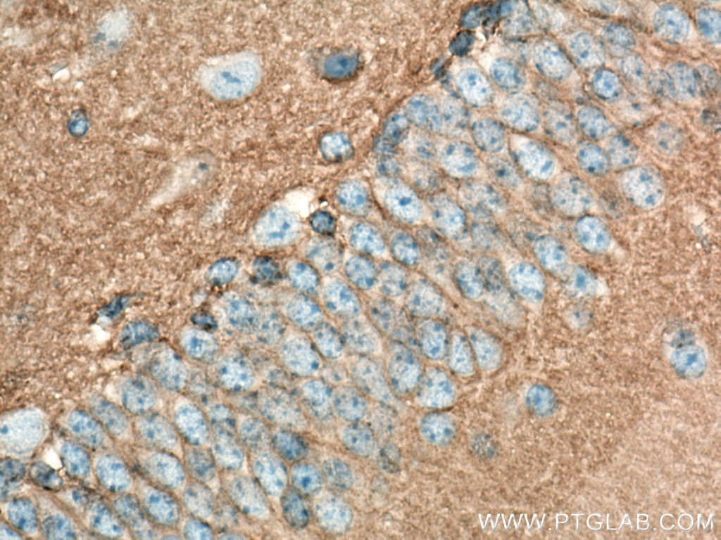
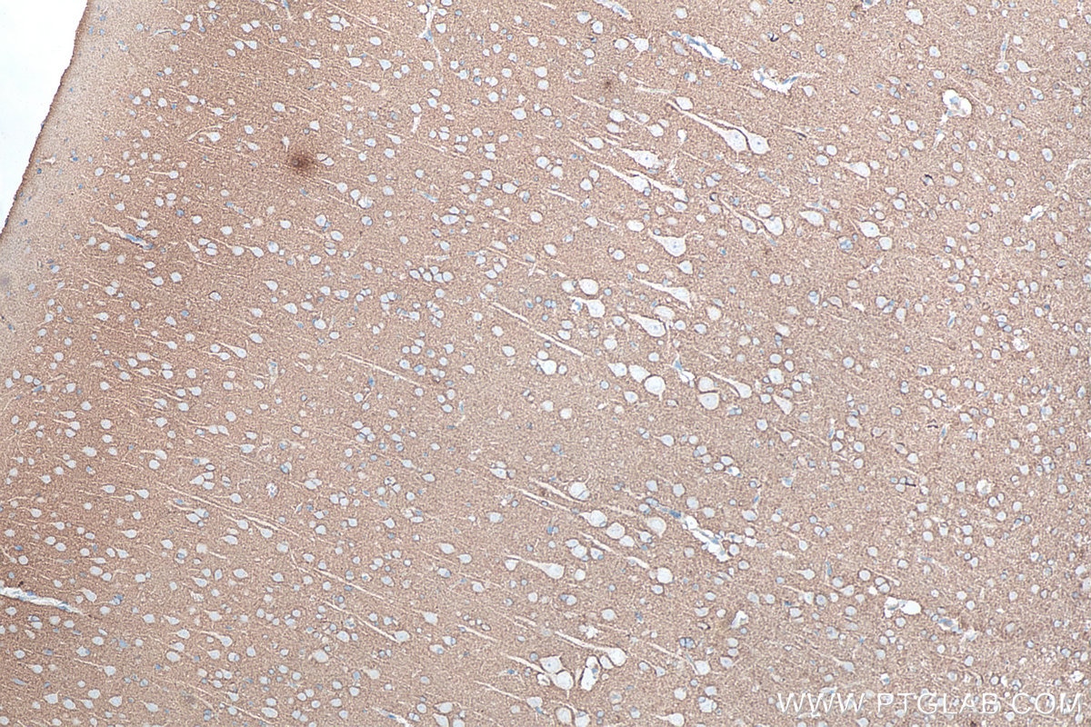
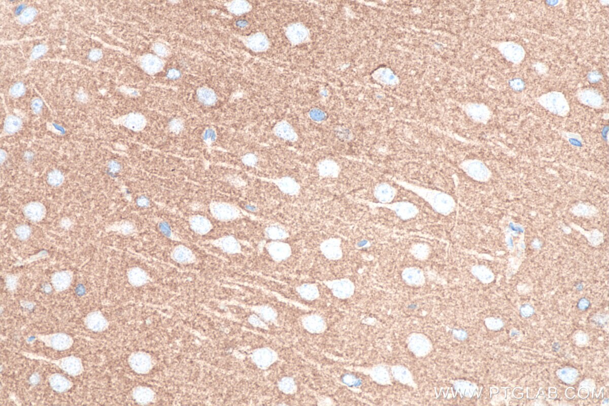
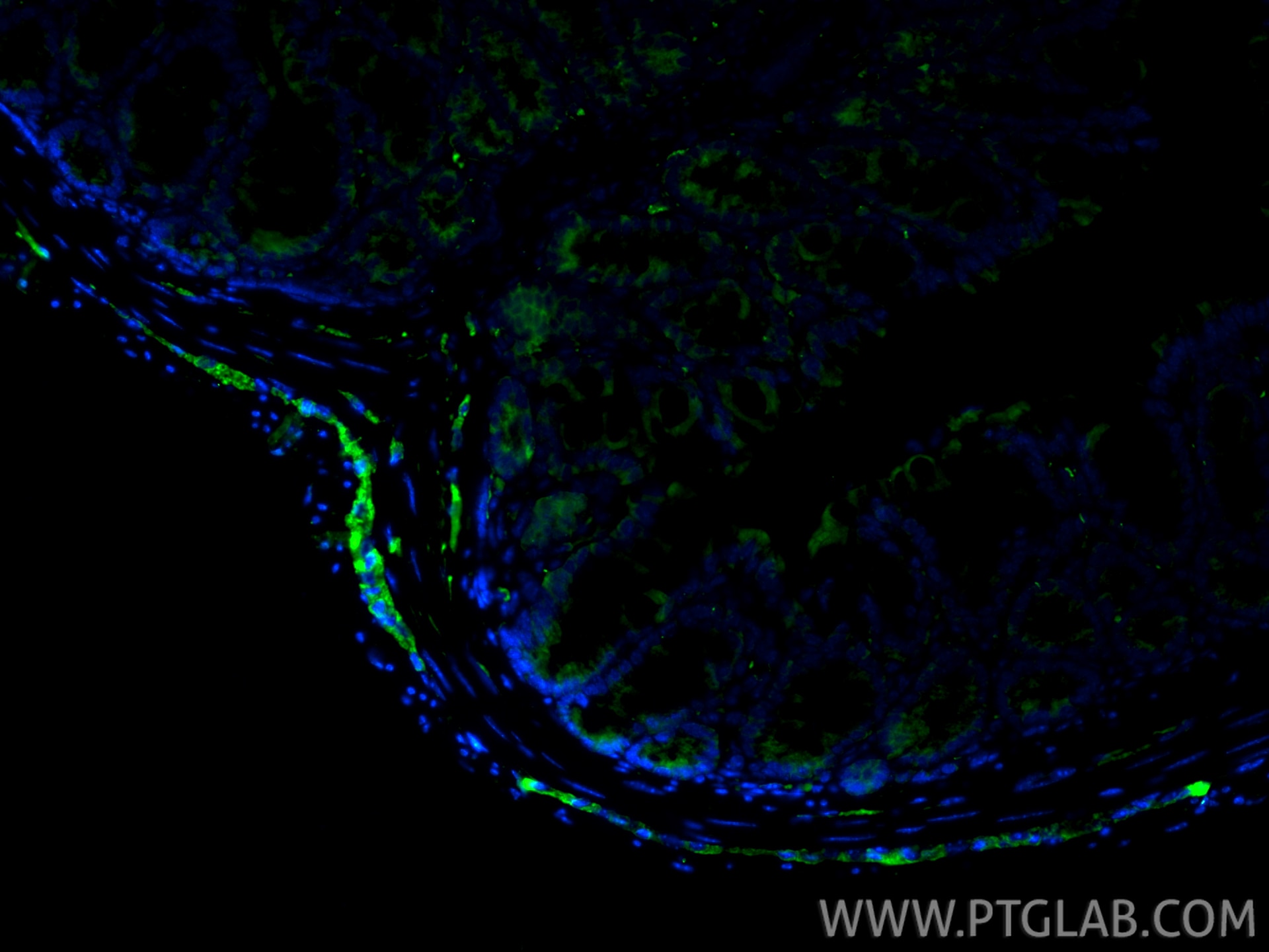
| Positive IHC detected in | human tonsillitis tissue, human appendicitis tissue, human colon tissue, human lung cancer tissue, Insulinoma tissue, mouse brain tissue, mouse colon tissue, rat brain tissue Note: suggested antigen retrieval with TE buffer pH 9.0; (*) Alternatively, antigen retrieval may be performed with citrate buffer pH 6.0 |
| Positive IF-P detected in | mouse colon tissue |
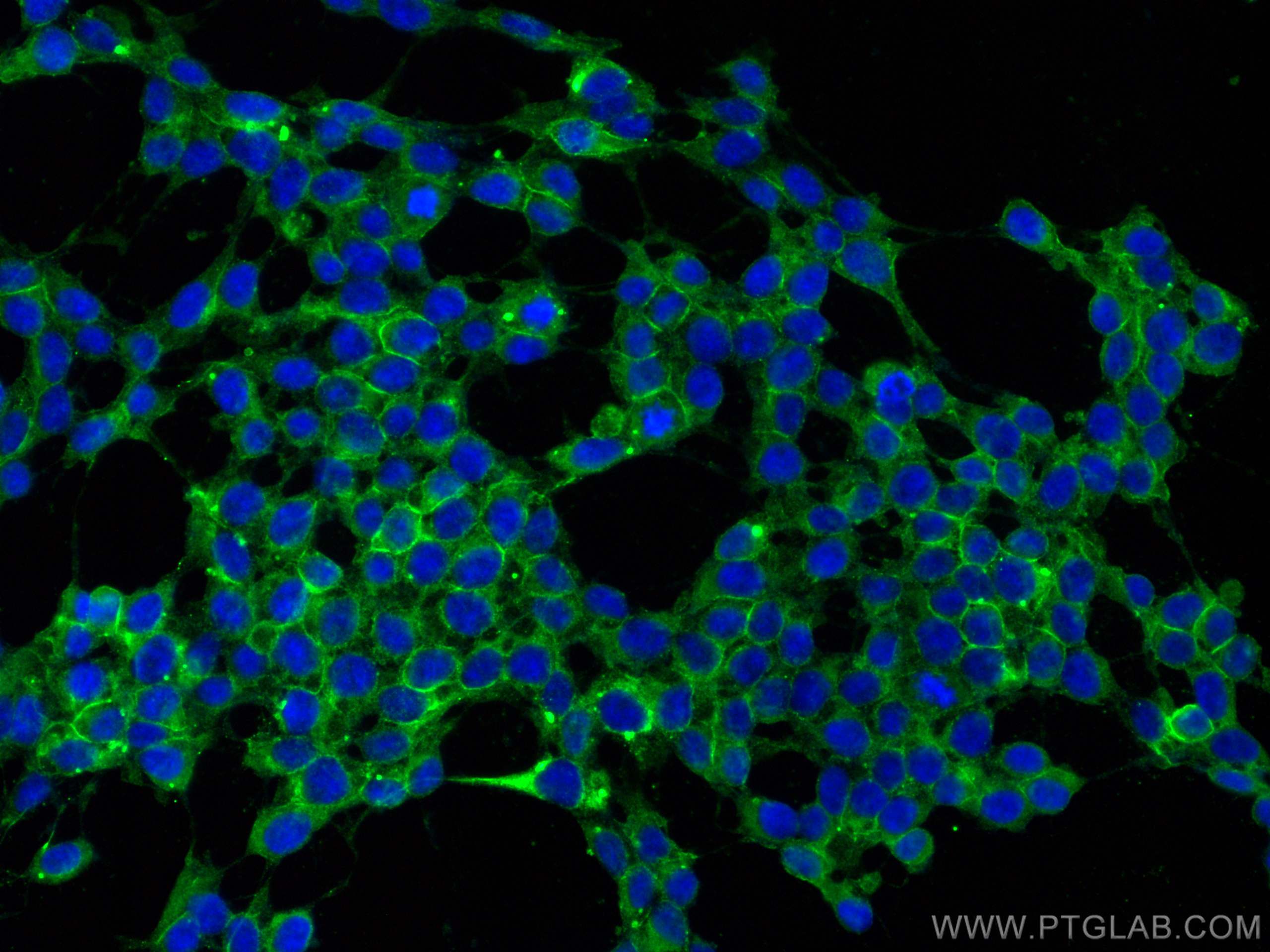
| Positive IF/ICC detected in | SH-SY5Y cells |
Recommended dilution
| Application | Dilution |
|---|---|
| Western Blot (WB) | WB : 1:5000-1:50000 |
| Immunohistochemistry (IHC) | IHC : 1:8000-1:32000 |
| Immunofluorescence (IF)-P | IF-P : 1:50-1:500 |
| Immunofluorescence (IF)/ICC | IF/ICC : 1:50-1:500 |
| It is recommended that this reagent should be titrated in each testing system to obtain optimal results. | |
| Sample-dependent, Check data in validation data gallery. | |
Published Applications
| KD/KO | See 3 publications below |
| WB | See 22 publications below |
| IHC | See 47 publications below |
| IF | See 31 publications below |
Product Information
14255-1-AP targets NCAM1/CD56 in WB, IHC, IF/ICC, IF-P, ELISA applications and shows reactivity with human, mouse, rat, pig samples.
| Tested Reactivity | human, mouse, rat, pig |
| Cited Reactivity | human, mouse, rat, pig |
| Host / Isotype | Rabbit / IgG |
| Class | Polyclonal |
| Type | Antibody |
| Immunogen |
CatNo: Ag5528 Product name: Recombinant human NCAM1/CD56 protein Source: e coli.-derived, PGEX-4T Tag: GST Domain: 30-352 aa of BC047244 Sequence: EISVGESKFFLCQVAGDAKDKDISWFSPNGEKLTPNQQRISVVWNDDSSSTLTIYNANIDDAGIYKCVVTGEDGSESEATVNVKIFQKLMFKNAPTPQEFREGEDAVIVCDVVSSLPPTIIWKHKGRDVILKKDVRFIVLSNNYLQIRGIKKTDEGTYRCEGRILARGEINFKDIQVIVNVPPTIQARQNIVNATANLGQSVTLVCDAEGFPEPTMSWTKDGEQIEQEEDDEKYIFSDDSSQLTIKKVDKNDEAEYICIAENKAGEQDATIHLKVFAKPKITYVENQTAMELEEQVTLTCEASGDPIPSITWRTSTRNISSEE Predict reactive species |
| Full Name | neural cell adhesion molecule 1 |
| Calculated Molecular Weight | 95 kDa |
| Observed Molecular Weight | 120 kDa, 140 kDa, 180 kDa |
| GenBank Accession Number | BC047244 |
| Gene Symbol | NCAM1 |
| Gene ID (NCBI) | 4684 |
| ENSEMBL Gene ID | ENSG00000149294 |
| RRID | AB_2149421 |
| Conjugate | Unconjugated |
| Form | Liquid |
| Purification Method | Antigen affinity purification |
| UNIPROT ID | P13591 |
| Storage Buffer | PBS with 0.02% sodium azide and 50% glycerol, pH 7.3. |
| Storage Conditions | Store at -20°C. Stable for one year after shipment. Aliquoting is unnecessary for -20oC storage. 20ul sizes contain 0.1% BSA. |
Background Information
Neural cell adhesion molecule 1 (NCAM1, also known as CD56) is a cell adhesion glycoprotein of the immunoglobulin (Ig) superfamily. It is a multifunction protein involved in synaptic plasticity, neurodevelopment, and neurogenesis. NCAM1 is expressed on human neurons, glial cells, skeletal muscle cells, NK cells and a subset of T cells, and the expression is observed in a wide variety of human tumors, including myeloma, myeloid leukemia, neuroendocrine tumors, Wilms' tumor, neuroblastoma, and NK/T cell lymphomas. Three major isoforms of NCAM1, with molecular masses of 120, 140, and 180 kDa, are generated by alternative splicing of mRNA (PMID: 9696812). The glycosylphosphatidylinositol (GPI)-anchored NCAM120 and the transmembrane NCAM140 and NCAM180 consist of five Ig-like domains and two fibronection-type III repeats (FNIII). All three forms can be posttranslationally modified by addition of polysialic acid (PSA) (PMID: 14976519). Several other isofroms have also been described (PMID: 1856291).
Protocols
| Product Specific Protocols | |
|---|---|
| IF protocol for NCAM1/CD56 antibody 14255-1-AP | Download protocol |
| IHC protocol for NCAM1/CD56 antibody 14255-1-AP | Download protocol |
| WB protocol for NCAM1/CD56 antibody 14255-1-AP | Download protocol |
| Standard Protocols | |
|---|---|
| Click here to view our Standard Protocols |
Publications
| Species | Application | Title |
|---|---|---|
Nat Med Selective modulation of the androgen receptor AF2 domain rescues degeneration in spinal bulbar muscular atrophy. | ||
Nat Immunol Short IL-18 generated by caspase-3 cleavage mobilizes NK cells to suppress tumor growth | ||
Cell Metab Multiplexed In Situ Imaging Mass Cytometry Analysis of the Human Endocrine Pancreas and Immune System in Type 1 Diabetes. | ||
Sci Adv Gene therapy with AR isoform 2 rescues spinal and bulbar muscular atrophy phenotype by modulating AR transcriptional activity. | ||
J Clin Invest Thioredoxin activity confers resistance against oxidative stress in tumor-infiltrating NK cells. |
Reviews
The reviews below have been submitted by verified Proteintech customers who received an incentive for providing their feedback.
FH Kenzo (Verified Customer) (01-09-2023) | Worked great. The staining pattern was consistent with what has been reported.
|
FH Emma (Verified Customer) (11-29-2021) | Works well by IF on FFPE tissue @ 1:1000. We used a Tris-EDTA antigen retrieval.
 |
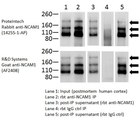
FH Toni (Verified Customer) (02-28-2019) | IP conditions:1ug of rbt anti-NCAM1 was added to 100ug human cortex homogenate (0.5ug/ul) in Tris-sucrose buffer with 1% SDS and 1% Triton X100 thenincubated rotating ON at 4C. Sample was then added to 50ul TBST-washed sheep anti-rabbit magnetic beads and incubated rotating 3hr at 4C. Following 4 TBST washes, bound proteins were eluted with 30ul of elution buffer containing SDS and BME. WB conditions:Samples subjected to SDS-PAGE on 4-12% Bis-Tris gels followed by semi-dry transfer to nitrocellulose membranes using standard conditions. Membranes were blocked for 1hr at RT in 50% LiCor Odyssey blocking buffer (TBS) then probed with either 1:5000 (v/v) rabbit anti-NCAM1 (Proteintech) or 1:1000 (v/v) goat anti-NCAM1 (R&D Systems) in 50% LiCor Odyssey blocking buffer (TBS + 0.05% Tween-20) ON at 4C. Following TBS+ 0.1% Tween-20 washes, membranes were incubated with the appropriate IR-dye labeled secondary antibody for 1hr at RT, TBST washed, then scanned.*you have my permission to edit the comments above and crop, but not alter, the image provided if you wish to remove reference to an antibody from another company.
 |