Tested Applications
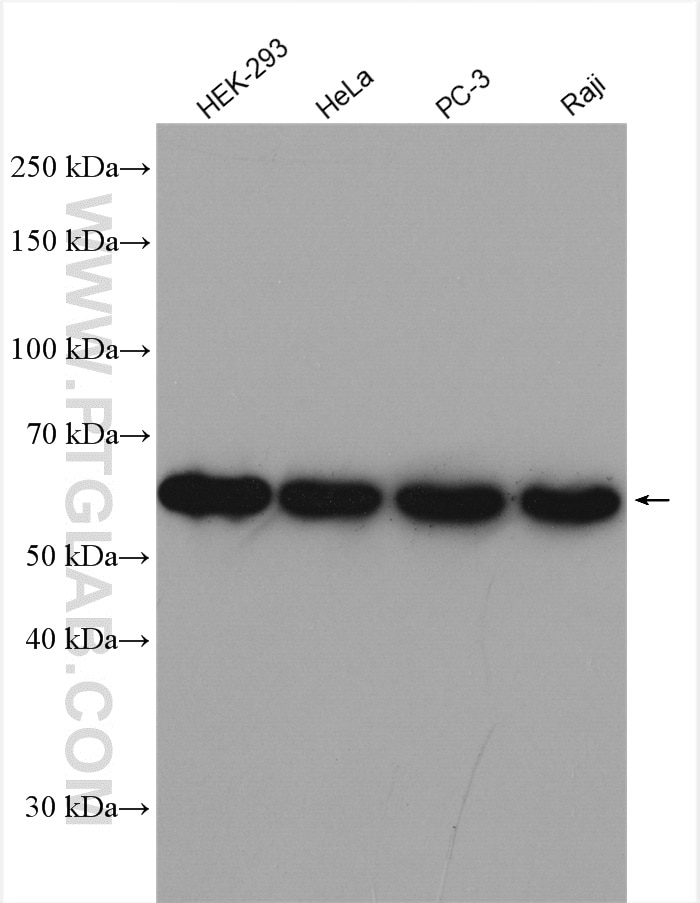
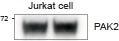
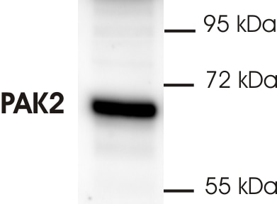
| Positive WB detected in | HEK-293 cells, human skeletal muscle tissue, Jurkat cells, HeLa cells, PC-3 cells, Raji cells |
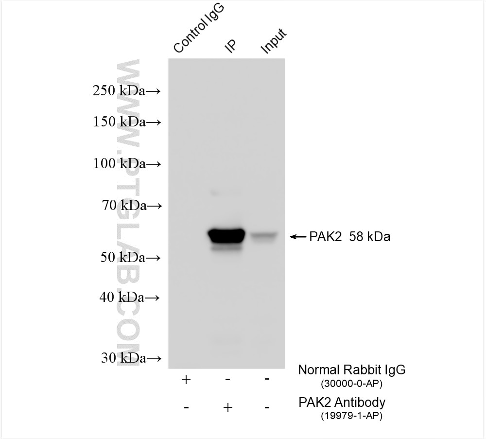
| Positive IP detected in | K-562 cells |
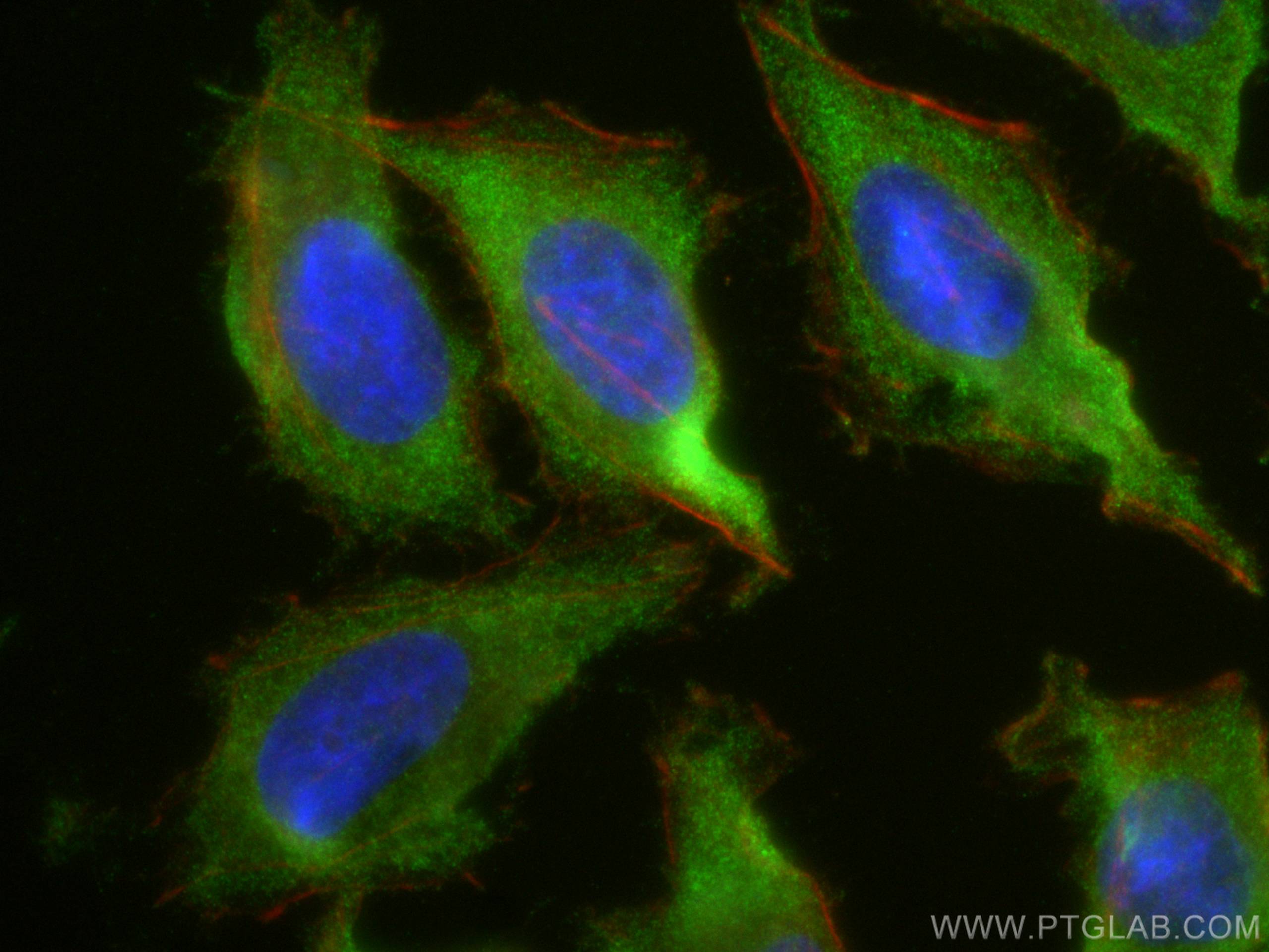
| Positive IF/ICC detected in | HeLa cells |
Recommended dilution
| Application | Dilution |
|---|---|
| Western Blot (WB) | WB : 1:1000-1:5000 |
| Immunoprecipitation (IP) | IP : 0.5-4.0 ug for 1.0-3.0 mg of total protein lysate |
| Immunofluorescence (IF)/ICC | IF/ICC : 1:50-1:500 |
| It is recommended that this reagent should be titrated in each testing system to obtain optimal results. | |
| Sample-dependent, Check data in validation data gallery. | |
Published Applications
| KD/KO | See 1 publications below |
| WB | See 3 publications below |
| IF | See 1 publications below |
| IP | See 1 publications below |
Product Information
19979-1-AP targets PAK2 in WB, IF/ICC, IP, ELISA applications and shows reactivity with human samples.
| Tested Reactivity | human |
| Cited Reactivity | human |
| Host / Isotype | Rabbit / IgG |
| Class | Polyclonal |
| Type | Antibody |
| Immunogen |
Peptide Predict reactive species |
| Full Name | p21 protein (Cdc42/Rac)-activated kinase 2 |
| Calculated Molecular Weight | 58 kDa |
| Observed Molecular Weight | 54 kDa |
| GenBank Accession Number | NM_002577 |
| Gene Symbol | PAK2 |
| Gene ID (NCBI) | 5062 |
| RRID | AB_10644144 |
| Conjugate | Unconjugated |
| Form | Liquid |
| Purification Method | Antigen affinity purification |
| UNIPROT ID | Q13177 |
| Storage Buffer | PBS with 0.02% sodium azide and 50% glycerol, pH 7.3. |
| Storage Conditions | Store at -20°C. Stable for one year after shipment. Aliquoting is unnecessary for -20oC storage. 20ul sizes contain 0.1% BSA. |
Background Information
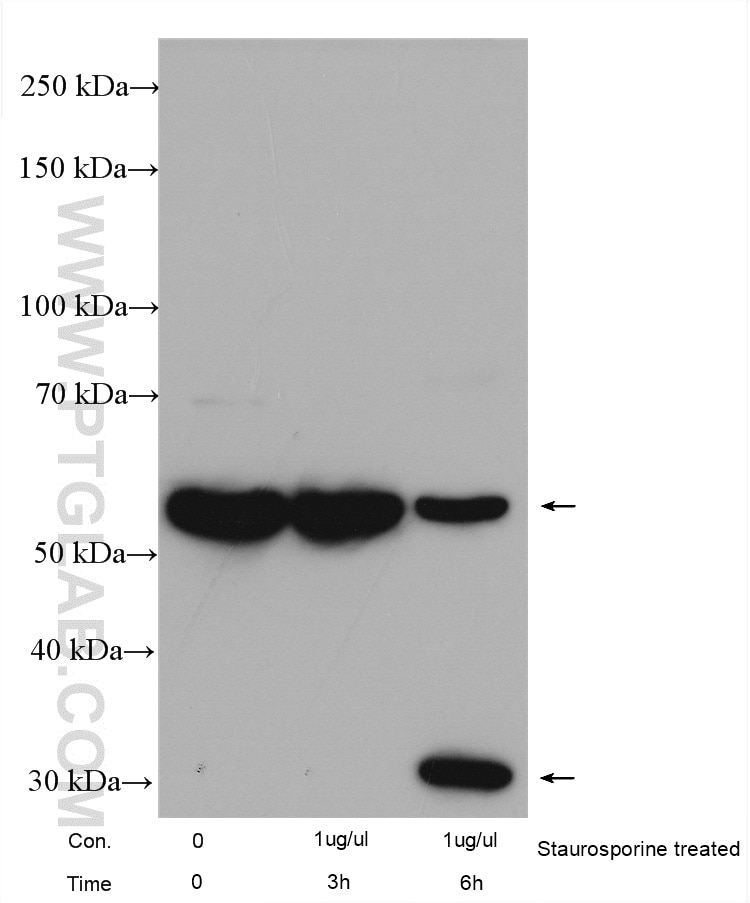
PAK2, also named as PAK65, PAKgamma, p58, PAK-2p27, PAK-2p24 and C-t-PAK2, belongs to the protein kinase superfamily, STE Ser/Thr protein kinase family and STE20 subfamily. Full length PAK 2 stimulates cell survival and cell growth. The process is, at least in part, mediated by phosphorylation and inhibition of pro-apoptotic BAD. PAK2 has several isoforms with the MW of 54-62 kDa and 41 kDa. The 62 kDa PAK2 is cleaved into a 34 kDa C terminal fragment and a 28 kDa N terminal fragment with a time course that parallels apoptotic death in Jurkat cells. (PMID:10200518). Caspase-activated PAK-2p34 is involved in cell death response, probably involving the JNK signaling pathway. Cleaved PAK-2p34 seems to have a higher activity than the CDC42-activated form. The antibody has no cross reaction to PAK1 and PAK3.
Protocols
| Product Specific Protocols | |
|---|---|
| IF protocol for PAK2 antibody 19979-1-AP | Download protocol |
| IP protocol for PAK2 antibody 19979-1-AP | Download protocol |
| WB protocol for PAK2 antibody 19979-1-AP | Download protocol |
| Standard Protocols | |
|---|---|
| Click here to view our Standard Protocols |
Publications
| Species | Application | Title |
|---|---|---|
Mol Genet Genomic Med Chinese Family With Knobloch Syndrome Associated With a Novel PAK2 Variant Leading to Reduced Phosphorylation Levels | ||
J Pharm Anal Aldolase A accelerates hepatocarcinogenesis by refactoring c-Jun transcription | ||
Front Cell Dev Biol PPP1R12B inhibits cell proliferation by inducing G0/G1 phase arrest via PAK2/β-catenin axis in hepatocellular carcinoma.
| ||
Genes Dis PAK1 inhibition synergistically enhances the anti-tumor efficacy of PARP inhibitors in ovarian cancers. |
Reviews
The reviews below have been submitted by verified Proteintech customers who received an incentive for providing their feedback.
FH Gaurav (Verified Customer) (02-27-2026) | Nice antibody working at room temperature
|
FH Titouan (Verified Customer) (11-12-2025) | works very well for WB
 |
FH Frédéric (Verified Customer) (02-18-2022) | Je suis satisfait du résultat car j’ai pu détecter par Western-blot une seule bande de forte intensité d’environ 69 kDa sur un lysat de plaquette humaine (voir figure ci-jointe).
 |