Tested Applications
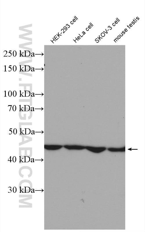
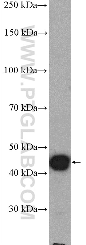
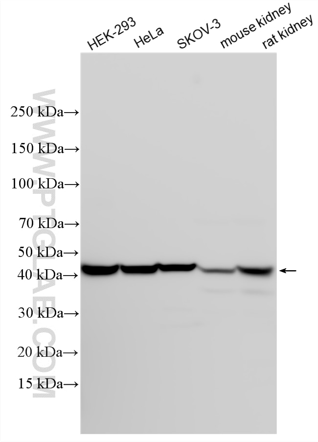
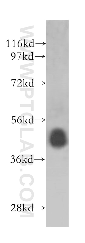
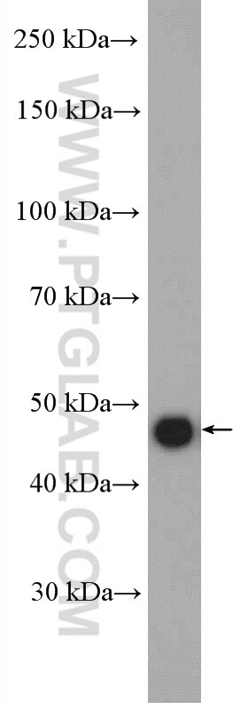
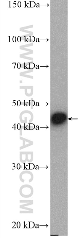
| Positive WB detected in | HEK-293 cells, HeLa cells, mouse testis tissue, SKOV-3 cells, mouse kidney tissue, rat kidney tissue |
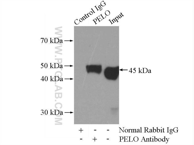
| Positive IP detected in | HEK-293 cells |
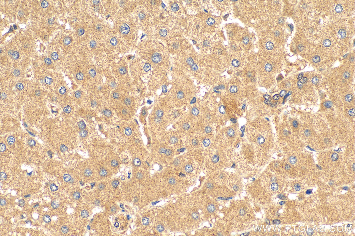
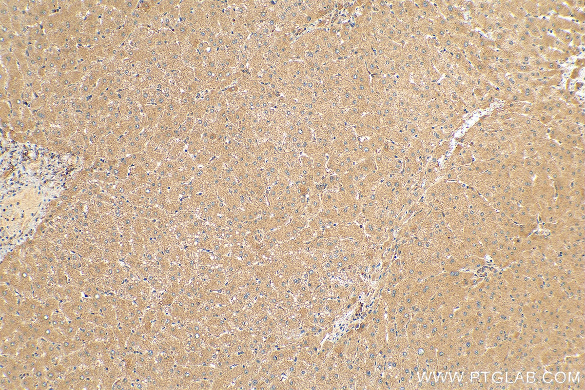
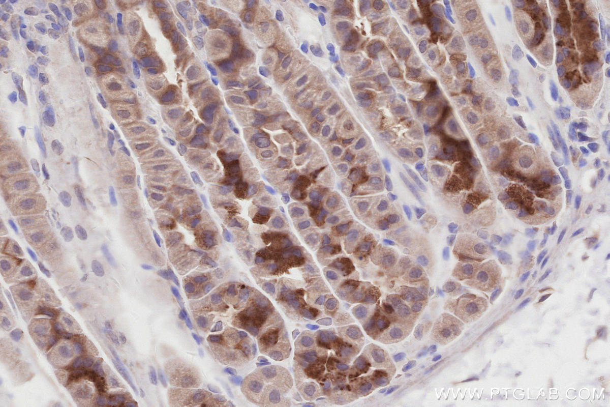
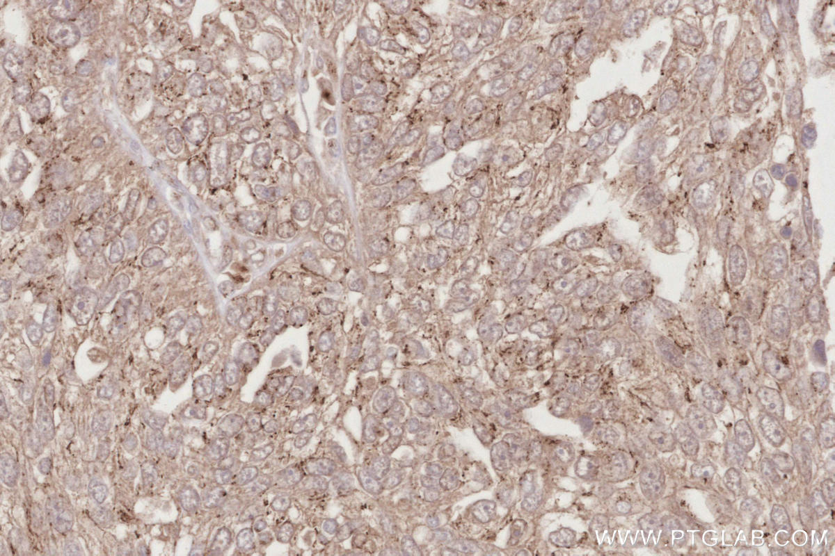
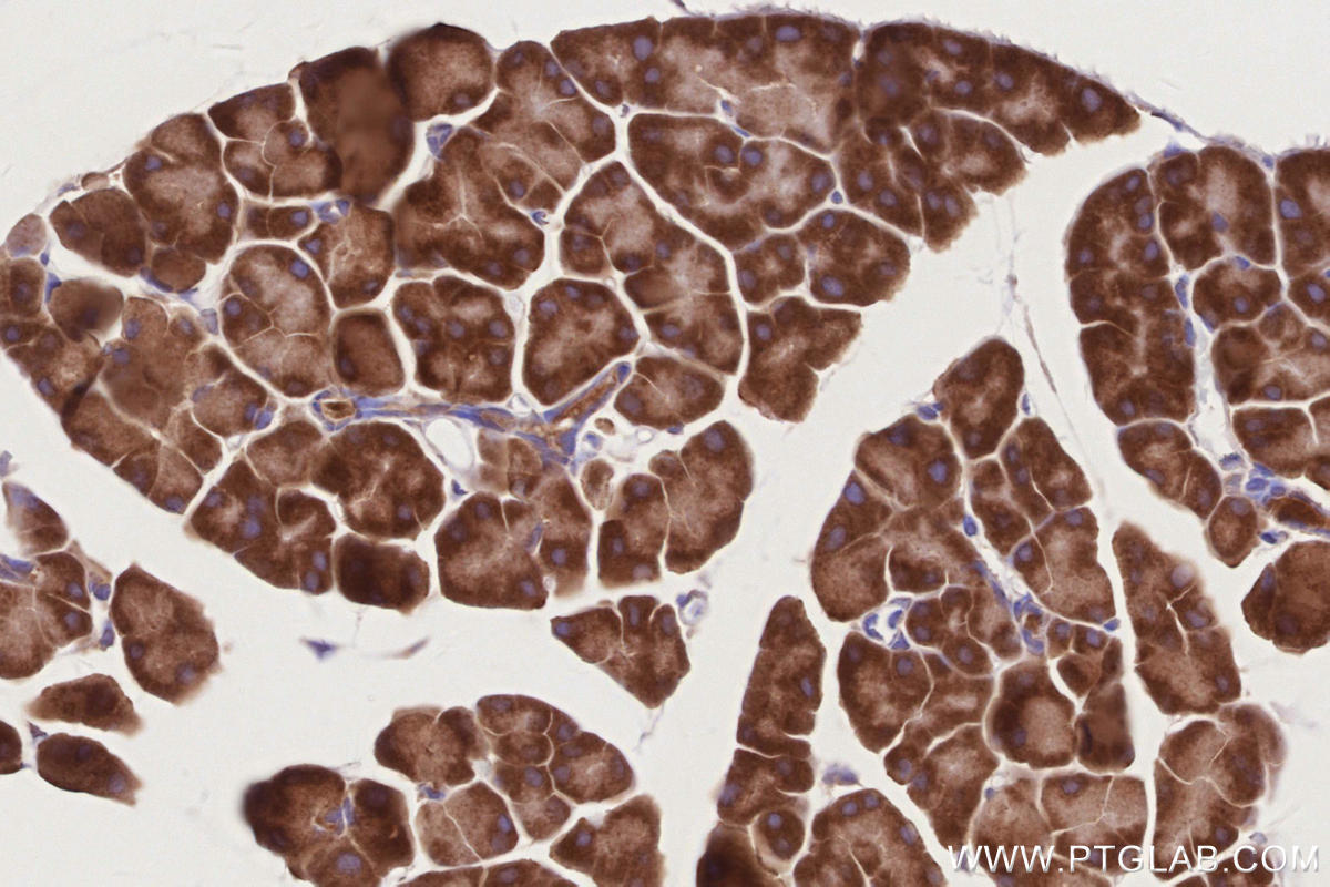
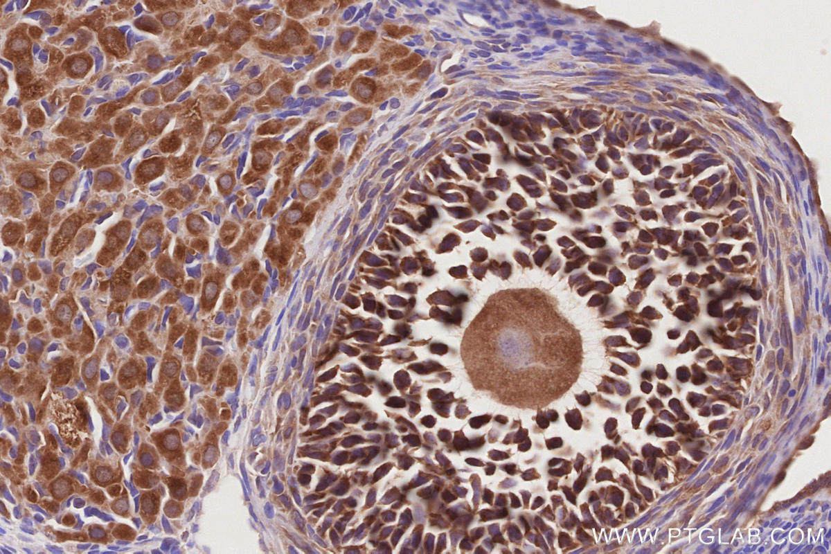
| Positive IHC detected in | human ovary cancer tissue, human liver tissue, mouse pancreas tissue, mouse stomach tissue, rat ovary tissue Note: suggested antigen retrieval with TE buffer pH 9.0; (*) Alternatively, antigen retrieval may be performed with citrate buffer pH 6.0 |
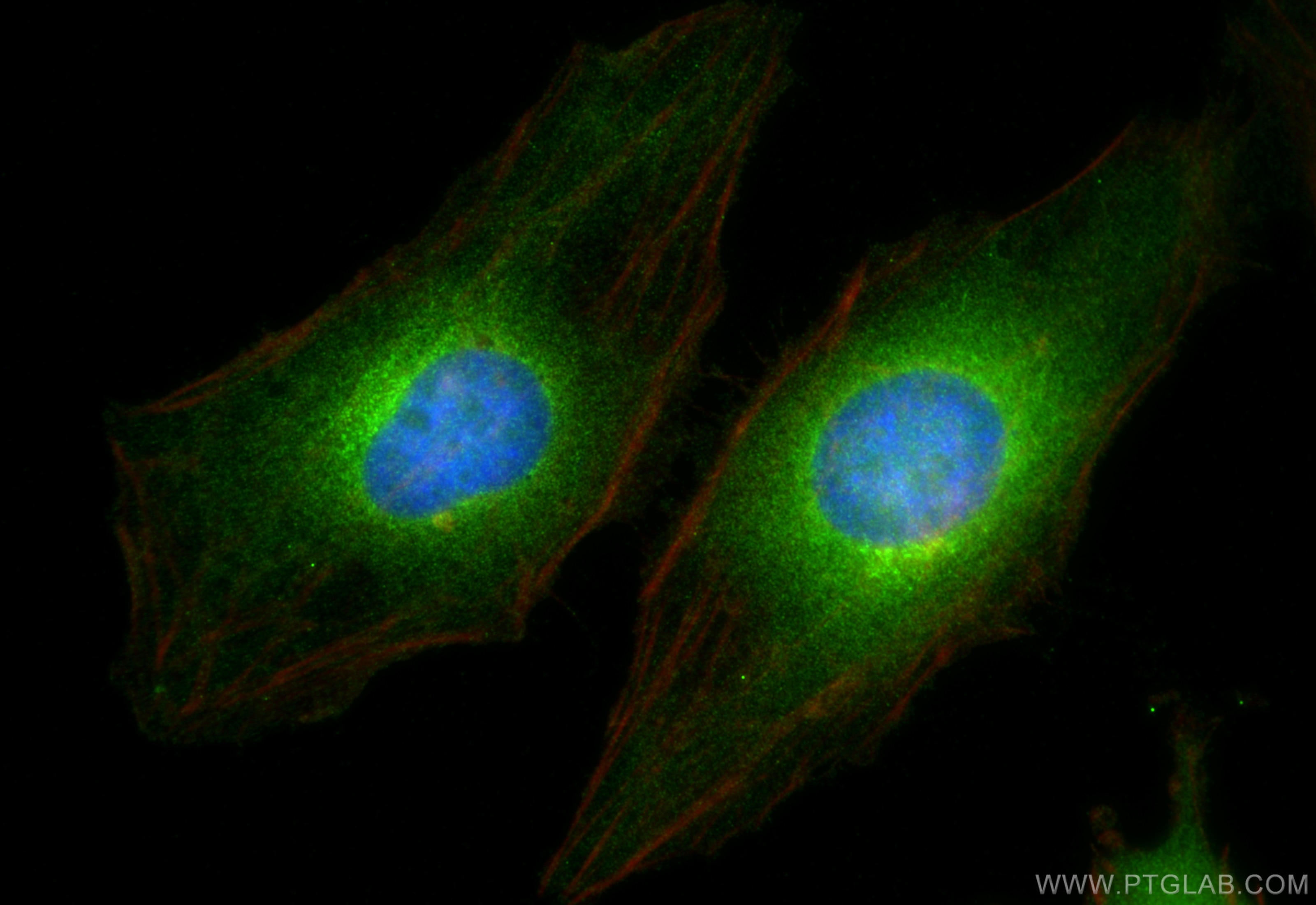
| Positive IF/ICC detected in | HeLa cells |
Recommended dilution
| Application | Dilution |
|---|---|
| Western Blot (WB) | WB : 1:2000-1:12000 |
| Immunoprecipitation (IP) | IP : 0.5-4.0 ug for 1.0-3.0 mg of total protein lysate |
| Immunohistochemistry (IHC) | IHC : 1:250-1:1000 |
| Immunofluorescence (IF)/ICC | IF/ICC : 1:200-1:800 |
| It is recommended that this reagent should be titrated in each testing system to obtain optimal results. | |
| Sample-dependent, Check data in validation data gallery. | |
Published Applications
| KD/KO | See 3 publications below |
| WB | See 13 publications below |
| IF | See 1 publications below |
| IP | See 1 publications below |
Product Information
10582-1-AP targets PELO in WB, IHC, IF/ICC, IP, ELISA applications and shows reactivity with human, mouse, rat samples.
| Tested Reactivity | human, mouse, rat |
| Cited Reactivity | human, mouse |
| Host / Isotype | Rabbit / IgG |
| Class | Polyclonal |
| Type | Antibody |
| Immunogen |
CatNo: Ag0873 Product name: Recombinant human PELO protein Source: e coli.-derived, PGEX-4T Tag: GST Domain: 42-384 aa of BC005889 Sequence: STIRKVQTESSTGSVGSNRVRTTLTLCVEAIDFDSQACQLRVKGTNIQENEYVKMGAYHTIELEPNRQFTLAKKQWDSVVLERIEQACDPAWSADVAAVVMQEGLAHICLVTPSMTLTRAKVEVNIPRKRKGNCSQHDRALERFYEQVVQAIQRHIHFDVVKCILVASPGFVREQFCDYMFQQAVKTDNKLLLENRSKFLQVHASSGHKYSLKEALCDPTVASRLSDTKAAGEVKALDDFYKMLQHEPDRAFYGLKQVEKANEAMAIDTLLISDELFRHQDVATRSRYVRLVDSVKENAGTVRIFSSLHVSGEQLSQLTGVAAILRFPVPELSDQEGDSSSEE Predict reactive species |
| Full Name | pelota homolog (Drosophila) |
| Calculated Molecular Weight | 43 kDa |
| Observed Molecular Weight | 43-45 kDa |
| GenBank Accession Number | BC005889 |
| Gene Symbol | PELO |
| Gene ID (NCBI) | 53918 |
| RRID | AB_2236833 |
| Conjugate | Unconjugated |
| Form | Liquid |
| Purification Method | Antigen affinity purification |
| UNIPROT ID | Q9BRX2 |
| Storage Buffer | PBS with 0.02% sodium azide and 50% glycerol, pH 7.3. |
| Storage Conditions | Store at -20°C. Stable for one year after shipment. Aliquoting is unnecessary for -20oC storage. 20ul sizes contain 0.1% BSA. |
Background Information
The Pelo gene was originally identified in a mutagenesis screen for spermatogenesis-specific genes of Drosophila melanogaster. The PELO is required for the meiotic division during the G2/M transition, and functions in recognizing stalled ribosomes and triggering endonucleolytic cleavage of the mRNA, a mechanism to release non-functional ribosomes and degrade damaged mRNAs [PMID:12556505]. It may participate in the machinery of protein synthesis or in the regulation of mRNA translation [PMID:9584085].
Protocols
| Product Specific Protocols | |
|---|---|
| IF protocol for PELO antibody 10582-1-AP | Download protocol |
| IHC protocol for PELO antibody 10582-1-AP | Download protocol |
| IP protocol for PELO antibody 10582-1-AP | Download protocol |
| WB protocol for PELO antibody 10582-1-AP | Download protocol |
| Standard Protocols | |
|---|---|
| Click here to view our Standard Protocols |
Publications
| Species | Application | Title |
|---|---|---|
Nat Commun Stalled translation by mitochondrial stress upregulates a CNOT4-ZNF598 ribosomal quality control pathway important for tissue homeostasis | ||
Mol Cell Unresolved stalled ribosome complexes restrict cell-cycle progression after genotoxic stress. | ||
Nucleic Acids Res XAB2 depletion induces intron retention in POLR2A to impair global transcription and promote cellular senescence. | ||
Elife Defects in translation-dependent quality control pathways lead to convergent molecular and neurodevelopmental pathology. | ||
Cell Rep A role for the ribosome-associated complex in activation of the IRE1 branch of UPR.
| ||
Res Sq Translation stalling induced mitochondrial entrapment of ribosomal quality control related proteins offers cancer cell vulnerability |
Reviews
The reviews below have been submitted by verified Proteintech customers who received an incentive for providing their feedback.
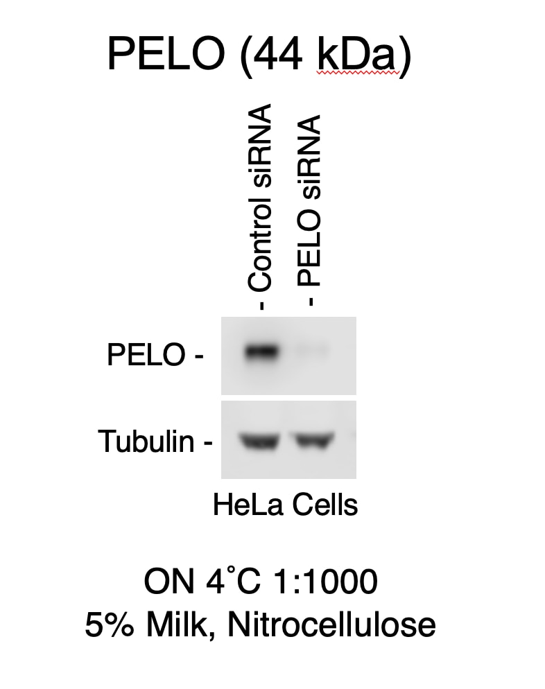
FH Daniel (Verified Customer) (10-26-2022) | This is a good antibody. It works well in western blot using chemiluminescence but not fluorescent secondary antibodies.
 |

