Tested Applications
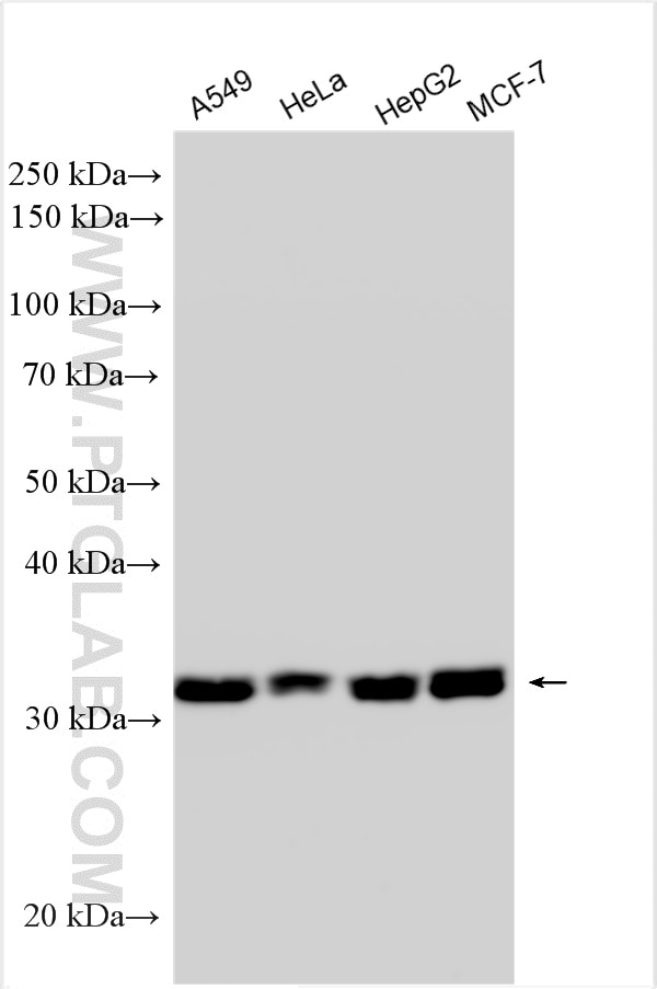
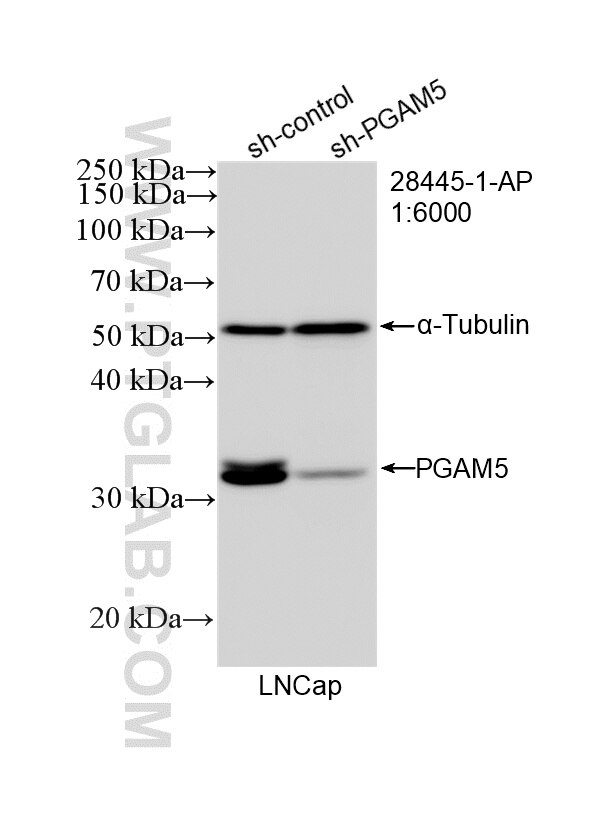
| Positive WB detected in | A549 cells, LNCaP cells, HeLa cells, HepG2 cells, MCF-7 cells |
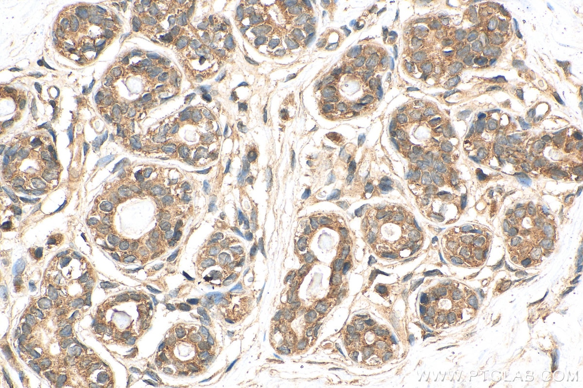
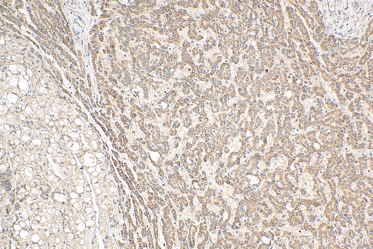
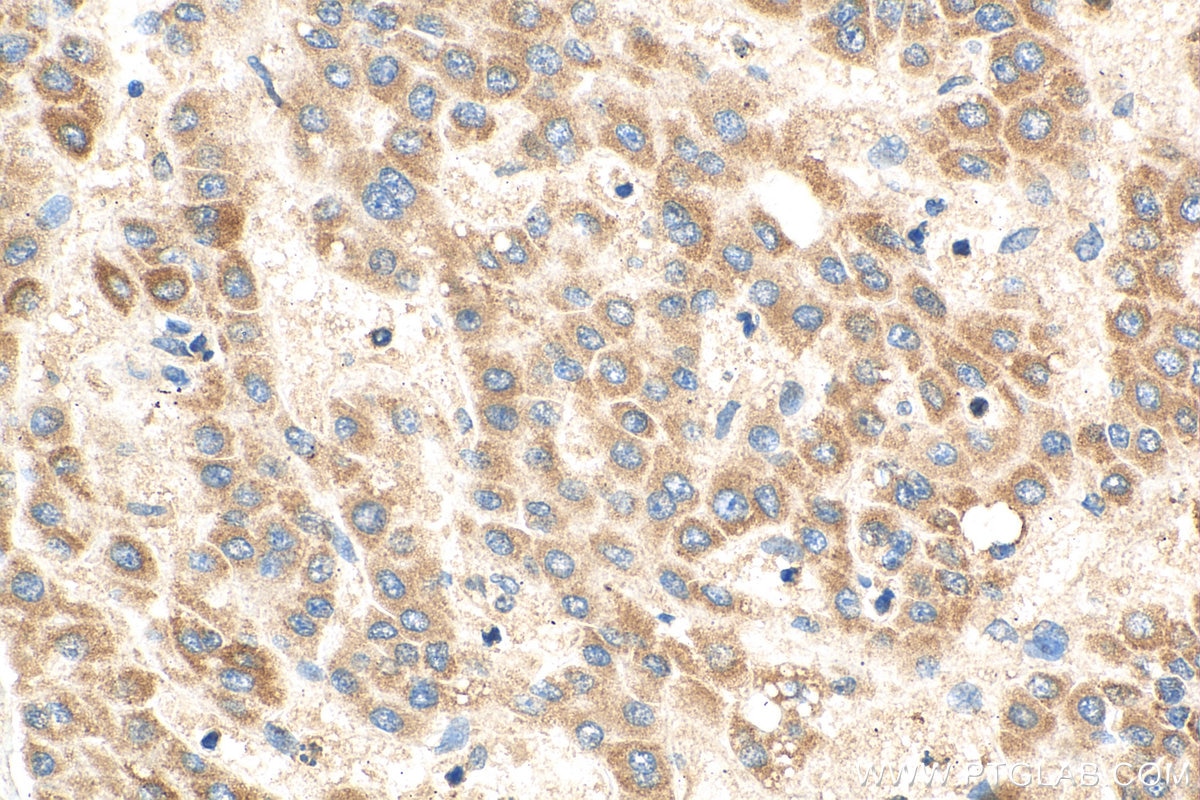
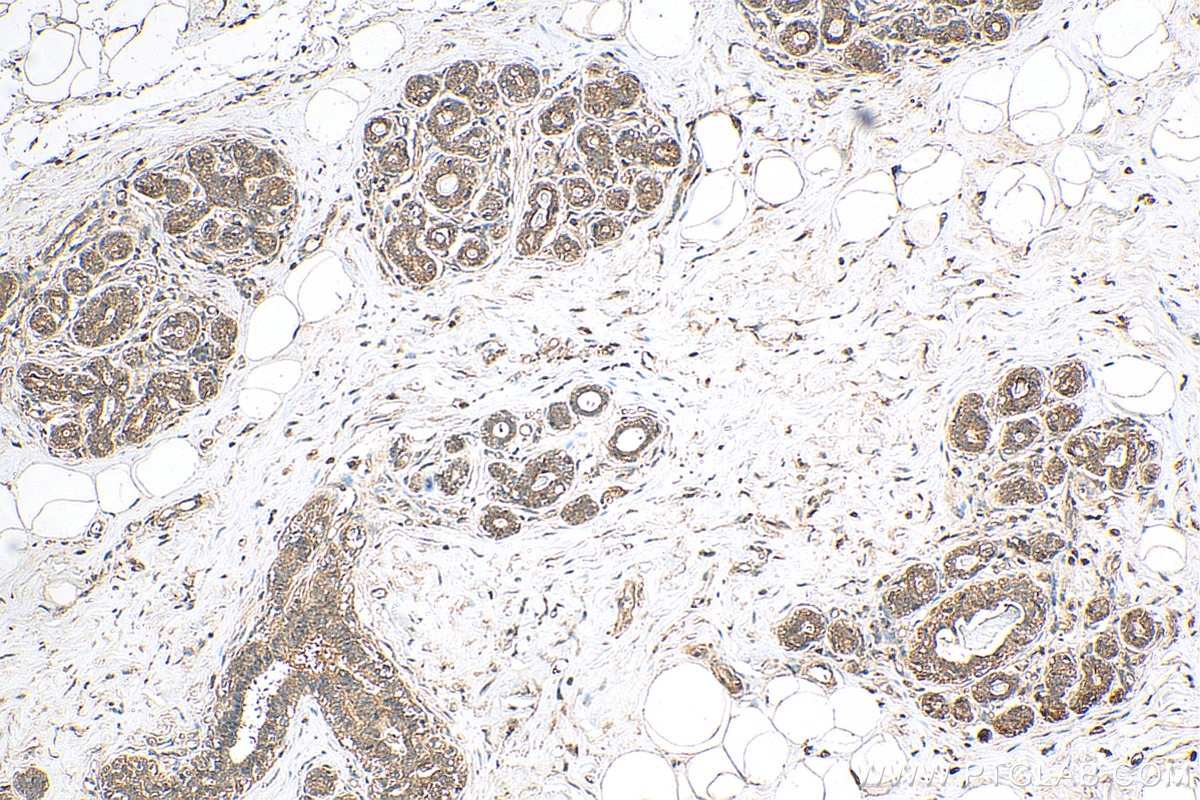
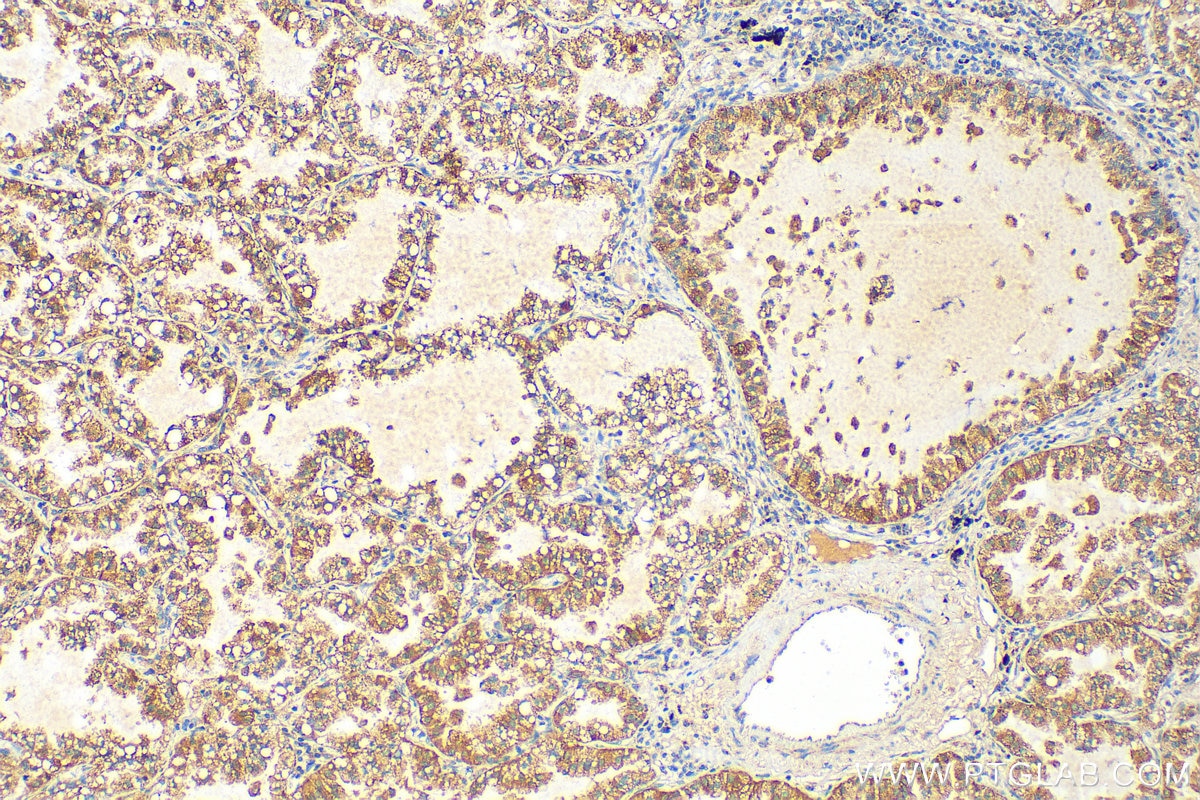
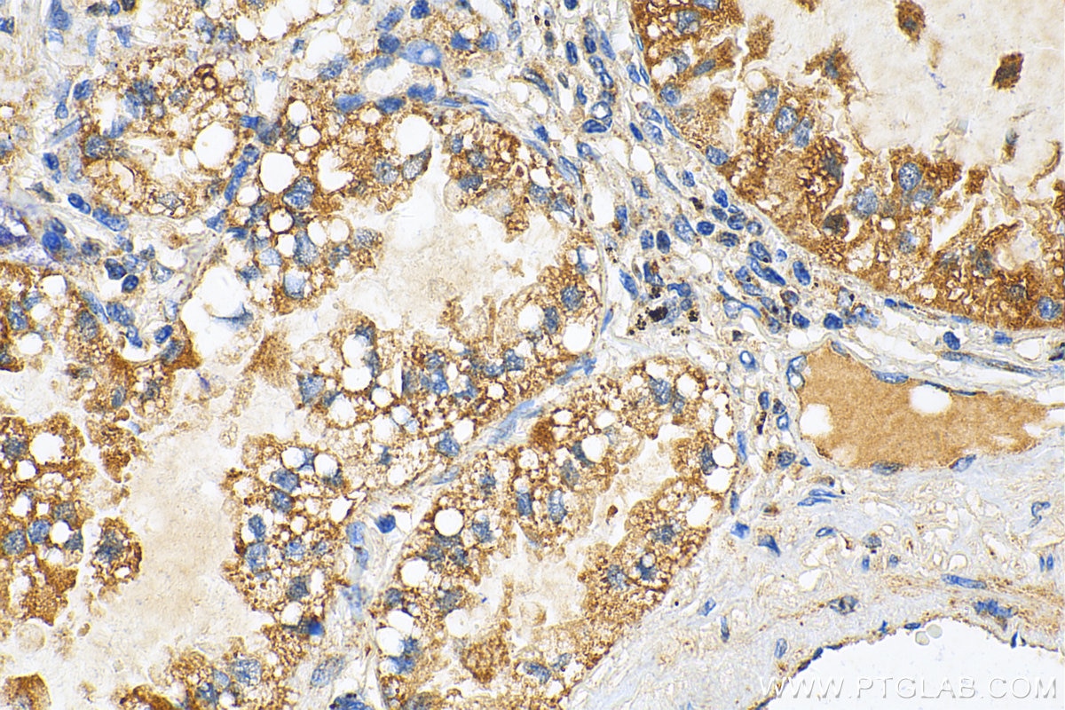
| Positive IHC detected in | human lung cancer tissue, human breast cancer tissue, human liver cancer tissue Note: suggested antigen retrieval with TE buffer pH 9.0; (*) Alternatively, antigen retrieval may be performed with citrate buffer pH 6.0 |
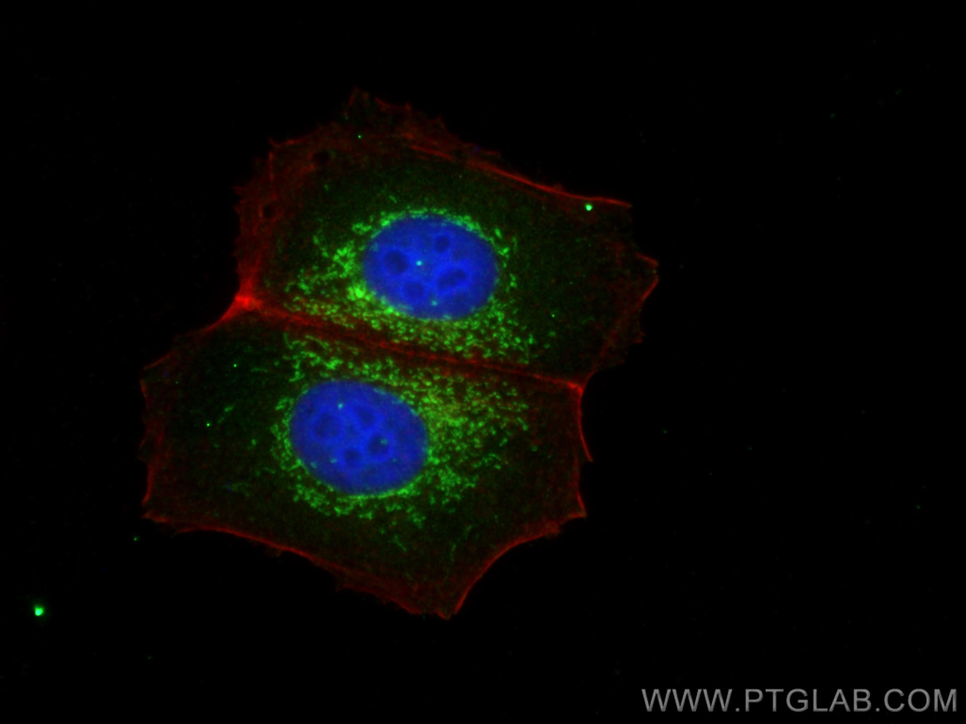
| Positive IF/ICC detected in | MCF-7 cells |
Recommended dilution
| Application | Dilution |
|---|---|
| Western Blot (WB) | WB : 1:2000-1:14000 |
| Immunohistochemistry (IHC) | IHC : 1:1000-1:4000 |
| Immunofluorescence (IF)/ICC | IF/ICC : 1:200-1:800 |
| It is recommended that this reagent should be titrated in each testing system to obtain optimal results. | |
| Sample-dependent, Check data in validation data gallery. | |
Published Applications
| KD/KO | See 4 publications below |
| WB | See 27 publications below |
| IHC | See 5 publications below |
| IF | See 7 publications below |
| CoIP | See 1 publications below |
Product Information
28445-1-AP targets PGAM5 in WB, IHC, IF/ICC, CoIP, ELISA applications and shows reactivity with human samples.
| Tested Reactivity | human |
| Cited Reactivity | human, mouse, rat, pig, sheep, goat |
| Host / Isotype | Rabbit / IgG |
| Class | Polyclonal |
| Type | Antibody |
| Immunogen |
CatNo: Ag28195 Product name: Recombinant human PGAM5 protein Source: e coli.-derived, PET28a Tag: 6*His Domain: 1-255 aa of BC008196 Sequence: MAFRQALQLAACGLAGGSAAVLFSAVAVGKPRAGGDAEPRPAEPPAWAGGARPGPGVWDPNWDRREPLSLINVRKRNVESGEEELASKLDHYKAKATRHIFLIRHSQYHVDGSLEKDRTLTPLGREQAELTGLRLASLGLKFNKIVHSSMTRAIETTDIISRHLPGVCKVSTDLLREGAPIEPDPPVSHWKPEAVQYYEDGARIEAAFRNYIHRADARQEEDSYEIFICHANVIRYIVCSIPPLLSAGDFVVLGS Predict reactive species |
| Full Name | phosphoglycerate mutase family member 5 |
| Calculated Molecular Weight | 32 kDa |
| Observed Molecular Weight | 32 kDa |
| GenBank Accession Number | BC008196 |
| Gene Symbol | PGAM5 |
| Gene ID (NCBI) | 192111 |
| RRID | AB_2881143 |
| Conjugate | Unconjugated |
| Form | Liquid |
| Purification Method | Antigen affinity purification |
| UNIPROT ID | Q96HS1 |
| Storage Buffer | PBS with 0.02% sodium azide and 50% glycerol, pH 7.3. |
| Storage Conditions | Store at -20°C. Stable for one year after shipment. Aliquoting is unnecessary for -20oC storage. 20ul sizes contain 0.1% BSA. |
Background Information
Phosphoglycerate mutase 5 (PGAM5) is a mitochondrial Serine (Ser)/Threonine (Thr) phosphatase normally located in the inner mitochondrial membrane. Upon mitochondrial dysfunction, PGAM5 recruits and dephosphorylates Drp1 at Ser-637, triggers its GTPase activity and promotes mitochondrial fission. PGAM5 regulates mitophagy by stabilizing PINK1 under stress conditions, which recruits E3 ubiquitin ligase PARKIN for degradation of the damaged mitochondria. PGAM5 can be cleaved and released to the cytoplasm through PARKIN, which activates Wnt signaling and induces mitochondrial biogenesis (PMID: 32439975). PGAM5 has 2 isoforms with the molecular mass of 28 and 32 kDa.
Protocols
| Product Specific Protocols | |
|---|---|
| IF protocol for PGAM5 antibody 28445-1-AP | Download protocol |
| IHC protocol for PGAM5 antibody 28445-1-AP | Download protocol |
| WB protocol for PGAM5 antibody 28445-1-AP | Download protocol |
| Standard Protocols | |
|---|---|
| Click here to view our Standard Protocols |
Publications
| Species | Application | Title |
|---|---|---|
Cell Biosci Differential effects of PGAM5 knockout on high fat high fructose diet and methionine choline-deficient diet induced non-alcoholic steatohepatitis (NASH) in mice | ||
Int J Mol Sci The Mitochondrial PHB2/OMA1/DELE1 Pathway Cooperates with Endoplasmic Reticulum Stress to Facilitate the Response to Chemotherapeutics in Ovarian Cancer. | ||
Int Immunopharmacol 4-Octyl itaconate inhibits inflammation to attenuate psoriasis as an agonist of oxeiptosis | ||
J Virol PGAM5 degrades PDCoV N protein and activates type I interferon to antagonize viral replication | ||
Biochim Biophys Acta Mol Cell Res Tau phosphorylation and OPA1 proteolysis are unrelated events: Implications for Alzheimer's Disease. | ||
Heliyon LFHP-1c improves cognitive function after TBI in mice by reducing oxidative stress through the PGAM5-NRF2-KEAP1 ternary complex |
Reviews
The reviews below have been submitted by verified Proteintech customers who received an incentive for providing their feedback.
FH Iram (Verified Customer) (07-03-2020) | 1:1000 dilution in 1%BSA giving a very clear results
|