

Tested Applications
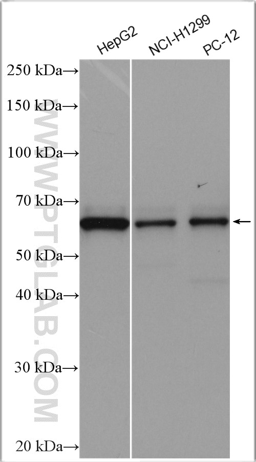
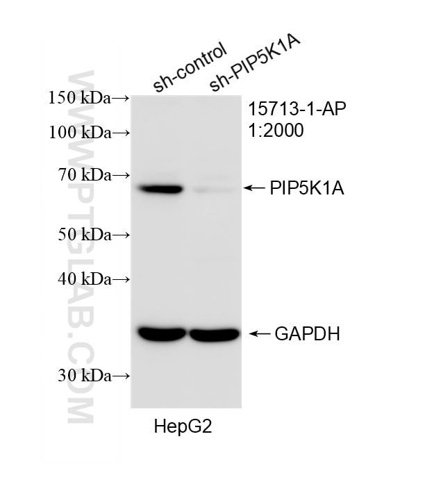
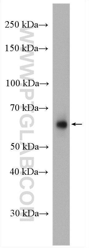
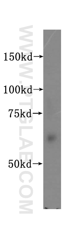
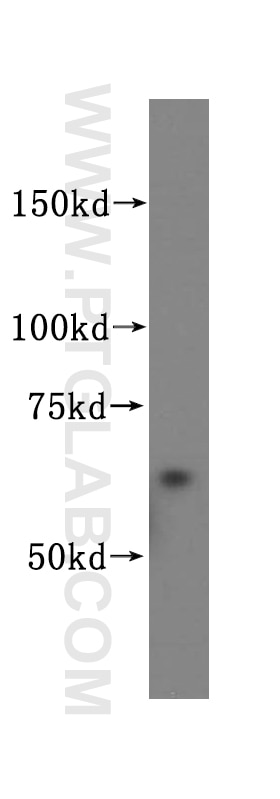
| Positive WB detected in | HepG2 cells, HeLa cells, human heart tissue, human liver tissue, NCI-H1299 cells, PC-12 cells |
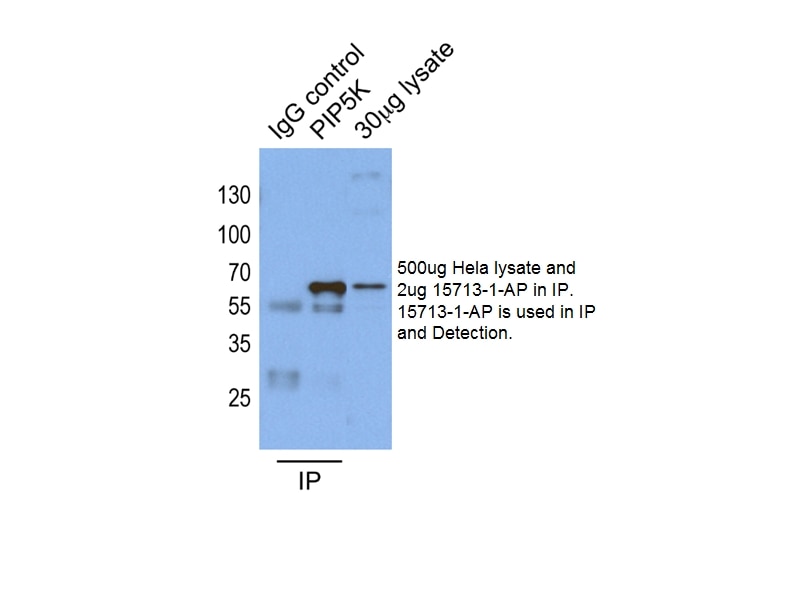
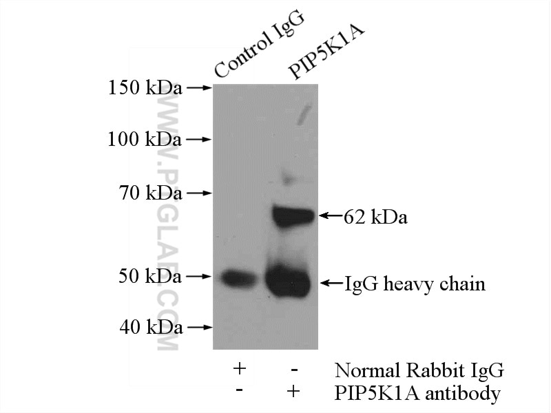
| Positive IP detected in | HeLa cells, mouse heart tissue |
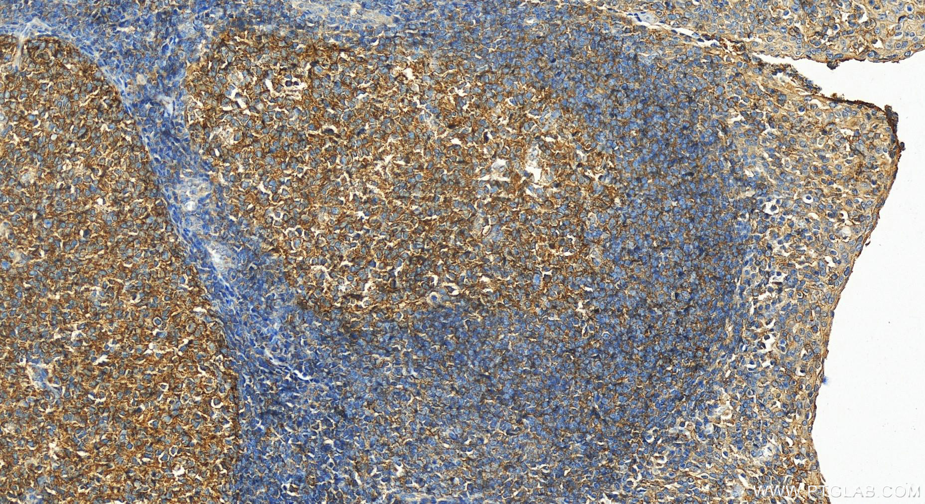
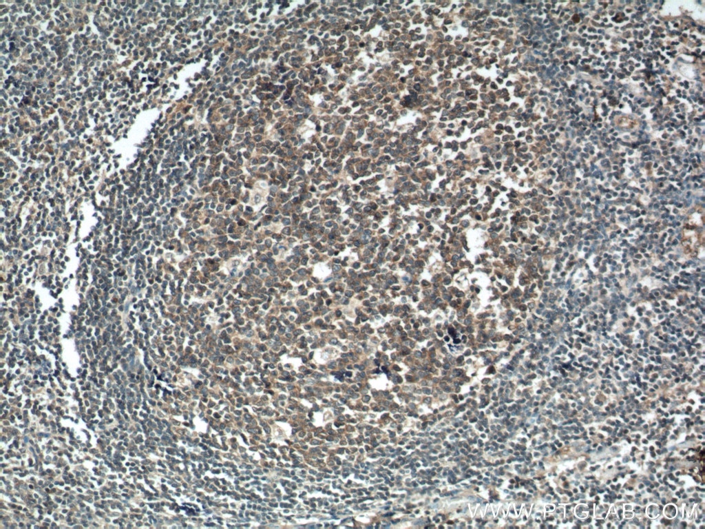
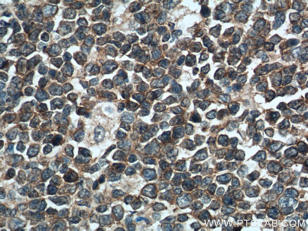
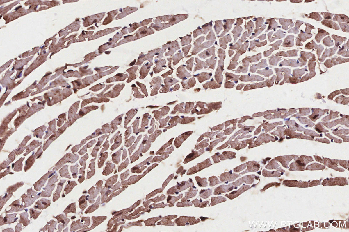
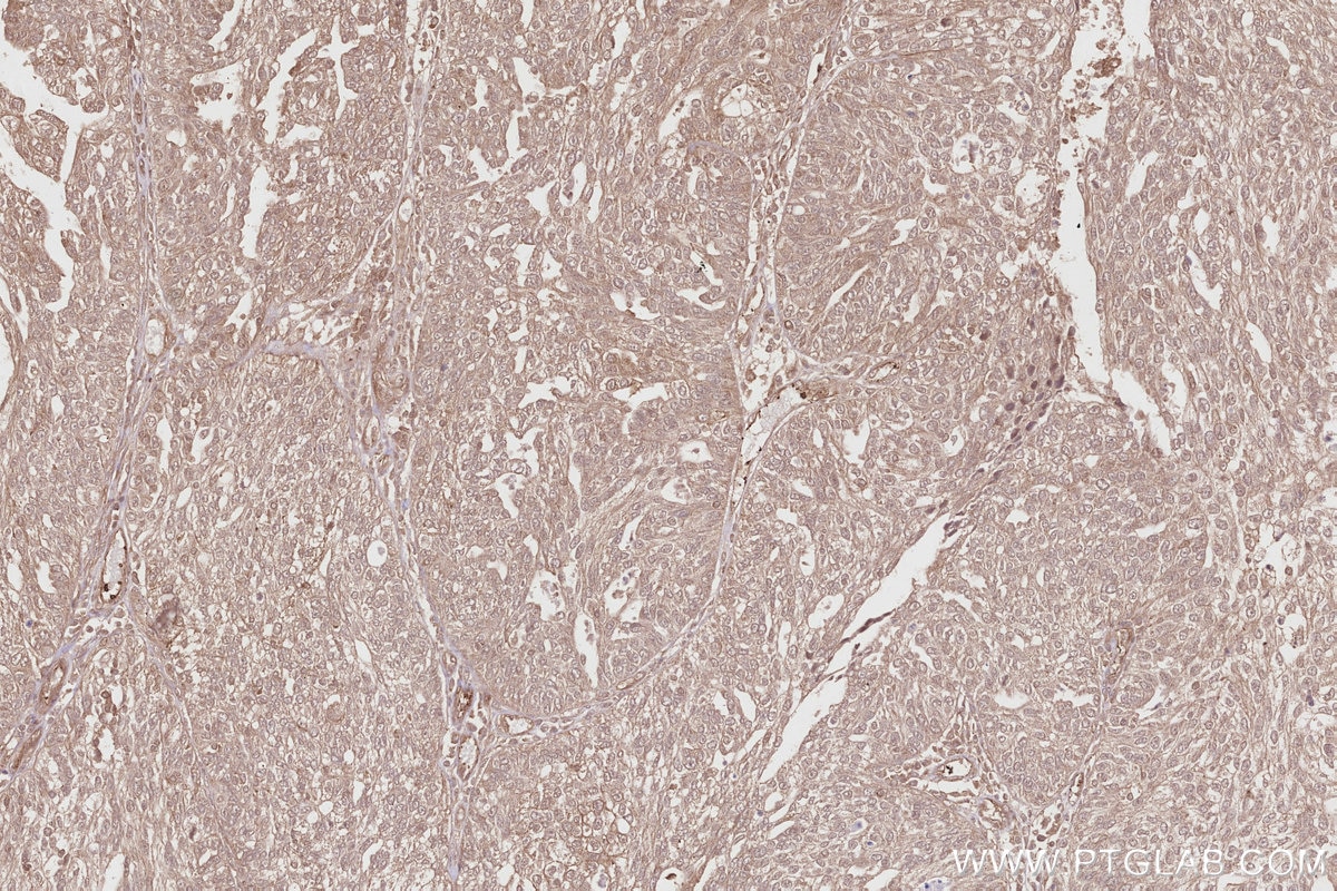
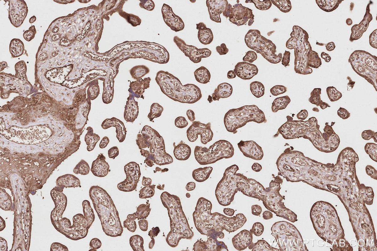
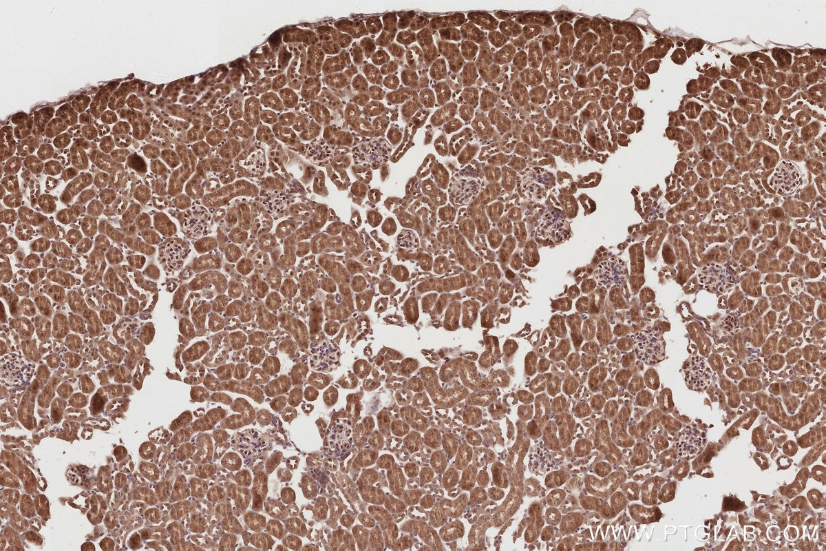
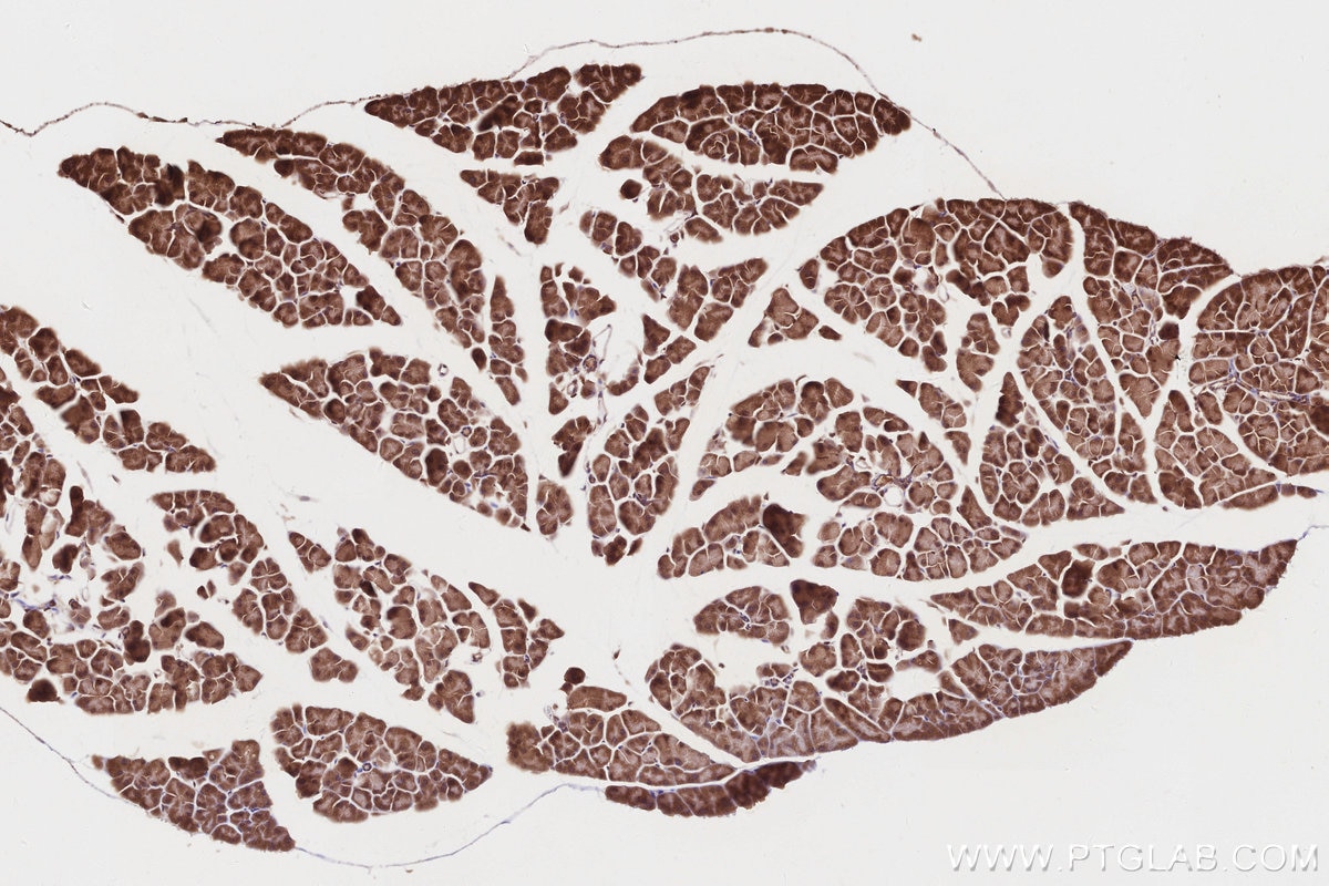
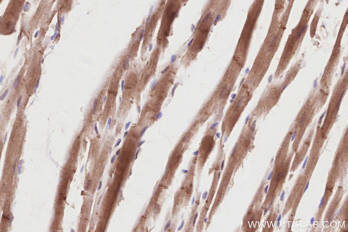
| Positive IHC detected in | human tonsillitis tissue, human placenta tissue, mouse pancreas tissue, rat heart tissue, mouse kidney tissue, mouse heart tissue, human ovary cancer tissue, human tonsillitis tissue Note: suggested antigen retrieval with TE buffer pH 9.0; (*) Alternatively, antigen retrieval may be performed with citrate buffer pH 6.0 |
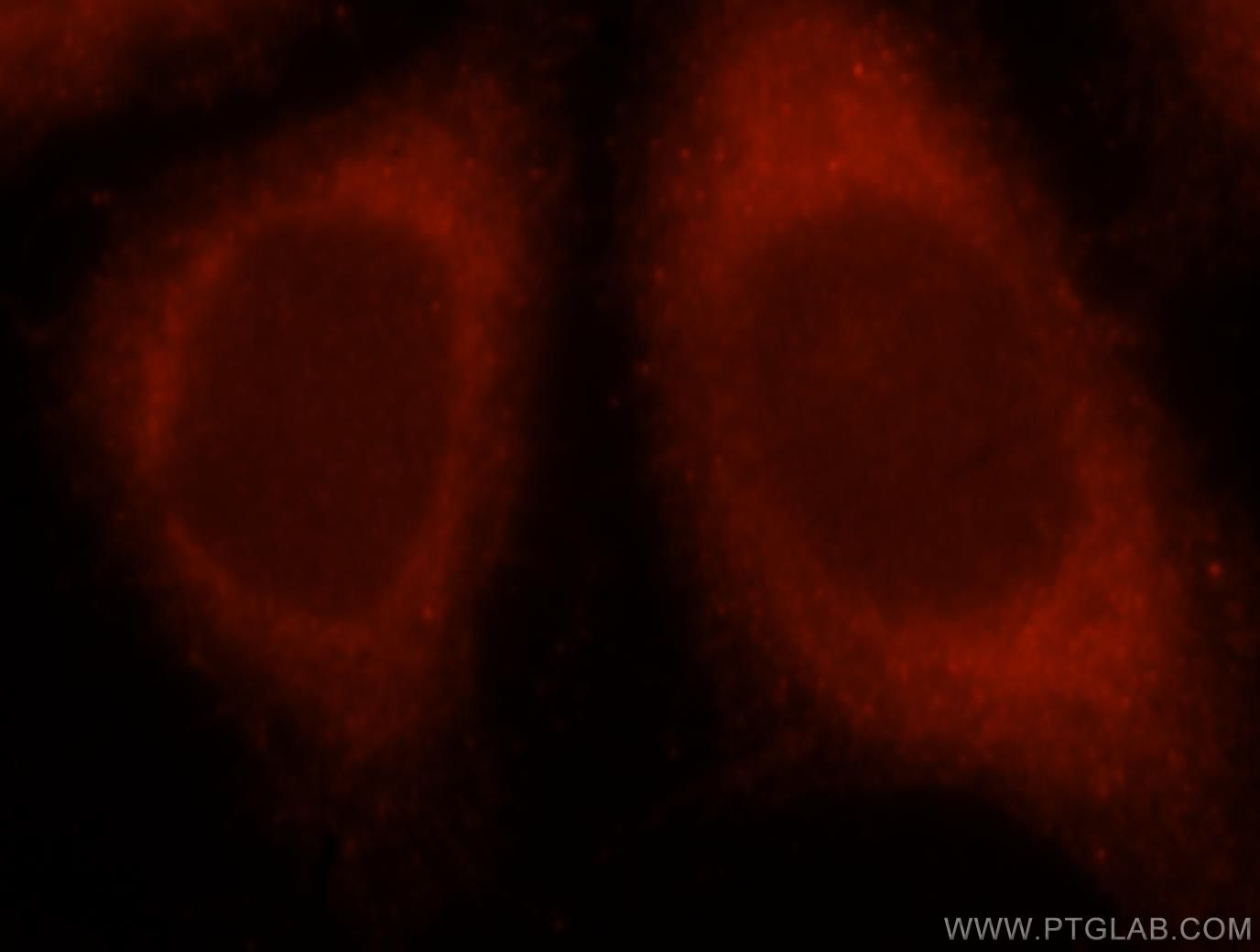
| Positive IF/ICC detected in | Hela cells |
Recommended dilution
| Application | Dilution |
|---|---|
| Western Blot (WB) | WB : 1:5000-1:50000 |
| Immunoprecipitation (IP) | IP : 0.5-4.0 ug for 1.0-3.0 mg of total protein lysate |
| Immunohistochemistry (IHC) | IHC : 1:50-1:500 |
| Immunofluorescence (IF)/ICC | IF/ICC : 1:10-1:100 |
| It is recommended that this reagent should be titrated in each testing system to obtain optimal results. | |
| Sample-dependent, Check data in validation data gallery. | |
Product Information
15713-1-AP targets PIP5K1A in WB, IHC, IF/ICC, IP, ELISA applications and shows reactivity with human, mouse, rat samples.
| Tested Reactivity | human, mouse, rat |
| Cited Reactivity | human, mouse, rat |
| Host / Isotype | Rabbit / IgG |
| Class | Polyclonal |
| Type | Antibody |
| Immunogen |
CatNo: Ag8340 Product name: Recombinant human PIP5K1A protein Source: e coli.-derived, PGEX-4T Tag: GST Domain: 151-500 aa of BC007833 Sequence: SLCSEPLIELCSSGASGSLFYVSSDDEFIIKTVQHKEAEFLQKLLPGYYMNLNQNPRTLLPKFYGLYCVQAGGKNIRIVVMNNLLPRSVKMHIKYDLKGSTYKRRASQKEREKPLPTFKDLDFLQDIPDGLFLDADMYNALCKTLQRDCLVLQSFKIMDYSLLMSIHNIDHAQREPLSSETQYSVDTRRPAPQKALYSTAMESIQGEARRGGTMETDDHMGGIPARNSKGERLLLYIGIIDILQSYRFVKKLEHSWKALVHDGDTVSVHRPGFYAERFQRFMCNTVFKKIPCVHLGRPDVLPQTPPLEEISEGSPIPDPSFSPLVGETLQMLTTSTTLEKLEVAESEFTH Predict reactive species |
| Full Name | phosphatidylinositol-4-phosphate 5-kinase, type I, alpha |
| Calculated Molecular Weight | 56 kDa, 63 kDa |
| Observed Molecular Weight | 63 kDa |
| GenBank Accession Number | BC007833 |
| Gene Symbol | PIP5K1A |
| Gene ID (NCBI) | 8394 |
| RRID | AB_2164696 |
| Conjugate | Unconjugated |
| Form | Liquid |
| Purification Method | Antigen affinity purification |
| UNIPROT ID | Q99755 |
| Storage Buffer | PBS with 0.02% sodium azide and 50% glycerol, pH 7.3. |
| Storage Conditions | Store at -20°C. Stable for one year after shipment. Aliquoting is unnecessary for -20oC storage. 20ul sizes contain 0.1% BSA. |
Background Information
Phosphatidylinositol 4-phosphate 5-kinases (PIP5Ks) play diverse roles in the cellular biology of many organisms, including signal transduction, secretion and vesicular trafficking, and regulation of cytoskeleton assembly(PMID:17688436). There are three PIP5K isoforms,α, β, and γ. The nomenclature for the α and β isoforms is switched between humans and mice(PMID:22096541). There are also several splicing variants of the γ isozyme have been identified.(PMID:20945365). PIP5K1A has some isoforms produced by alternative splicing with the molecular mass of 56-63 kDa.
Protocols
| Product Specific Protocols | |
|---|---|
| IF protocol for PIP5K1A antibody 15713-1-AP | Download protocol |
| IHC protocol for PIP5K1A antibody 15713-1-AP | Download protocol |
| IP protocol for PIP5K1A antibody 15713-1-AP | Download protocol |
| WB protocol for PIP5K1A antibody 15713-1-AP | Download protocol |
| Standard Protocols | |
|---|---|
| Click here to view our Standard Protocols |
Publications
What published studies show
The PIP5K1α antibody (catalog no. 15713-1-AP) has 15 citations. These appear in journals including Nature Cell Biology, Cancer Research, Autophagy, Journal of Clinical Investigation, Advanced Science, Cell Death & Disease, EMBO Journal, Oncogene, Cell Reports, Nutrients, Frontiers in Cell and Developmental Biology, Oncotarget, Virus Research, and Experimental Hematology & Oncology. Research fields span autophagy and lysosome reformation, phosphoinositide/Akt signaling, cancer (glioblastoma, breast, prostate, pancreatic), neurodegeneration (Huntington's, α-synuclein), and viral infection.
| Species | Application | Title |
|---|---|---|
Nat Cell Biol Clathrin and phosphatidylinositol-4,5-bisphosphate regulate autophagic lysosome reformation. | ||
Cancer Res β2-Microglobulin Maintains Glioblastoma Stem Cells and Induces M2-like Polarization of Tumor-Associated Macrophages
| ||
Autophagy IL17A disrupts autophagy-lysosomal function and lysosome reformation through the GSK3B-TFE3 signaling pathway in Huntington disease. | ||
J Clin Invest Spastic paraplegia proteins spastizin and spatacsin mediate autophagic lysosome reformation. | ||
Adv Sci (Weinh) Cell Cycle Control of Nuclear Metabolism Couples Phosphatidylinositol Signaling to Histone Methylation. | ||
Cell Death Dis Bacterial protein MakA causes suppression of tumour cell proliferation via inhibition of PIP5K1α/Akt signalling |
Reviews
What customers say
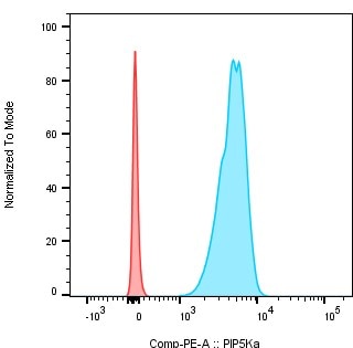
The single review for 15713-1-AP reports a 4-star rating for flow cytometry. The user stained B cells from mouse bone marrow after cytoplasmic permeabilization, fixing and permeabilizing cells with an intracellular fix/perm kit before staining with 0.25 µl per million cells for 50 minutes. A secondary staining with a PE-conjugated anti-rabbit IgG (H+L) F(ab')2 was then performed at 0.1 µl per million cells. The reviewer included images comparing secondary antibody alone versus primary plus secondary antibody staining.
The reviews below have been submitted by verified Proteintech customers who received an incentive for providing their feedback.
Bastien (Verified Customer) (08-19-2020) | Staining of B cells from mice bone marrow after cytoplasmic permezbiliation. The cells were first fixed and permeabilized with intracellular fix and perm set from ebioscience and then stained with 0,25 µl/ million of cells during 50min After, a seconde staining was performed with 0,1µl/million cells of F(ab')2-Donkey anti-Rabbit IgG (H+L), PE, Secondary Antibody from invitrogen. In red secondary antibody alone and in blue primary + secondary antibody
 |


















