



Tested Applications
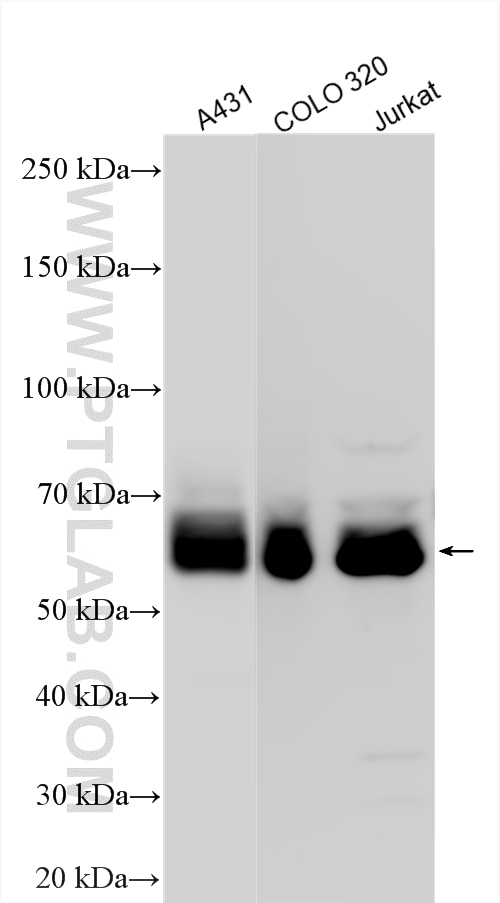
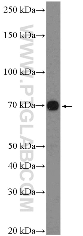
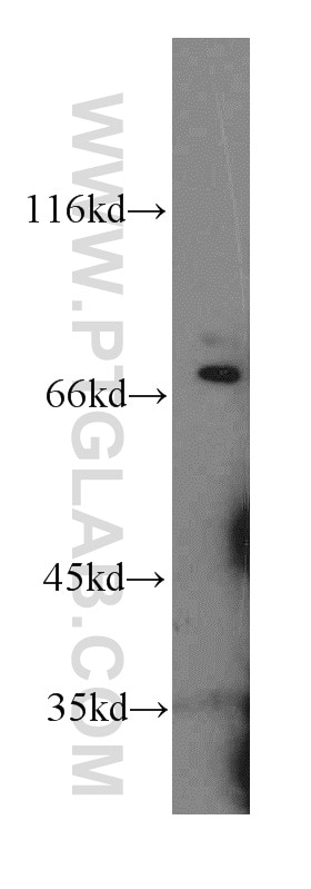
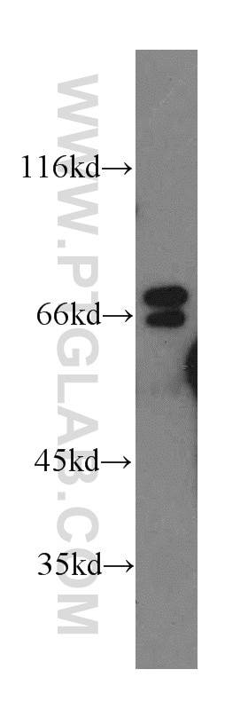
| Positive WB detected in | A431 cells, Jurkat cells, MCF-7 cells, COLO 320 cells |
| Positive IP detected in | A431 cells |
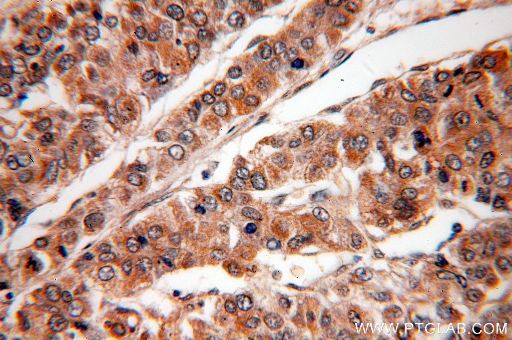
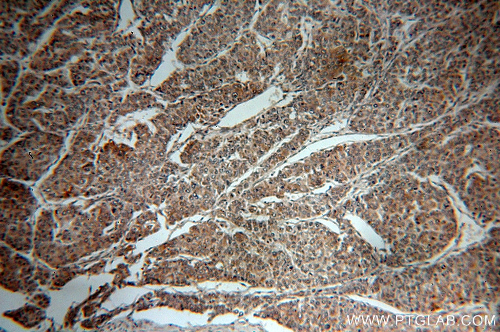
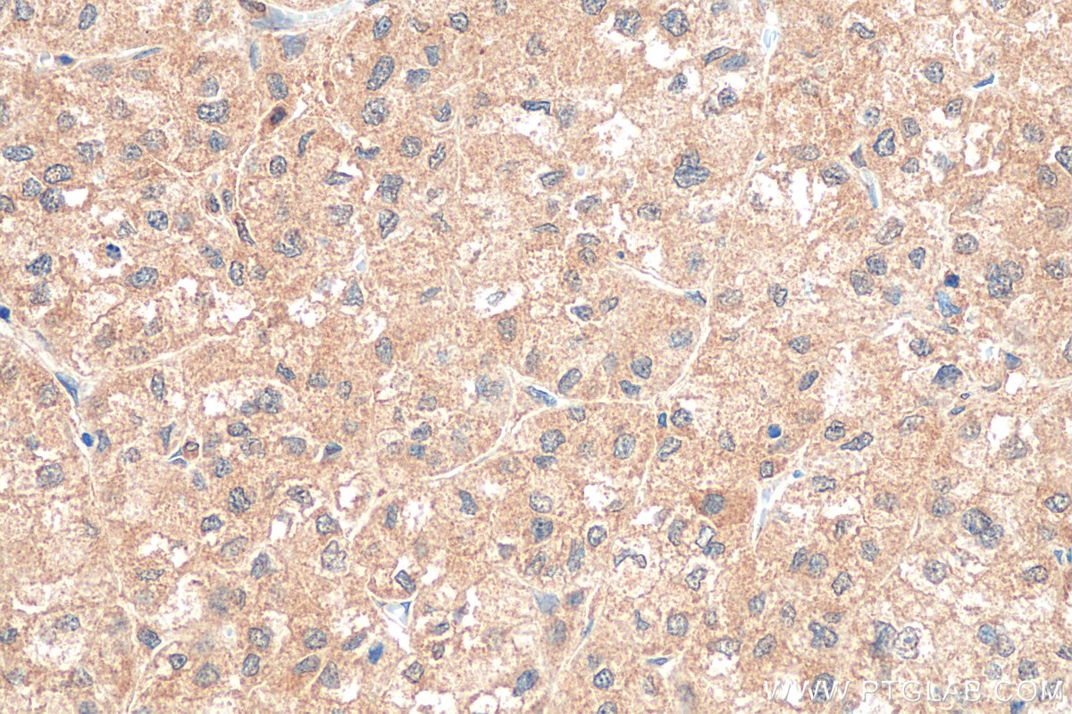
| Positive IHC detected in | human liver cancer tissue Note: suggested antigen retrieval with TE buffer pH 9.0; (*) Alternatively, antigen retrieval may be performed with citrate buffer pH 6.0 |
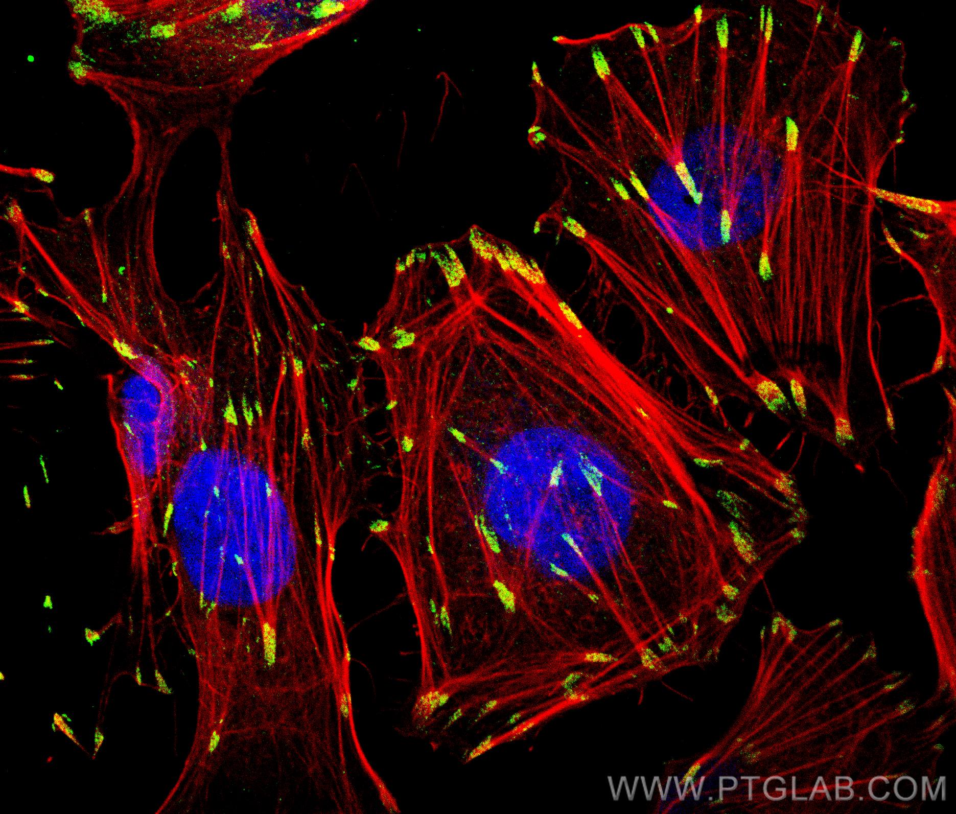
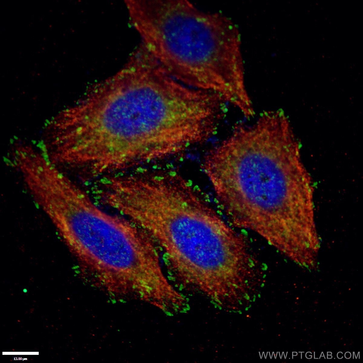
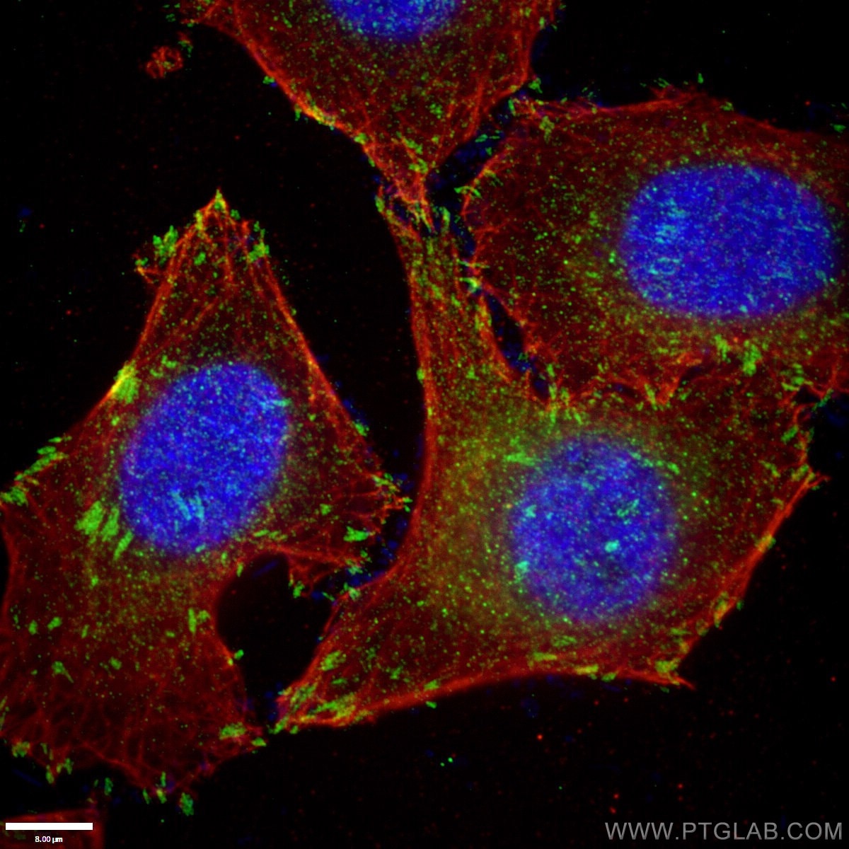
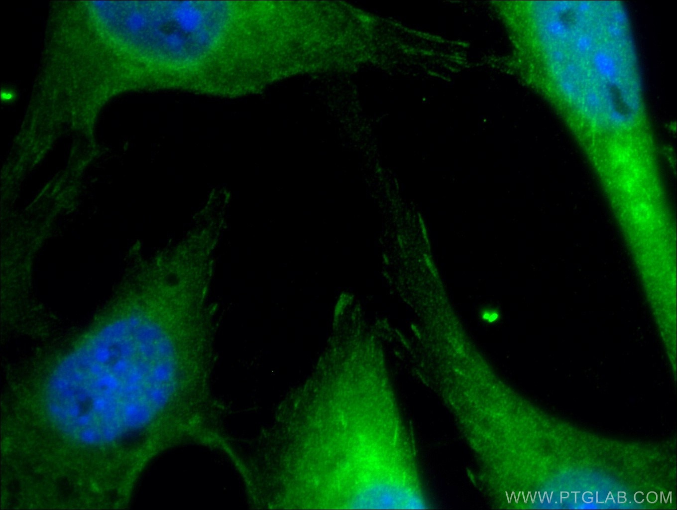
| Positive IF/ICC detected in | HUVEC cells, NIH/3T3 cells, HepG2 cells |
Recommended dilution
| Application | Dilution |
|---|---|
| Western Blot (WB) | WB : 1:5000-1:50000 |
| Immunoprecipitation (IP) | IP : 0.5-4.0 ug for 1.0-3.0 mg of total protein lysate |
| Immunohistochemistry (IHC) | IHC : 1:50-1:500 |
| Immunofluorescence (IF)/ICC | IF/ICC : 1:50-1:500 |
| It is recommended that this reagent should be titrated in each testing system to obtain optimal results. | |
| Sample-dependent, Check data in validation data gallery. | |
Product Information
10029-1-Ig targets Paxillin in WB, IHC, IF/ICC, IP, ELISA applications and shows reactivity with human, mouse samples.
| Tested Reactivity | human, mouse |
| Cited Reactivity | human, mouse, rat |
| Host / Isotype | Rabbit / IgG |
| Class | Polyclonal |
| Type | Antibody |
| Immunogen |
Peptide Predict reactive species |
| Full Name | paxillin |
| Calculated Molecular Weight | 68 kDa |
| Observed Molecular Weight | 68 kDa |
| GenBank Accession Number | NM_002859 |
| Gene Symbol | Paxillin |
| Gene ID (NCBI) | 5829 |
| RRID | AB_513929 |
| Conjugate | Unconjugated |
| Form | Liquid |
| Purification Method | Protein A purification |
| UNIPROT ID | P49023 |
| Storage Buffer | PBS with 0.02% sodium azide and 50% glycerol, pH 7.3. |
| Storage Conditions | Store at -20°C. Stable for one year after shipment. Aliquoting is unnecessary for -20oC storage. 20ul sizes contain 0.1% BSA. |
Background Information
PXN (paxillin) is a 68 kDa scaffold protein that interacts with multiple structural and signaling proteins and regulates cell adhesion, migration, proliferation, and apoptosis. PXN is thought to play an important role in tumor migration, invasion, and metastasis (21045234). PXN has been identified as a direct substrate of protein tyrosine phosphatase receptor-type T (PTPRT), a potent tumor suppressor gene. Increased phospho-PXN at tyrosine residue 88 (Y88) has been found as a common feature of human colon cancers (20133777).
Protocols
| Product Specific Protocols | |
|---|---|
| IF protocol for Paxillin antibody 10029-1-Ig | Download protocol |
| IHC protocol for Paxillin antibody 10029-1-Ig | Download protocol |
| IP protocol for Paxillin antibody 10029-1-Ig | Download protocol |
| WB protocol for Paxillin antibody 10029-1-Ig | Download protocol |
| Standard Protocols | |
|---|---|
| Click here to view our Standard Protocols |
Publications
What published studies show
Able is busy answering your previous question. Please try again later.
| Species | Application | Title |
|---|---|---|
Cancer Cell Mapping cell type-resolved transcriptomic profiles to patient survival in pancreatic cancer. | ||
Drug Resist Updat Exosome-derived lnc-FAM72D-3 promotes lenvatinib resistance by remodeling hepatocellular carcinoma cytoskeleton via MBNL1/FAK axis | ||
Bone Res Impairment of rigidity sensing caused by mutant TP53 gain of function in osteosarcoma | ||
Nat Commun TM9SF4 is an F-actin disassembly factor that promotes tumor progression and metastasis | ||
Nat Commun CCDC120 phase separation contributes to desmosomal integrity and cardiac function | ||
Nat Commun Mechanically heterogeneous hydrogel with cell-programmed network restructuring promotes tissue regeneration by mechano-epigenetic modulation. |
Reviews
What customers say
Two reviewers rated 10029-1-Ig (an antibody targeting PAK1) 5 and 4 out of 5. One reviewer used it in Western Blot and Immunofluorescence on a human brain microvascular endothelial cell line (1:500 WB, 1:100 IF), finding it worked well for WB (pulldown via PAK1) and IF co-staining with F-Actin. Another reviewer tested it on MEF cells, noting it recognized some non-specific bands in WB beyond the expected band, but successfully labeled cell cortex localization in IF, partially co-localizing with Actin.
The reviews below have been submitted by verified Proteintech customers who received an incentive for providing their feedback.
Thomas (Verified Customer) (09-22-2022) | Works in WB (pulldown via PAK1) and IF (costained with F-Actin in cyan)
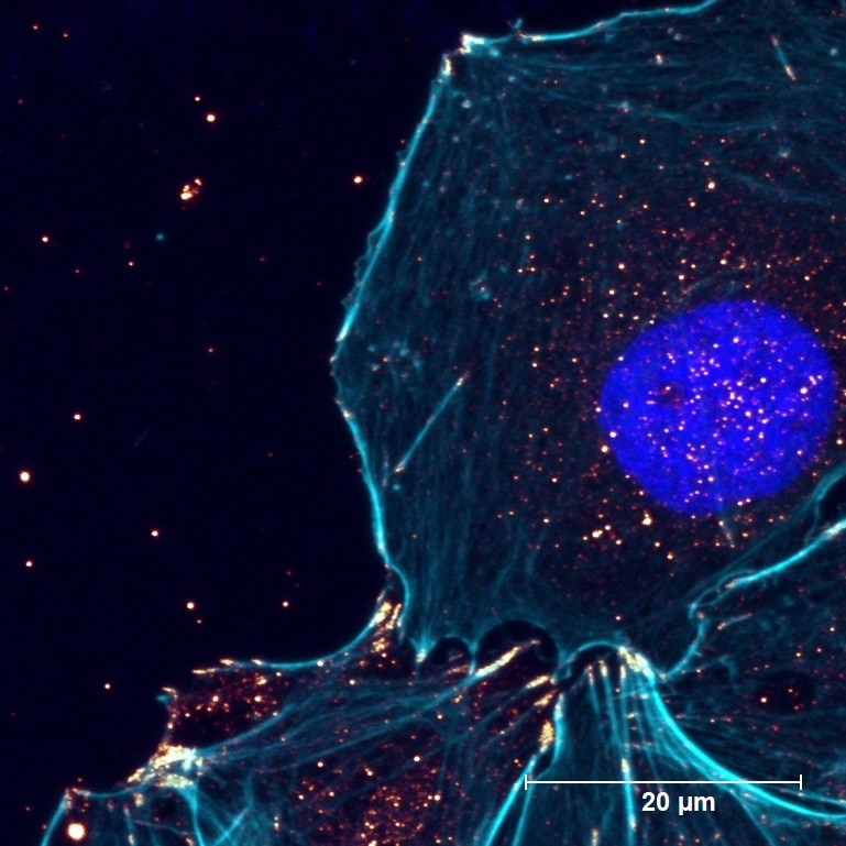 |
Boyan (Verified Customer) (03-11-2019) | For WB, besides the expected band, it also recognised some other non-specific bands; for IF, it could label the cell cortex localisation, partially co-localised with Actin.
|














