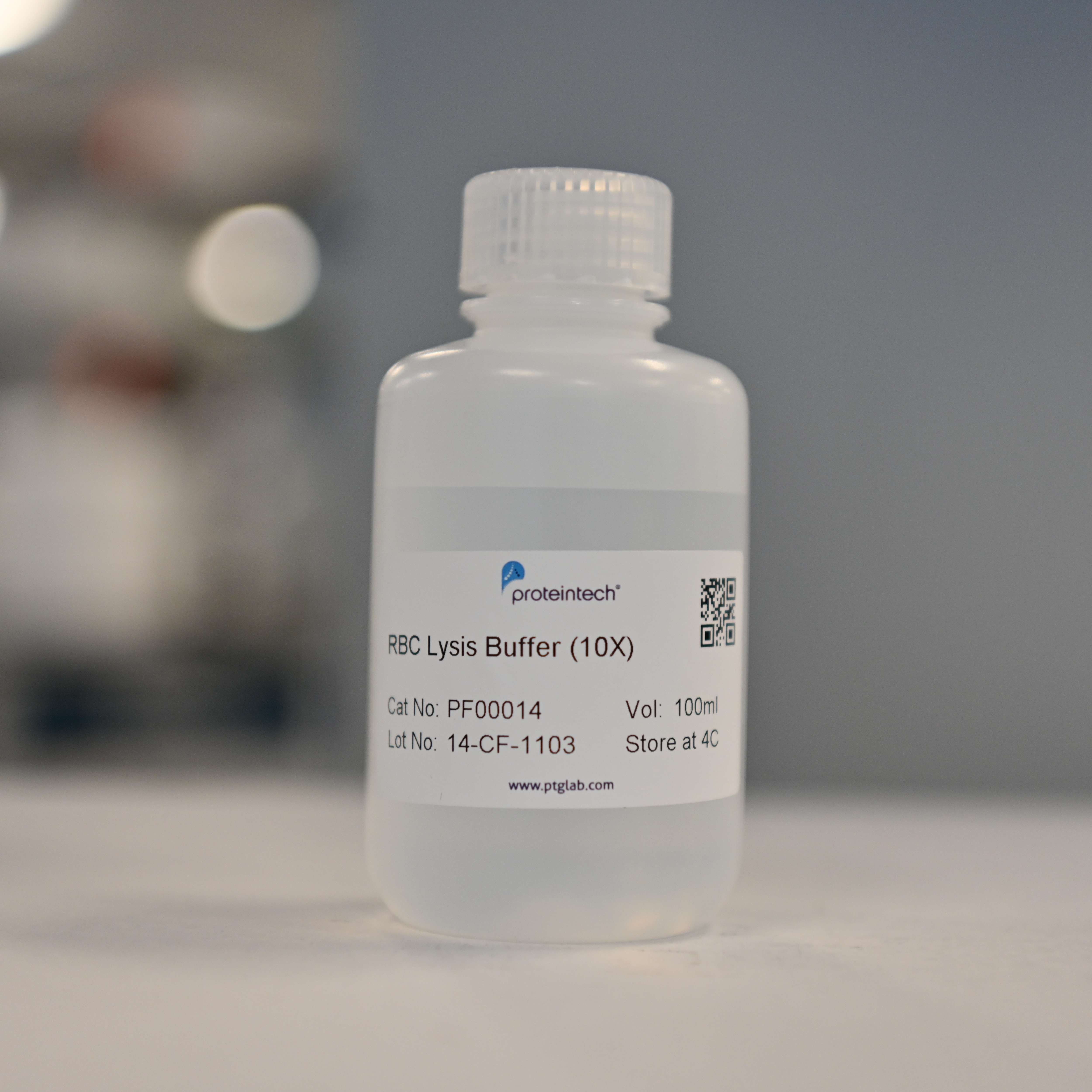
Product Information
Proteintech's RBC Lysis Buffer (10X) is suitable for lysing red blood cells following immunofluorescent staining of whole blood with fluorescently labelled antibodies prior to flow cytometry analysis, with minimal damage to white blood cells. This buffer is supplied as a 10× concentrate and must be diluted to 1× with deionised water before use.
This product is suitable for lysing red blood cells from human, rabbit, mouse, and rat samples. It has not been tested on other species.
This product is for research use only and must not be used in humans or animals.
Components
The primary active ingredient in this lysis buffer is ammonium chloride. It does not contain azide compounds.
Storage
Store sealed at 4°C. Valid for one year.
Usage
1. At room temperature (18-25°C), dilute 1 part of 10× red cell lysis concentrate with 9 parts deionised water to obtain 1× working solution.
2. Following incubation under conditions specified in the antibody manual, proceed with red cell lysis.
3. Gently mix whole blood before red cell lysis.
4. Add 2 mL working solution to each 100 uL fresh anticoagulated blood sample. Invert to mix thoroughly, then lyse at room temperature for 5 minutes.
5. Centrifuge at 300-400 g for 5 minutes at room temperature. Discard the red supernatant. (If incomplete lysis occurs, repeat step 2. Normally, slight amounts of red blood cells do not affect subsequent testing.)
6. Add 2 mL PBS, mix thoroughly, then centrifuge at 300-400 g for 5 minutes at room temperature. Discard the supernatant.
7. Add 200 uL PBS, mix thoroughly, and proceed to instrument analysis promptly.
Notes
1. The 1× working solution must be prepared freshly before use.
2. Blood samples exhibit considerable inter-individual variation, potentially leading to slight variations in lysis time. If whole blood exhibits a high red blood cell content, it may be diluted with an appropriate volume of PBS.
3. Following antibody incubation, red blood cells may settle at the bottom. Failure to mix thoroughly may result in incomplete lysis.
4. Lysis protocols may be adjusted as required, either by modifying the working solution volume or altering the lysis time.
5. Assess lysis efficacy visually. A cloudy appearance or abnormal light scatter histogram may indicate incomplete lysis.
Cited in Article as
PF00014, RBC Lysis Buffer (10X), Proteintech, IL, USA
Documentation
| Datasheet |
|---|
| RBC Lysis Buffer (10X) Datasheet |


