

Tested Applications
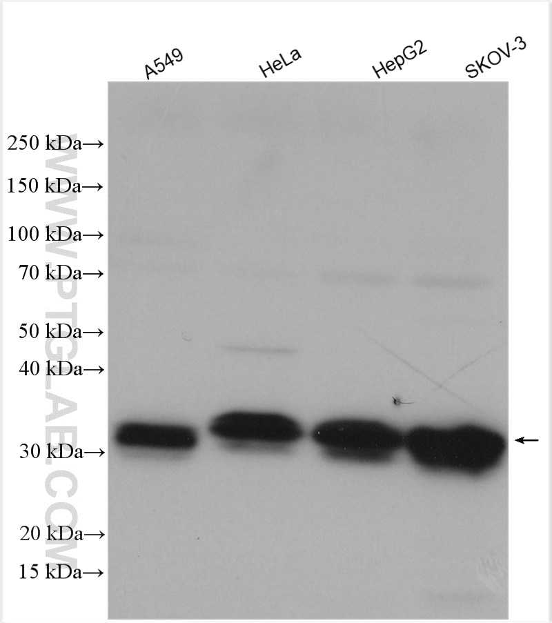
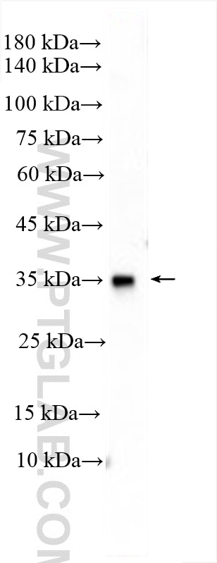
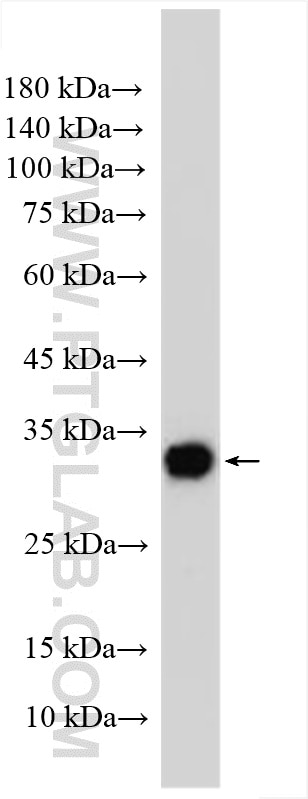
| Positive WB detected in | A549 cells, mouse testis tissue, rat testis tissue, HeLa cells, HepG2 cells, SKOV-3 cells |
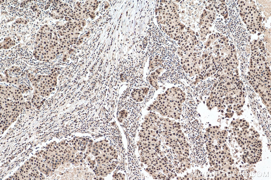
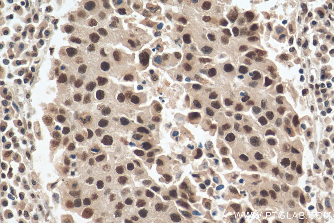
| Positive IHC detected in | human breast cancer tissue Note: suggested antigen retrieval with TE buffer pH 9.0; (*) Alternatively, antigen retrieval may be performed with citrate buffer pH 6.0 |
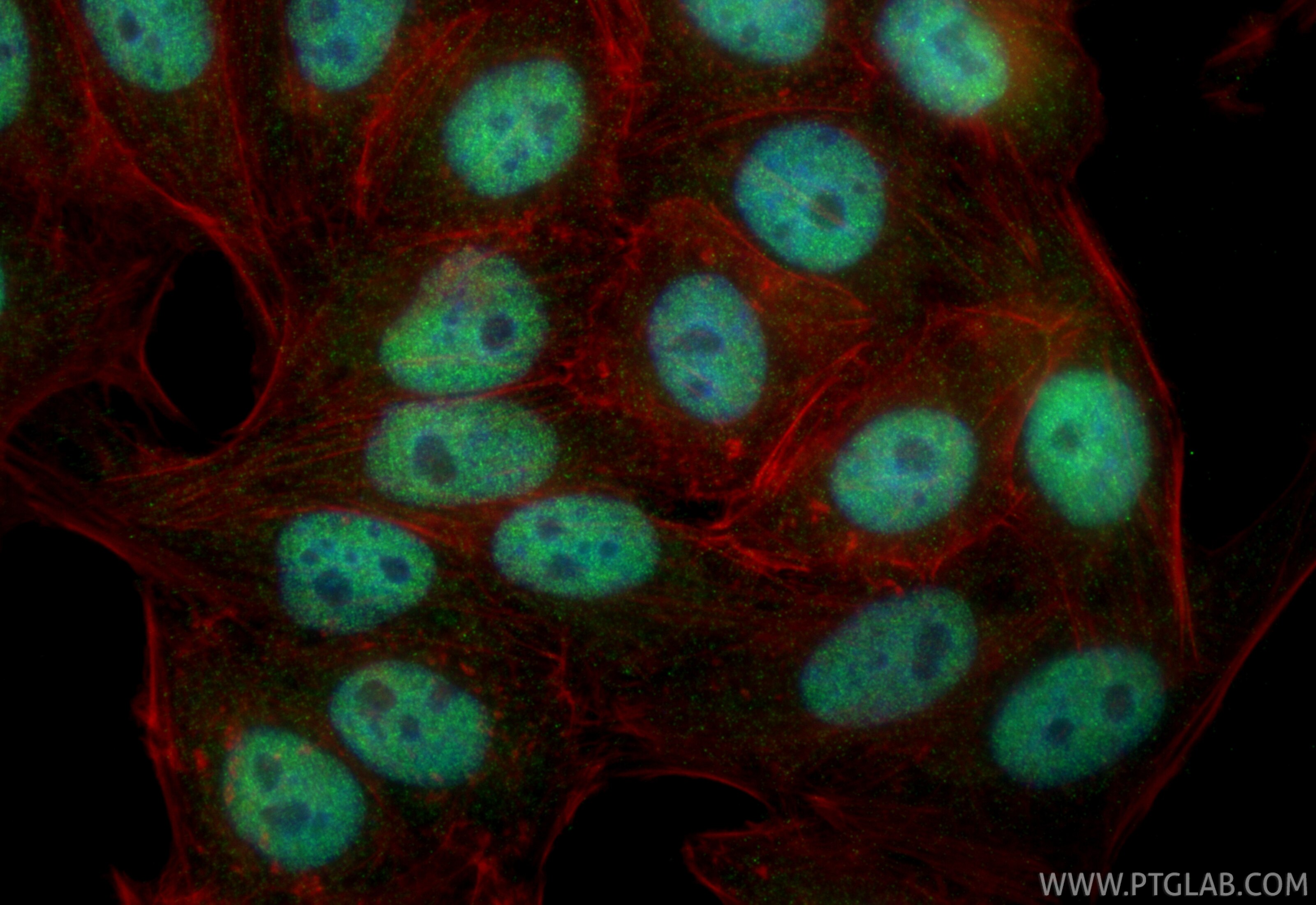
| Positive IF/ICC detected in | HepG2 cells |
Recommended dilution
| Application | Dilution |
|---|---|
| Western Blot (WB) | WB : 1:1000-1:8000 |
| Immunohistochemistry (IHC) | IHC : 1:50-1:500 |
| Immunofluorescence (IF)/ICC | IF/ICC : 1:50-1:500 |
| It is recommended that this reagent should be titrated in each testing system to obtain optimal results. | |
| Sample-dependent, Check data in validation data gallery. | |
Product Information
11044-1-AP targets SFRS7 in WB, IHC, IF/ICC, IP, ELISA applications and shows reactivity with human, mouse, rat samples.
| Tested Reactivity | human, mouse, rat |
| Cited Reactivity | human |
| Host / Isotype | Rabbit / IgG |
| Class | Polyclonal |
| Type | Antibody |
| Immunogen |
CatNo: Ag1526 Product name: Recombinant human SFRS7 protein Source: e coli.-derived, PGEX-4T Tag: GST Domain: 1-238 aa of BC000997 Sequence: MSRYGRYGGETKVYVGNLGTGAGKGELERAFSYYGPLRTVWIARNPPGFAFVEFEDPRDAEDAVRGLDGKVICGSRVRVELSTGMPRRSRFDRPPARRPFDPNDRCYECGEKGHYAYDCHRYSRRRRSRSRSRSHSRSRGRRYSRSRSRSRGRRSRSASPRRSRSISLRRSRSASLRRSRSGSIKGSRYFQSPSRSRSRSRSISRPRSSRSKSRSPSPKRSRSPSGSPRRSASPERMD Predict reactive species |
| Full Name | splicing factor, arginine/serine-rich 7, 35kDa |
| Calculated Molecular Weight | 27 kDa |
| Observed Molecular Weight | 35 kDa |
| GenBank Accession Number | BC000997 |
| Gene Symbol | SFRS7 |
| Gene ID (NCBI) | 6432 |
| RRID | AB_2185229 |
| Conjugate | Unconjugated |
| Form | Liquid |
| Purification Method | Antigen affinity purification |
| UNIPROT ID | Q16629 |
| Storage Buffer | PBS with 0.02% sodium azide and 50% glycerol, pH 7.3. |
| Storage Conditions | Store at -20°C. Stable for one year after shipment. Aliquoting is unnecessary for -20oC storage. 20ul sizes contain 0.1% BSA. |
Background Information
SFRS7 is a member of the serine/arginine (SR) splicing factor family, which mediated the alternative splicing events target the resulting mRNAs for degradation by means of an RNA surveillance pathway called nonsense-mediated mRNA decay. It was found to repress the splicing of MAPT/Tau exon 10
Protocols
| Product Specific Protocols | |
|---|---|
| IF protocol for SFRS7 antibody 11044-1-AP | Download protocol |
| IHC protocol for SFRS7 antibody 11044-1-AP | Download protocol |
| WB protocol for SFRS7 antibody 11044-1-AP | Download protocol |
| Standard Protocols | |
|---|---|
| Click here to view our Standard Protocols |
Publications
What published studies show
The SRSF7 antibody (Cat# 11044-1-AP) has 15 total citations. Citations appear in 14 journals including Cell, Gastroenterology, Nucleic Acids Research, Acta Pharmaceutica Sinica B, J Med Chem, J Biol Chem, and Genes Genomics. Research fields span alternative splicing regulation, cancer (pancreatic, glioblastoma, hepatocellular carcinoma, GIST), neurodegeneration (ALS/FTD), pulmonary and cardiac fibrosis, viral replication (influenza A, HBV), and m6A RNA modification.
| Species | Application | Title |
|---|---|---|
Cell C9orf72 Dipeptide Repeats Impair the Assembly, Dynamics, and Function of Membrane-Less Organelles. | ||
Gastroenterology Proteomic characterization identifies clinically relevant subgroups of gastrointestinal stromal tumors | ||
Drug Resist Updat Targeting CLK1/SRSF7 axis-dependent alternative splicing sensitizes pancreatic ductal adenocarcinoma to chemotherapy and immunotherapy | ||
Nucleic Acids Res Sequence-dependent recruitment of SRSF1 and SRSF7 to intronless lncRNA NKILA promotes nuclear export via the TREX/TAP pathway.
| ||
Nucleic Acids Res Exonic splicing code and protein binding sites for calcium.
| ||
Acta Pharm Sin B SRSF7 promotes pulmonary fibrosis through regulating PKM alternative splicing in lung fibroblasts.
|
Reviews
What customers say
Both reviewers rated this antibody 5 stars for immunofluorescence. One user (2025) used a 1:100 dilution on human Alzheimer's disease tissue and reported strong staining with minimal background, showing clear differences between AD and control tissues. The other (2021) used a 1:150 dilution on a human cancer cell line and confirmed nuclear staining. Overall, users find it produces specific, high-signal results with low background across different sample types.
The reviews below have been submitted by verified Proteintech customers who received an incentive for providing their feedback.
Madison (Verified Customer) (07-24-2025) | The antibody stained well with little background interference and overall showed a nice and noticeable pattern within the tissue and between the control and AD tissues.
|
Jingwen (Verified Customer) (04-28-2021) | The staining signal showed up inside the nucleus.
|







