
Use Able AI chat for product recommendations

TBP Monoclonal antibody

TBP Monoclonal Antibody for WB, IHC, IF/ICC, IP, ELISA

Cat No. 66166-1-Ig
Clone No. 2H3B2
Publications (34)
Reviews (1)
Download datasheet

Host / Isotype

Mouse / IgG2a

Reactivity

human, mouse, rat, pig

Applications

WB, IHC, IF/ICC, IP, ELISA
For FC, select conjugated version.

2H3B2, GTF2D1, TATA binding factor, TATA box binding protein, TATA box factor

Formulation:  PBS, Azide, Glycerol
PBS, Azide, Glycerol
PBS Only
Size/Concentration: 
SKU: 

-/ -

Freight/Packing: -

Quantity

Please visit your regions distributor:

Filter:
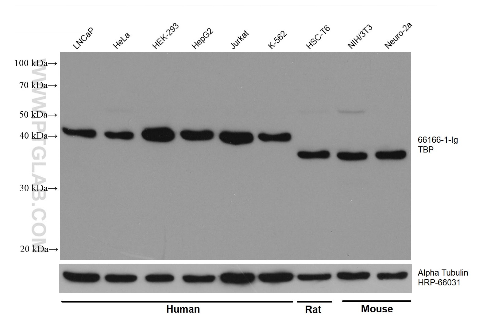
  • Western Blot (WB) analysis of various lysates using TBP Monoclonal antibody (66166-1-Ig)

    WB analysis using 66166-1-Ig

    Various lysates were subjected to SDS PAGE followed by western blot with 66166-1-Ig (TBP antibody) at dilution of 1:200000 incubated at room temperature for 1.5 hours. The membrane was stripped and reblotted with HRP-conjugated Alpha Tubulin Monoclonal antibody (HRP-66031) as loading control.

    This validation data has been generated in-house and reviewed by Proteintech scientists. Find out more on our antibody validation policy
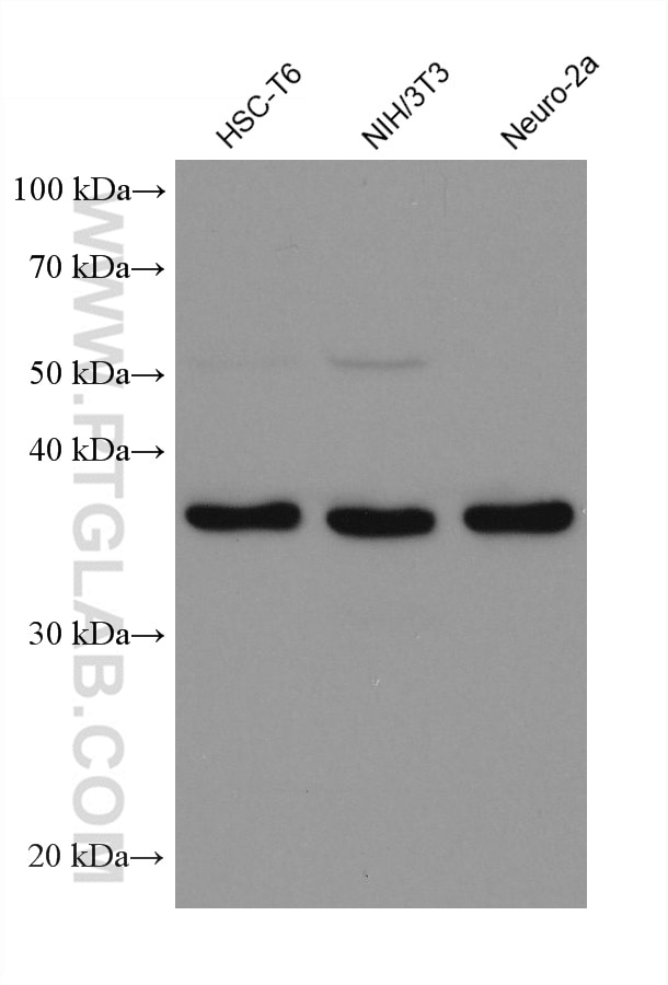
  • Western Blot (WB) analysis of various lysates using TBP Monoclonal antibody (66166-1-Ig)

    WB analysis using 66166-1-Ig

    Various lysates were subjected to SDS PAGE followed by western blot with 66166-1-Ig (TBP antibody) at dilution of 1:200000 incubated at room temperature for 1.5 hours.

    This validation data has been generated in-house and reviewed by Proteintech scientists. Find out more on our antibody validation policy
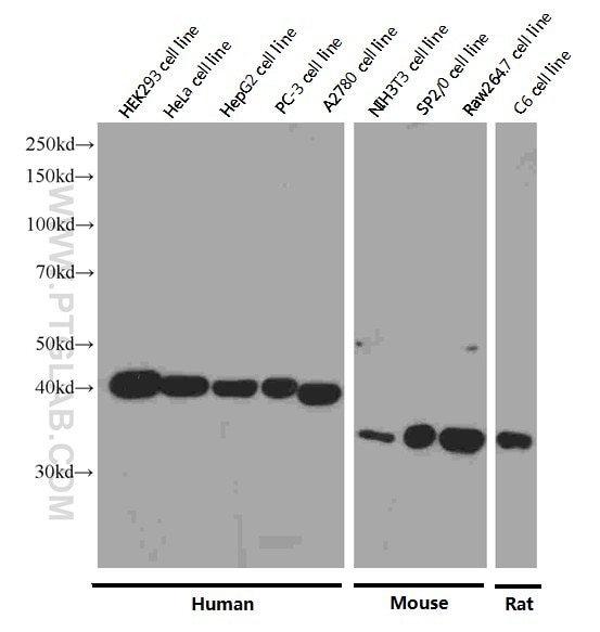
  • Western Blot (WB) analysis of HEK-293 cells using TBP Monoclonal antibody (66166-1-Ig)

    WB analysis of HEK-293 using 66166-1-Ig

    Western blot analysis of TBP in various cell lines using Proteintech antibody 66166-1-Ig at a dilution of 1:8000 incubated at room temperature for 1.5 hours.

    This validation data has been generated in-house and reviewed by Proteintech scientists. Find out more on our antibody validation policy
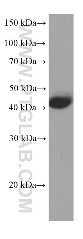
  • Western Blot (WB) analysis of pig liver tissue using TBP Monoclonal antibody (66166-1-Ig)

    WB analysis of pig liver using 66166-1-Ig

    pig liver tissue were subjected to SDS PAGE followed by western blot with 66166-1-Ig (TBP Antibody) at dilution of 1:5000 incubated at room temperature for 1.5 hours.

    This validation data has been generated in-house and reviewed by Proteintech scientists. Find out more on our antibody validation policy
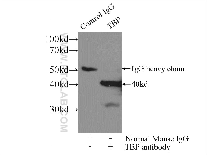
  • Immunoprecipitation (IP) experiment of HEK-293 cells using TBP Monoclonal antibody (66166-1-Ig)

    IP experiment of HEK-293 using 66166-1-Ig

    IP result of anti-TBP (IP:66166-1-Ig, 3ug; Detection:66166-1-Ig 1:800) with HEK-293 cells lysate 1800ug.

    This validation data has been generated in-house and reviewed by Proteintech scientists. Find out more on our antibody validation policy
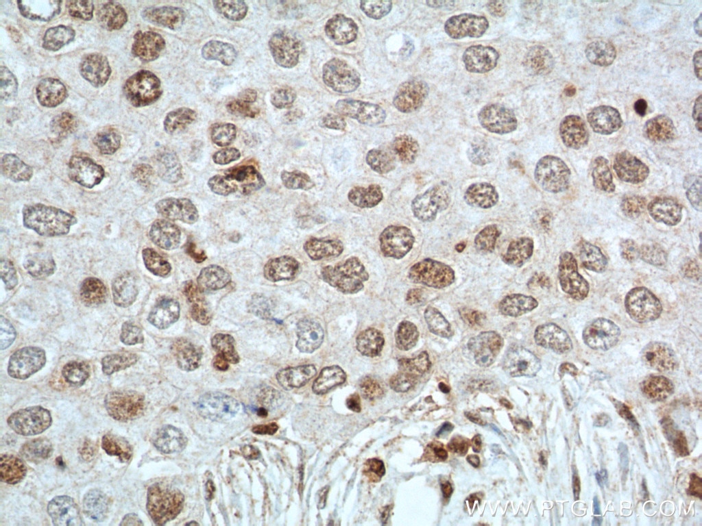
  • Immunohistochemistry (IHC) staining of human liver cancer tissue using TBP Monoclonal antibody (66166-1-Ig)

    IHC staining of human liver cancer using 66166-1-Ig

    Immunohistochemical analysis of paraffin-embedded human liver cancer tissue slide using 66166-1-Ig (TBP Antibody) at dilution of 1:650 (under 40x lens). Heat mediated antigen retrieval with Tris-EDTA buffer (pH 9.0).

    This validation data has been generated in-house and reviewed by Proteintech scientists. Find out more on our antibody validation policy
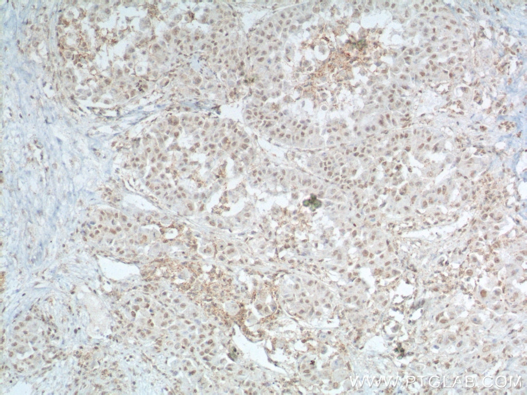
  • Immunohistochemistry (IHC) staining of human breast cancer tissue using TBP Monoclonal antibody (66166-1-Ig)

    IHC staining of human breast cancer using 66166-1-Ig

    Immunohistochemical analysis of paraffin-embedded human breast cancer tissue slide using 66166-1-Ig (TBP Antibody) at dilution of 1:200 (under 10x lens).

    This validation data has been generated in-house and reviewed by Proteintech scientists. Find out more on our antibody validation policy
  • Immunohistochemistry (IHC) staining of human breast cancer tissue using TBP Monoclonal antibody (66166-1-Ig)

    IHC staining of human breast cancer using 66166-1-Ig

    Immunohistochemical analysis of paraffin-embedded human breast cancer tissue slide using 66166-1-Ig (TBP Antibody) at dilution of 1:200 (under 40x lens).

    This validation data has been generated in-house and reviewed by Proteintech scientists. Find out more on our antibody validation policy
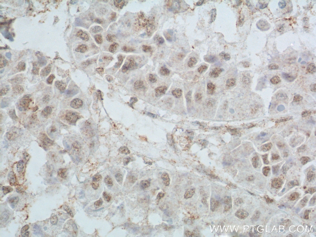
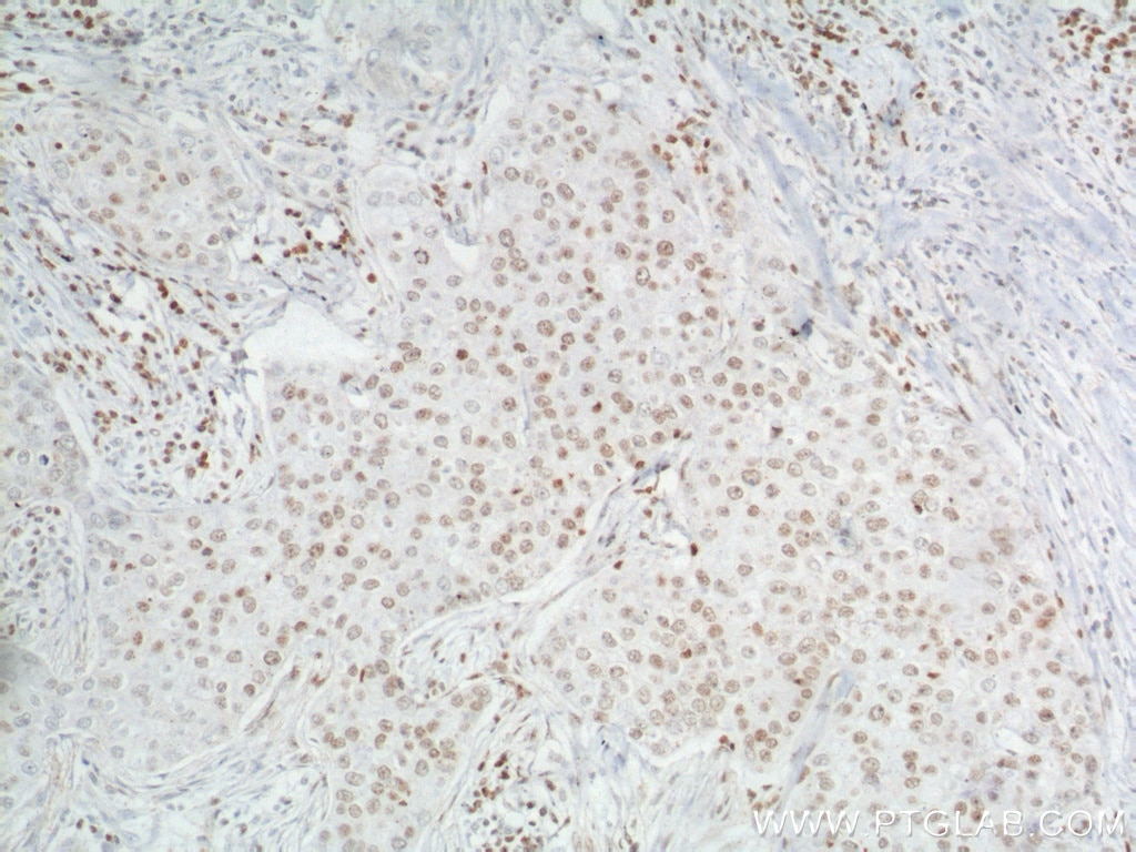
  • Immunohistochemistry (IHC) staining of human breast cancer tissue using TBP Monoclonal antibody (66166-1-Ig)

    IHC staining of human breast cancer using 66166-1-Ig

    After antigen unmasking with Tris-EDTA buffer (pH 9.0) for 15 mins and 2M HCL treatment for 30mins, Immunohistochemistry of paraffin-embedded human breast cancer tissue slideusing 66166-1-Ig (TBP Antibody) at dilution of 1:400 (under 10x lens).

    This validation data has been generated in-house and reviewed by Proteintech scientists. Find out more on our antibody validation policy
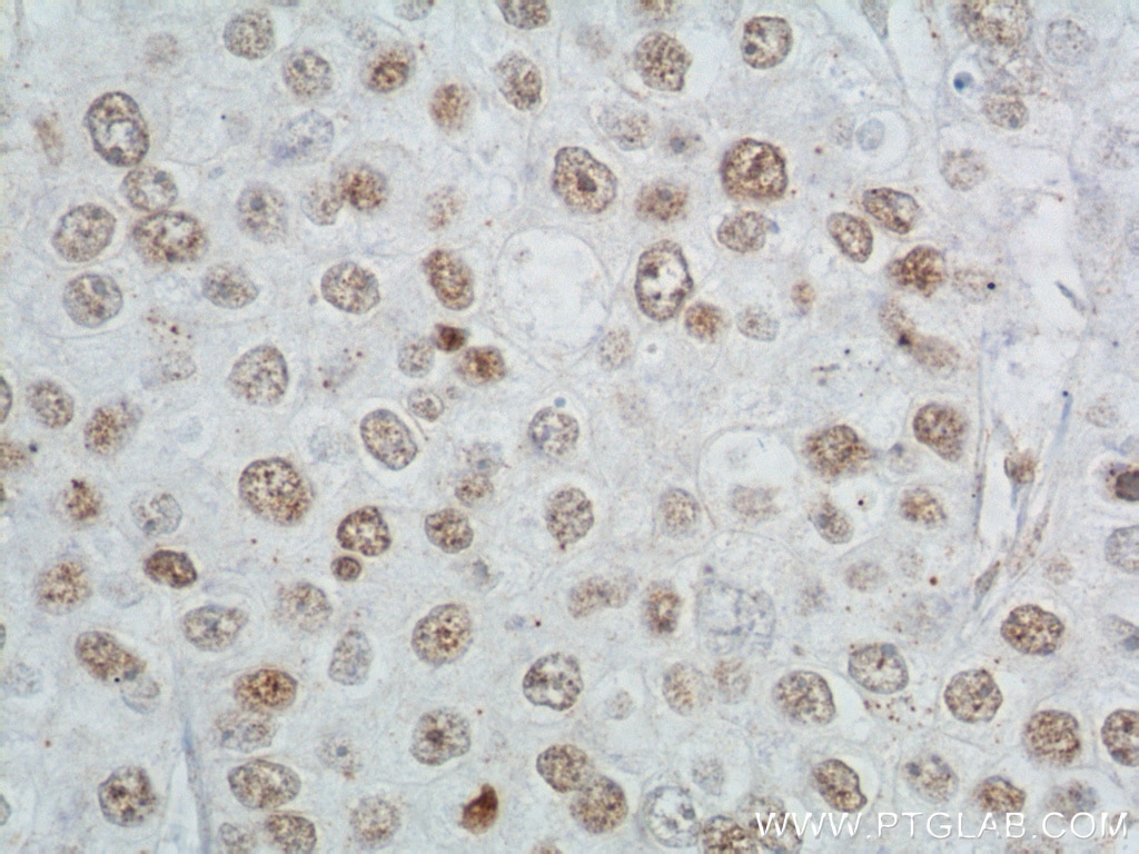
  • Immunohistochemistry (IHC) staining of human breast cancer tissue using TBP Monoclonal antibody (66166-1-Ig)

    IHC staining of human breast cancer using 66166-1-Ig

    I After antigen unmasking with Tris-EDTA buffer (pH 9.0) for 15 mins and 2M HCL treatment for 30mins, Immunohistochemistry of paraffin-embedded human breast cancer tissue slideusing 66166-1-Ig (TBP Antibody) at dilution of 1:400 (under 40x lens).

    This validation data has been generated in-house and reviewed by Proteintech scientists. Find out more on our antibody validation policy
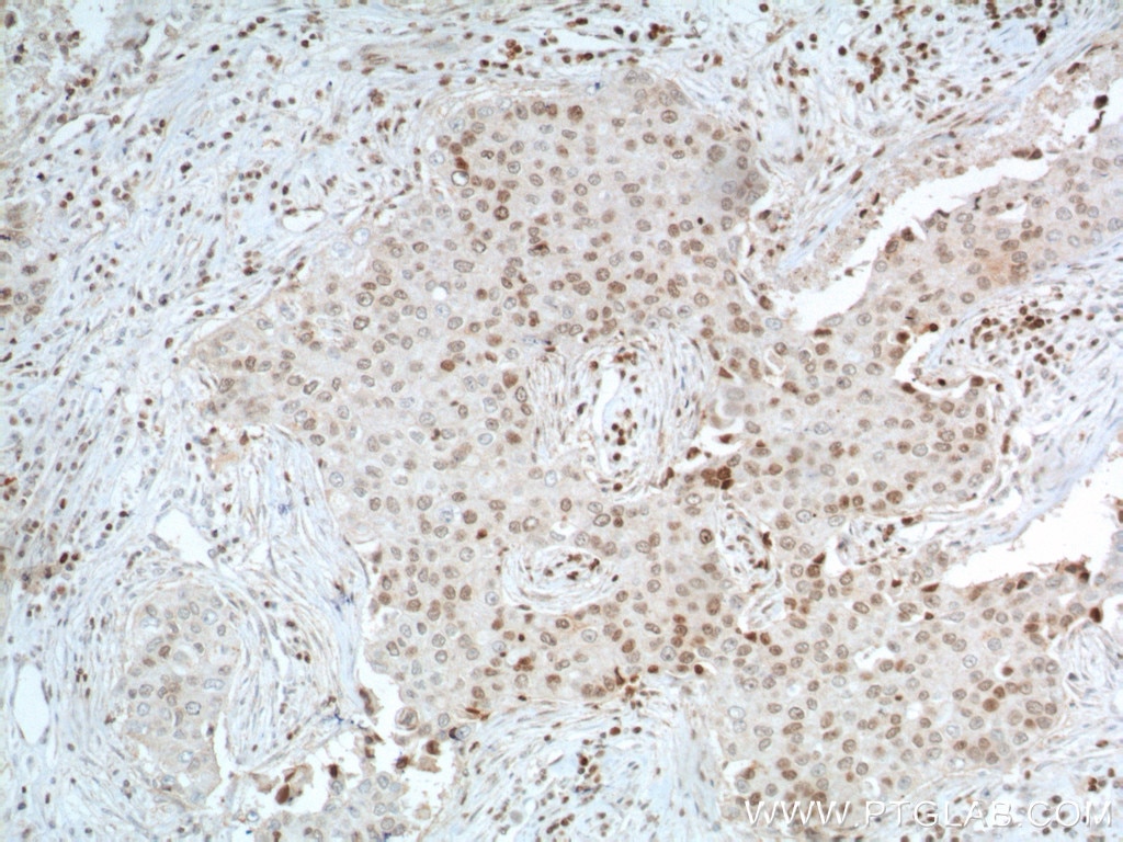
  • Immunohistochemistry (IHC) staining of human liver cancer tissue using TBP Monoclonal antibody (66166-1-Ig)

    IHC staining of human liver cancer using 66166-1-Ig

    Immunohistochemical analysis of paraffin-embedded human liver cancer tissue slide using 66166-1-Ig (TBP Antibody) at dilution of 1:650 (under 10x lens). Heat mediated antigen retrieval with Tris-EDTA buffer (pH 9.0).

    This validation data has been generated in-house and reviewed by Proteintech scientists. Find out more on our antibody validation policy
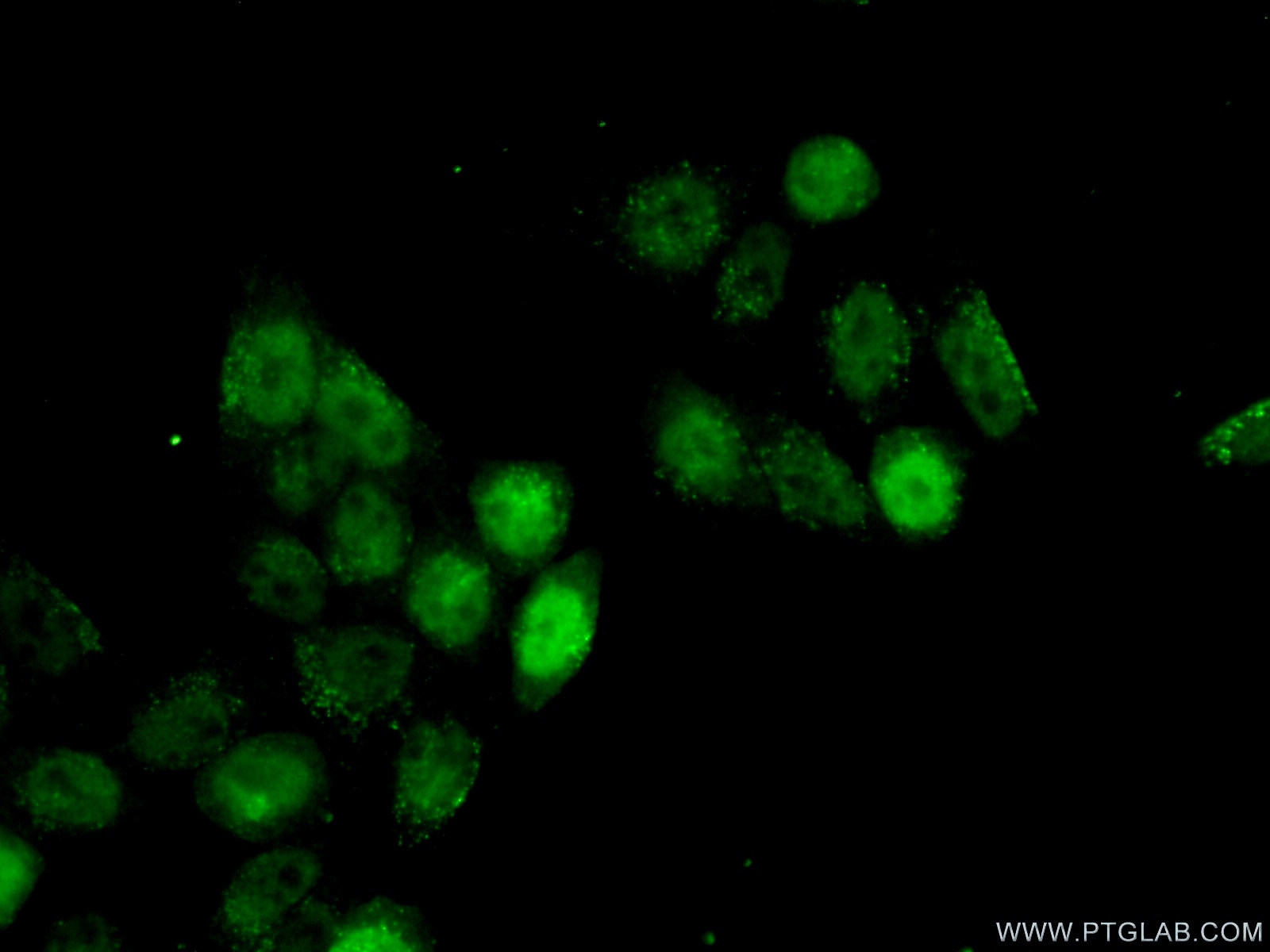
  • Immunofluorescence (IF) / fluorescent staining of HeLa cells using TBP Monoclonal antibody (66166-1-Ig)

    IF Staining of HeLa using 66166-1-Ig

    Immunofluorescent analysis of (10% Formaldehyde ) fixed HeLa cells using 66166-1-Ig(TBP antibody) at dilution of 1:100 and Alexa Fluor 488-conjugated AffiniPure Goat Anti-Mouse IgG(H+L).

    This validation data has been generated in-house and reviewed by Proteintech scientists. Find out more on our antibody validation policy
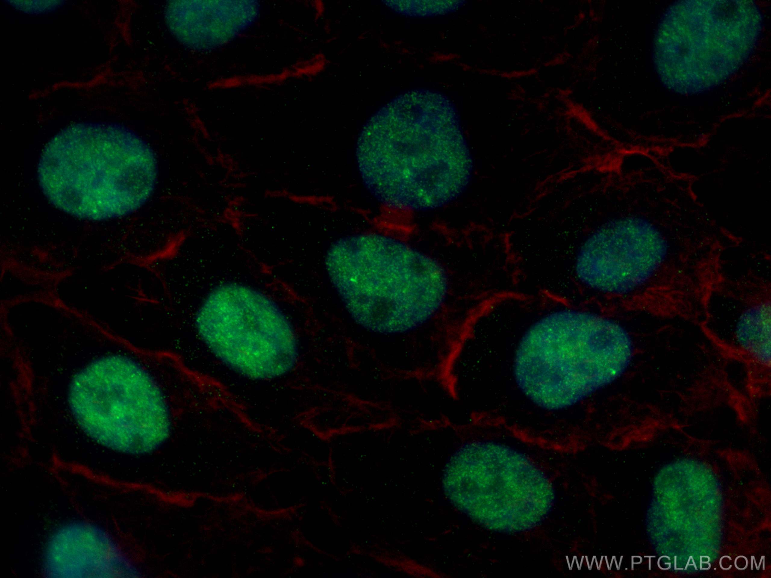
  • Immunofluorescence (IF) / fluorescent staining of A431 cells using TBP Monoclonal antibody (66166-1-Ig)

    IF Staining of A431 using 66166-1-Ig

    Immunofluorescent analysis of (4% PFA) fixed A431 cells using TBP antibody (66166-1-Ig, Clone: 2H3B2 ) at dilution of 1:2000 and CoraLite®488-Conjugated AffiniPure Goat Anti-Mouse IgG(H+L), CL594-phalloidin (red).

    This validation data has been generated in-house and reviewed by Proteintech scientists. Find out more on our antibody validation policy

"TBP Antibodies" Comparison

View side-by-side comparison of TBP antibodies from other vendors to find the one that best suits your research needs.

Able AI
Able AI
What's the best dilution for TBP in ?

Tested Applications

Positive WB detected inLNCaP cells, HSC-T6 cells, HEK-293 cells, pig liver tissue, HeLa cells, HepG2 cells, Jurkat cells, K-562 cells, NIH/3T3 cells, Neuro-2a cells
Positive IP detected inHEK-293 cells
Positive IHC detected inhuman liver cancer tissue, human breast cancer tissue
Note: suggested antigen retrieval with TE buffer pH 9.0; (*) Alternatively, antigen retrieval may be performed with citrate buffer pH 6.0
Positive IF/ICC detected inA431 cells, HeLa cells

Recommended dilution

ApplicationDilution
Western Blot (WB)WB : 1:20000-1:100000
Immunoprecipitation (IP)IP : 0.5-4.0 ug for 1.0-3.0 mg of total protein lysate
Immunohistochemistry (IHC)IHC : 1:325-1:1300
Immunofluorescence (IF)/ICCIF/ICC : 1:1000-1:4000
It is recommended that this reagent should be titrated in each testing system to obtain optimal results.
Sample-dependent, Check data in validation data gallery.

Product Information

66166-1-Ig targets TBP in WB, IHC, IF/ICC, IP, ELISA applications and shows reactivity with human, mouse, rat, pig samples.

Tested Reactivity human, mouse, rat, pig
Cited Reactivityhuman, mouse, rat, pig
Host / Isotype Mouse / IgG2a
Class Monoclonal
Type Antibody
Immunogen

CatNo: Ag12383

Product name: Recombinant human TBP protein

Source: e coli.-derived, PET28a

Tag: 6*His

Domain: 1-338 aa of BC110341

Sequence: MDQNNSLPPYAQGLASPQGAMTPGIPIFSPMMPYGTGLTPQPIQNTNSLSILEEQQRQQQQQQQQQQQQQQQQQQQQQQQQQQQQQQQQQQQQQAVAAAAVQQSTSQQATQGTSGQAPQLFHSQTLTTAPLPGTTPLYPSPMTPMTPITPATPASESSGIVPQLQNIVSTVNLGCKLDLKTIALRARNAEYNPKRFAAVIMRIREPRTTALIFSSGKMVCTGAKSEEQSRLAARKYARVVQKLGFPAKFLDFKIQNMVGSCDVKFPIRLEGLVLTHQQFSSYEPELFPGLIYRMIKPRIVLLIFVSGKVVLTGAKVRAEIYEAFENIYPILKGFRKTT

Predict reactive species
Full Name TATA box binding protein
Calculated Molecular Weight 338 aa, 38 kDa
Observed Molecular Weight mouse/rat 33-36 kDa and human 37-43kDa
GenBank Accession NumberBC110341
Gene Symbol TBP
Gene ID (NCBI) 6908
RRIDAB_2881562
Conjugate Unconjugated
FormLiquid
Purification MethodProtein A purification
UNIPROT IDP20226
Storage Buffer PBS with 0.02% sodium azide and 50% glycerol, pH 7.3.
Storage ConditionsStore at -20°C. Stable for one year after shipment. Aliquoting is unnecessary for -20oC storage. 20ul sizes contain 0.1% BSA.

Background Information

The TATA binding protein (TBP) is a transcription factor that binds specifically to a DNA sequence TATA box. This DNA sequence is found about 25-30 base pairs upstream of the transcription start site in some eukaryotic gene promoters. TBP, along with a variety of TBP-associated factors, make up the TFIID, a general transcription factor that in turn makes up part of the RNA polymerase II preinitiation complex. As one of the few proteins in the preinitation complex that binds DNA in a sequence-specific manner, it helps position RNA polymerase II over the transcription start site of the gene. However, it is estimated that only 10-20% of human promoters have TATA boxes. Therefore, TBP is probably not the only protein involved in positioning RNA polymerase II. This antibody detects human TBP (~40 kDa) and mouse/rat Tbp (~35 kDa).

Protocols

Product Specific Protocols
IF protocol for TBP antibody 66166-1-IgDownload protocol
IHC protocol for TBP antibody 66166-1-IgDownload protocol
IP protocol for TBP antibody 66166-1-IgDownload protocol
WB protocol for TBP antibody 66166-1-IgDownload protocol
Standard Protocols
Click here to view our Standard Protocols

Publications

What published studies show

The TBP Monoclonal antibody (2H3B2), catalog #66166-1-Ig, has 34 citations published in journals including Nature Cell Biology, Nature Communications, Cell Metabolism, Cell Reports, Genome Biology, PLoS Biology, Free Radical Biology and Medicine, Biochemical Pharmacology, and Journal of Nanobiotechnology. Research fields span transcription regulation, chromatin biology, cancer (pancreatic, prostate, glioblastoma, CML), ferroptosis, inflammation, drug metabolism, tissue repair, aging/NAD⁺ homeostasis, and neuroscience.

SpeciesApplicationTitle
mouse

Cell Metab

Nicotinic acid riboside maintains NAD+ homeostasis and ameliorates aging-associated NAD+ decline

Authors - Won-Suk Song
mouseWB

Nat Cell Biol

Nuclear N-glycosylation maintains H3K9me3 heterochromatin and genomic stability.

Authors - Xiuxiao Tang
WB

Nat Commun

Reprogramming gut microenvironment for the treatment of acute severe ulcerative colitis via a synergistic therapy of necroptosis blockade and organoid transplantation.

Authors - Qifeng Deng
ratWB,IF

J Nanobiotechnology

3D-printed hydrogel particles containing PRP laden with TDSCs promote tendon repair in a rat model of tendinopathy

Authors - Congsun Li
mouseWB

Burns Trauma

Histone lactylation exacerbates acute lung injury in septic mice by promoting ferroptosis in pulmonary microvascular endothelial cells.

Authors - Pu Fang
mouseWB

Biomater Res

Tryptophan-Enriched Lactobacillus rhamnosus GG-derived Nanovesicles Promote Alveolar Bone Regeneration through Macrophage Fatty Acid Oxidation.

Authors - Zhendong Huang

Reviews

    5.0 out of 5
Add a review

1 verified customer reviews

What customers say

The single review for this product is highly positive. Aditya Chandru rated it 5 stars in July 2025, using it for Western Blot with lot 10002500. The reviewer described the antibody as "excellent" and noted that it "works as advertised," indicating reliable performance and consistent results matching the manufacturer's claims. Overall, the feedback reflects complete satisfaction with both the quality and functionality of the reagent.

The reviews below have been submitted by verified Proteintech customers who received an incentive for providing their feedback.


Aditya (Verified Customer) (07-15-2025)

Excellent, works as advertised

  • Applications: Western Blot
Loading...
||
New chat

Able™

正在加载,请稍候...












