

Tested Applications
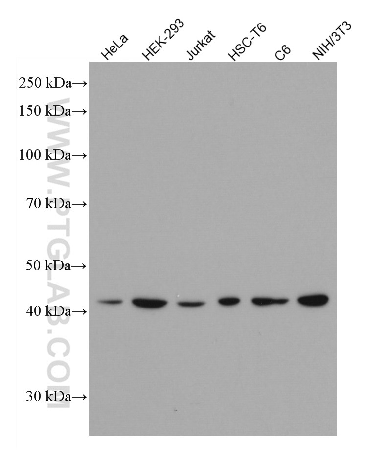
| Positive WB detected in | HeLa cells, HEK-293 cells, Jurkat cells, HSC-T6 cells, C6 cells, NIH/3T3 cells |
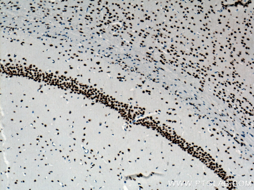
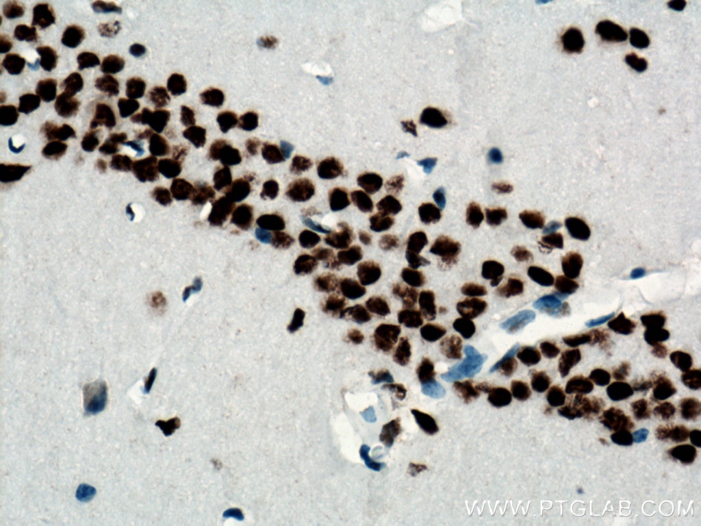
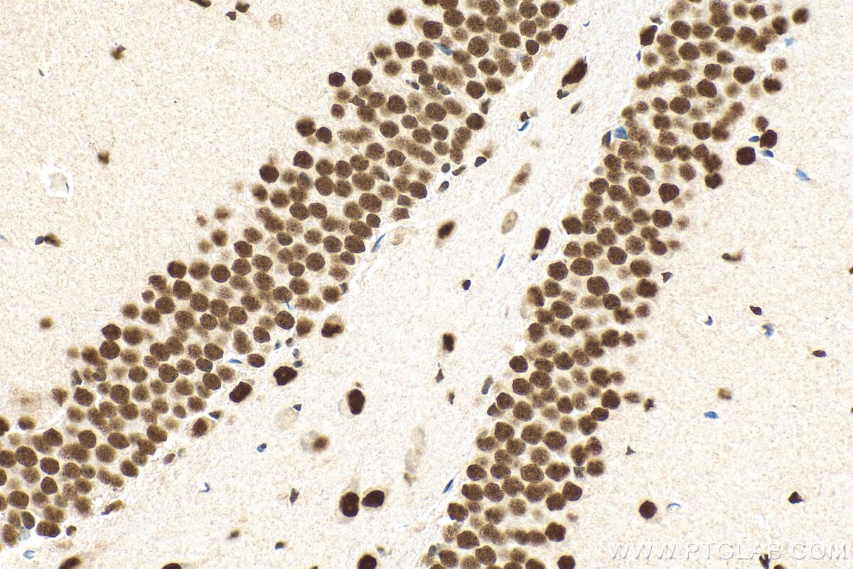
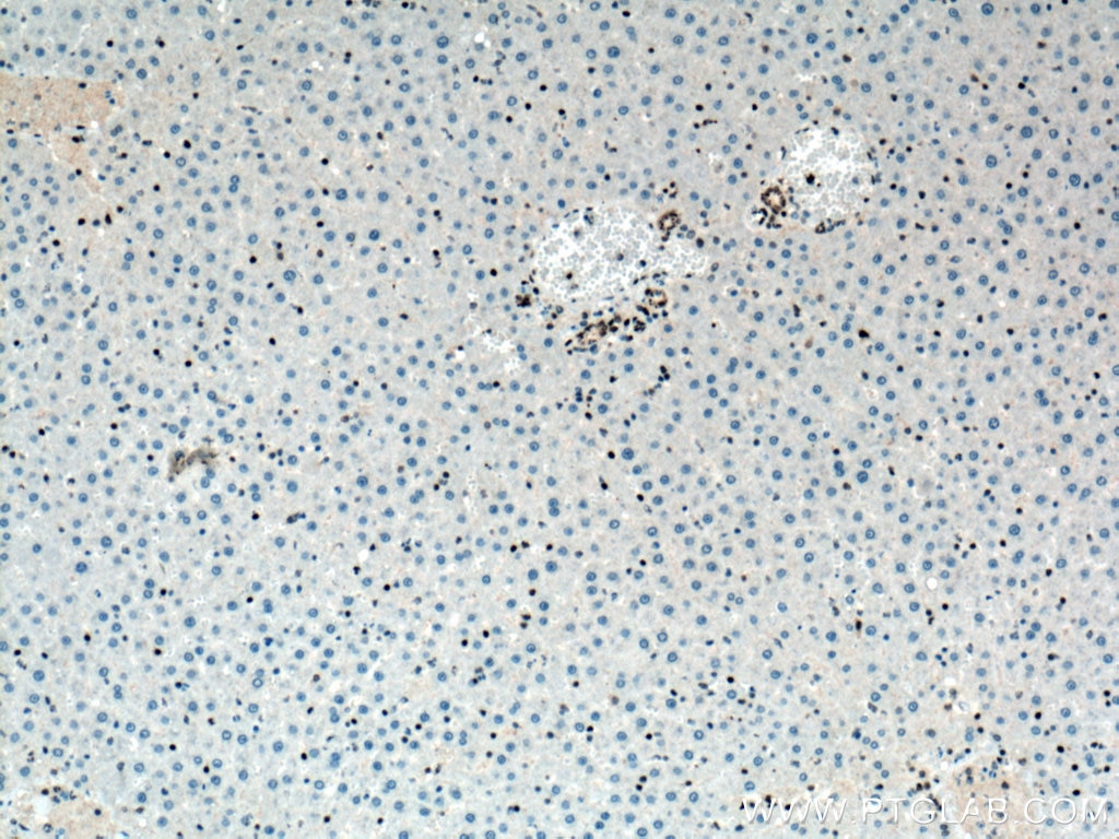
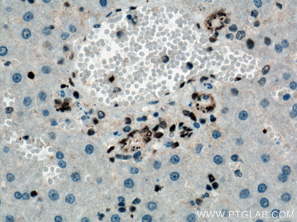
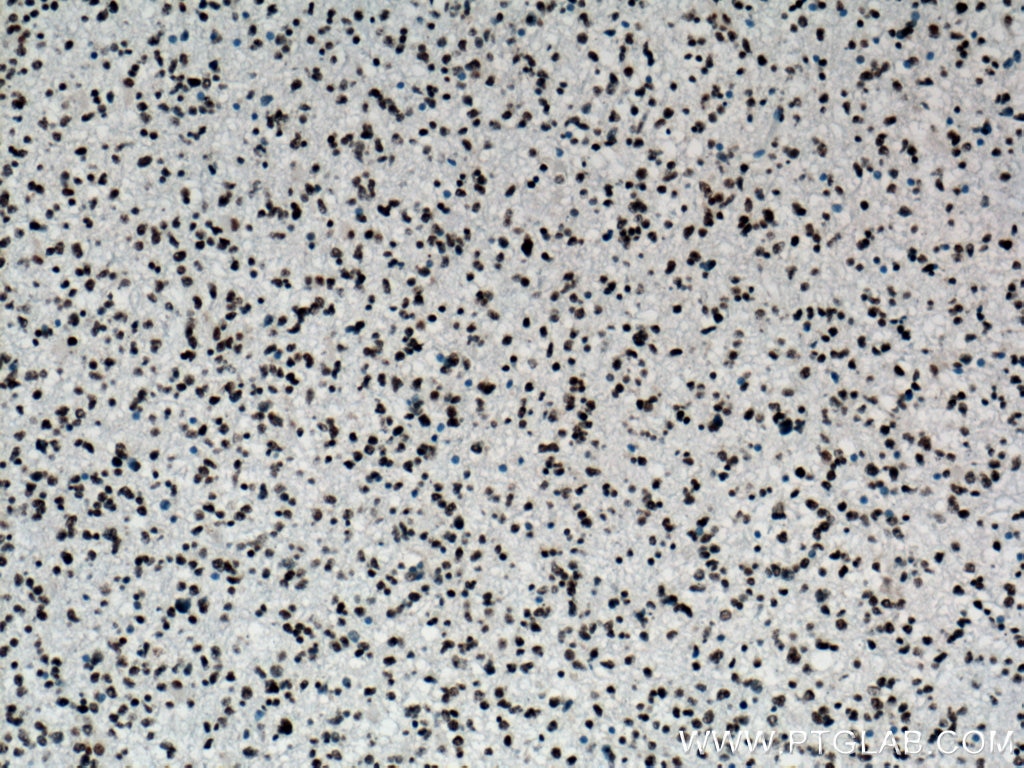
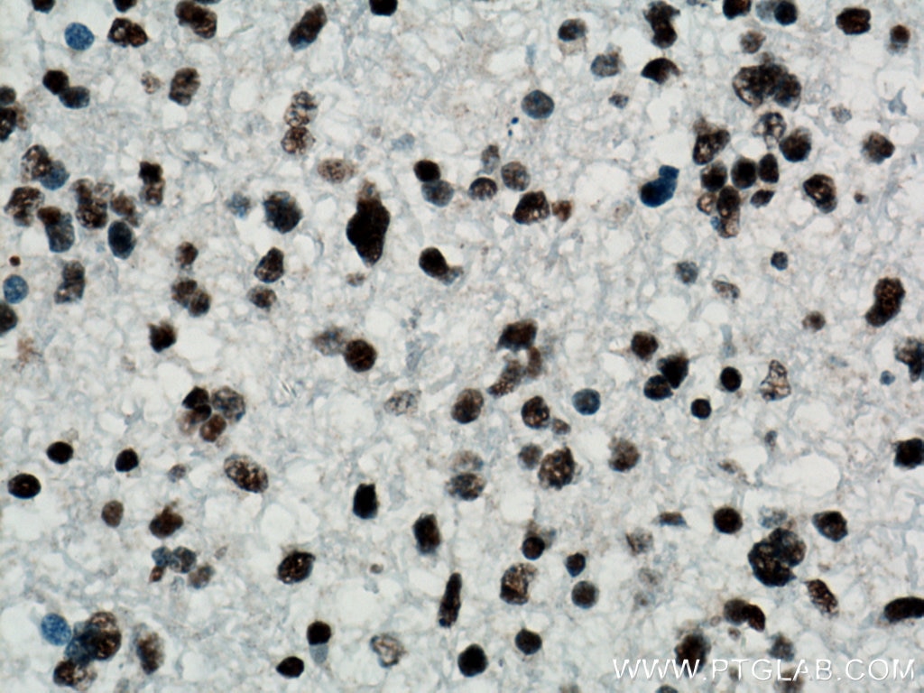
| Positive IHC detected in | mouse brain tissue, human gliomas tissue, rat liver tissue Note: suggested antigen retrieval with TE buffer pH 9.0; (*) Alternatively, antigen retrieval may be performed with citrate buffer pH 6.0 |
Recommended dilution
| Application | Dilution |
|---|---|
| Western Blot (WB) | WB : 1:5000-1:50000 |
| Immunohistochemistry (IHC) | IHC : 1:1000-1:4000 |
| It is recommended that this reagent should be titrated in each testing system to obtain optimal results. | |
| Sample-dependent, Check data in validation data gallery. | |
Product Information
66734-1-Ig targets TDP-43 in WB, IHC, ELISA applications and shows reactivity with human, mouse, rat samples.
| Tested Reactivity | human, mouse, rat |
| Cited Reactivity | human, mouse |
| Host / Isotype | Mouse / IgG1 |
| Class | Monoclonal |
| Type | Antibody |
| Immunogen |
Recombinant protein Predict reactive species |
| Full Name | TAR DNA binding protein |
| Calculated Molecular Weight | 43 kDa |
| Observed Molecular Weight | 43 kDa |
| GenBank Accession Number | BC001487 |
| Gene Symbol | TDP-43 |
| Gene ID (NCBI) | 23435 |
| RRID | AB_2882084 |
| Conjugate | Unconjugated |
| Form | Liquid |
| Purification Method | Protein G purification |
| UNIPROT ID | Q13148 |
| Storage Buffer | PBS with 0.02% sodium azide and 50% glycerol, pH 7.3. |
| Storage Conditions | Store at -20°C. Stable for one year after shipment. Aliquoting is unnecessary for -20oC storage. 20ul sizes contain 0.1% BSA. |
Background Information
Transactivation response (TAR), DNA-binding protein of 43 kDa (also known as TARDBP or TDP-43), was first isolated as a transcriptional inactivator binding to the TAR DNA element of the HIV-1 virus. Neumann et al. (2006) found that a hyperphosphorylated, ubiquitinated, and cleaved form of TARDBP, known as pathologic TDP-43, is the major component of the tau-negative and ubiquitin-positive inclusions that characterize amyotrophic lateral sclerosis (ALS) and the most common pathological subtype of frontotemporal lobar degeneration (FTLD-U). 66734-1-Ig is a mouse monoclonal antibody recognizing the intact protein of TDP-43.
Protocols
| Product Specific Protocols | |
|---|---|
| IHC protocol for TDP-43 antibody 66734-1-Ig | Download protocol |
| WB protocol for TDP-43 antibody 66734-1-Ig | Download protocol |
| Standard Protocols | |
|---|---|
| Click here to view our Standard Protocols |
Publications
What published studies show
The TDP-43 antibody (Cat# 66734-1-Ig) has 9 total citations. These appear in journals including Nature Communications, Journal of the American Chemical Society, Acta Neuropathologica, International Journal of Biological Sciences, International Immunopharmacology, Toxicology, Scientific Reports, Experimental Neurology, and Journal of Natural Products. The research fields span amyotrophic lateral sclerosis (ALS), frontotemporal dementia, TDP-43 protein aggregation, neurodegeneration, environmental toxicology, and hepatoprotection.
| Species | Application | Title |
|---|---|---|
Nat Commun TDP-43 proteinopathy in ALS is triggered by loss of ASRGL1 and associated with HML-2 expression | ||
Int J Biol Sci Spreading of pathological TDP-43 along corticospinal tract axons induces ALS-like phenotypes in Atg5+/- mice. | ||
Acta Neuropathol Neurons selectively targeted in frontotemporal dementia reveal early stage TDP-43 pathobiology. | ||
Int Immunopharmacol Paraquat exposure triggers amyloid-β and α-synuclein aggregation in the prefrontal cortex of mice: Suppression of microglial phagocytosis via IL-17A | ||
Toxicology Atrazine Exposure Induces TDP-43 Protein Translocation: A Potential Mechanism for Prefrontal Cortical Neurodegeneration Induced by Environmental Pollutants |









