Tested Applications
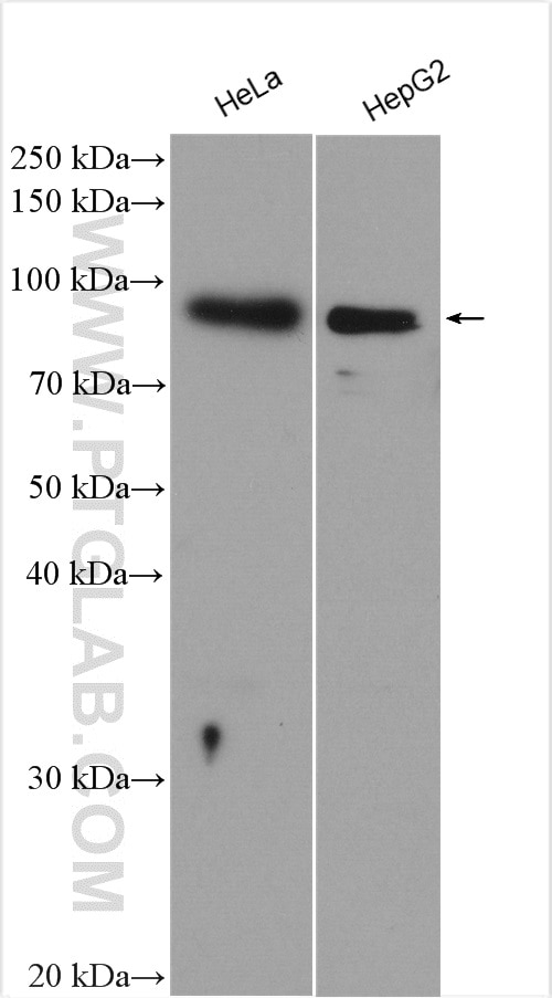
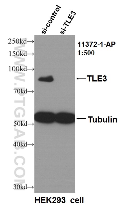
| Positive WB detected in | HeLa cells, HEK-293 cells |
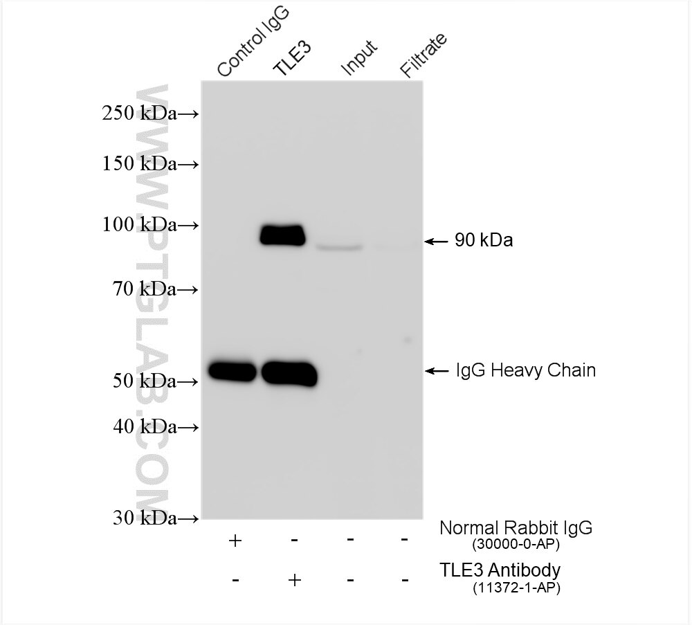
| Positive IP detected in | HepG2 cells |
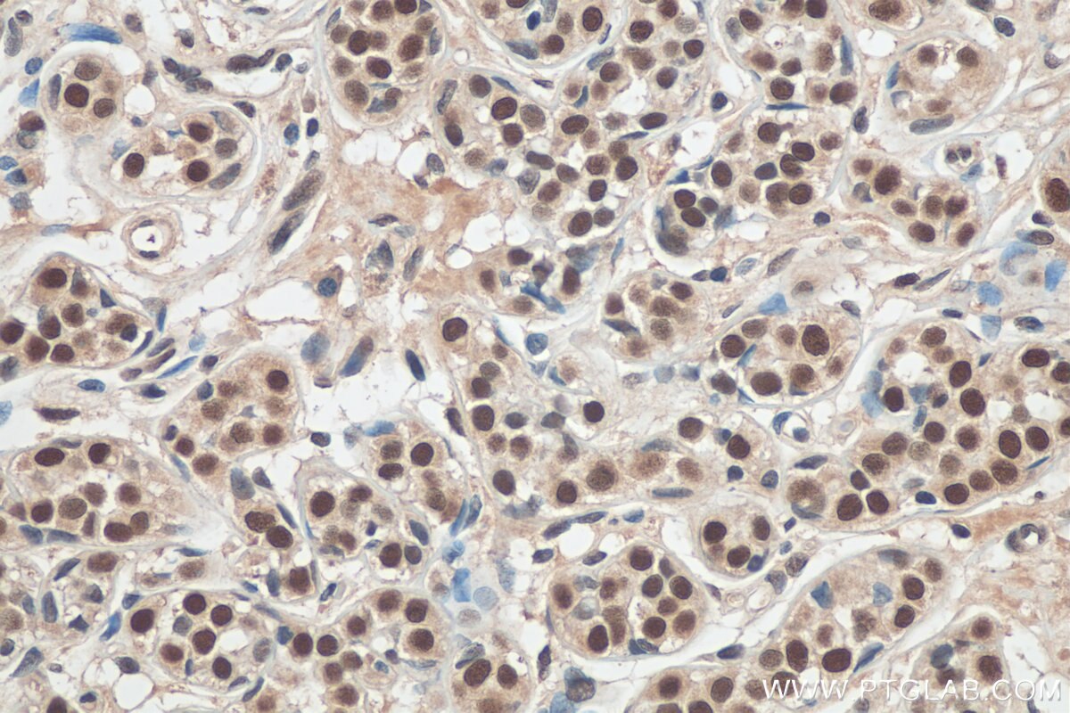
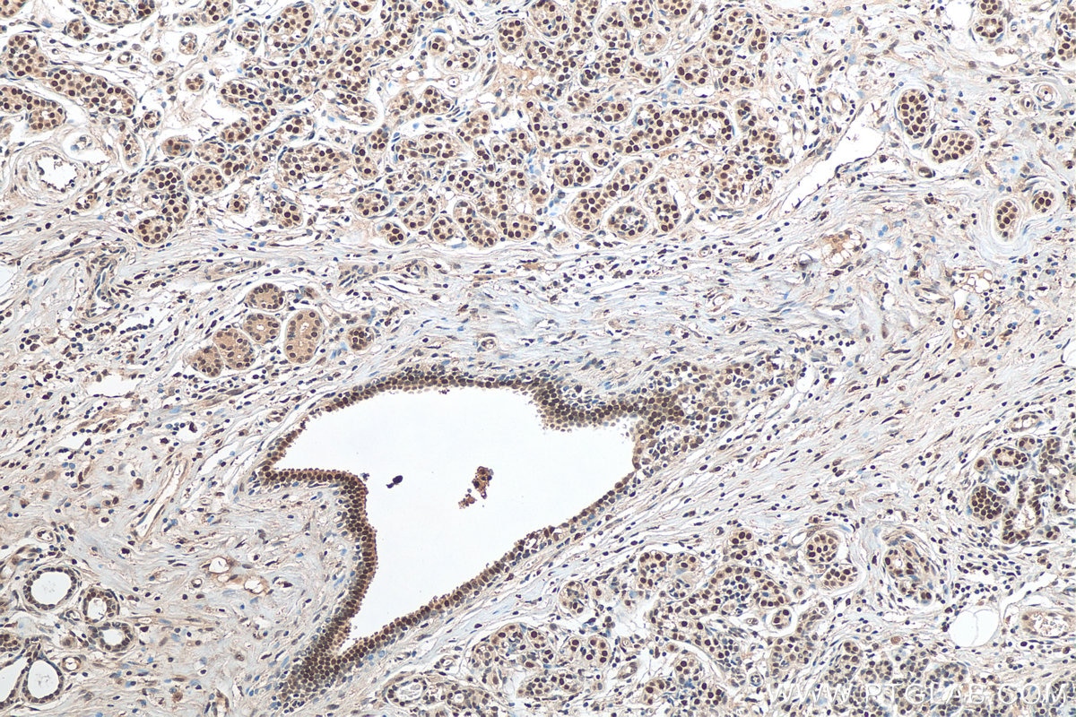
| Positive IHC detected in | human breast cancer tissue Note: suggested antigen retrieval with TE buffer pH 9.0; (*) Alternatively, antigen retrieval may be performed with citrate buffer pH 6.0 |
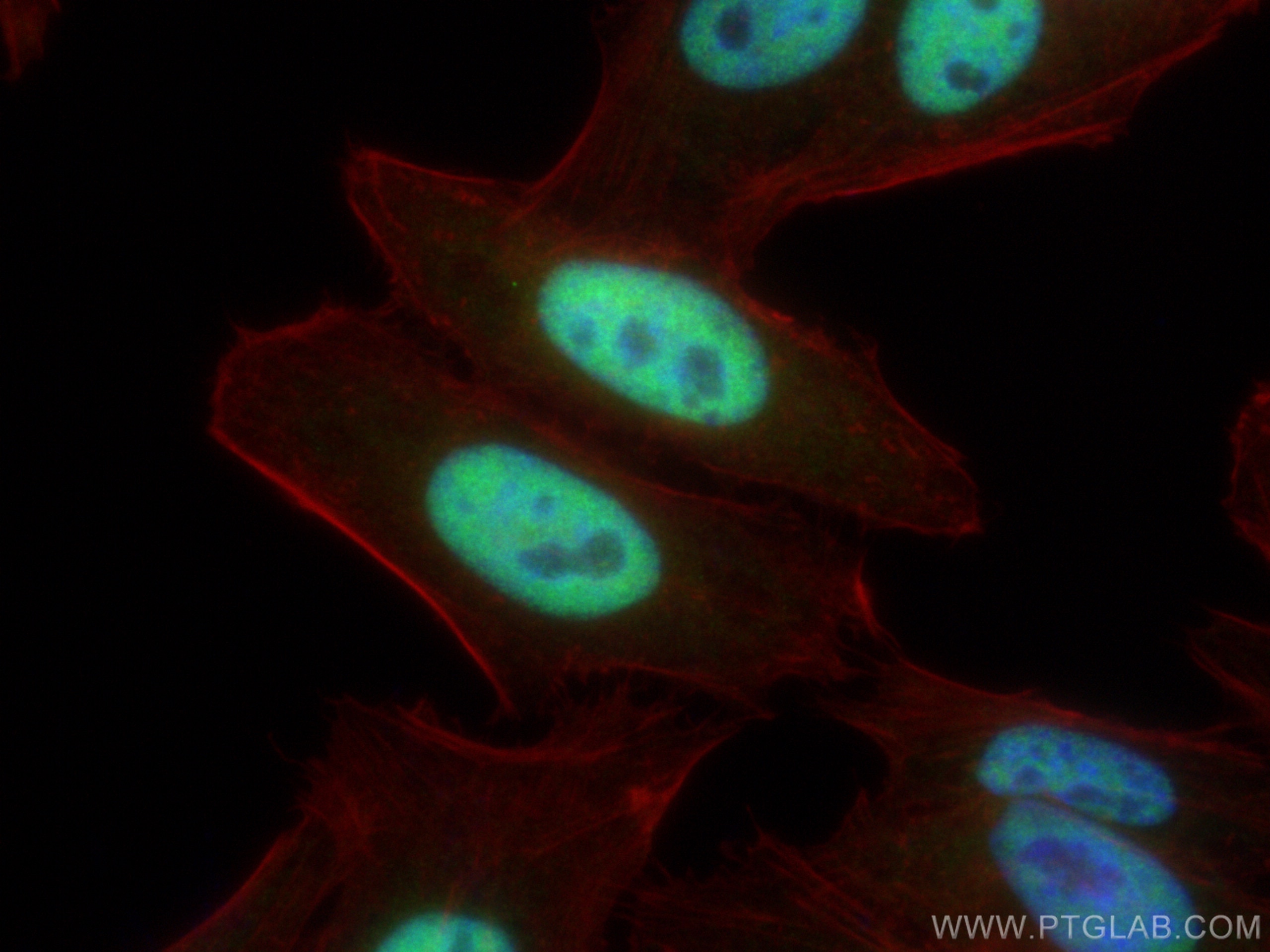
| Positive IF/ICC detected in | HepG2 cells |
Recommended dilution
| Application | Dilution |
|---|---|
| Western Blot (WB) | WB : 1:1000-1:6000 |
| Immunoprecipitation (IP) | IP : 0.5-4.0 ug for 1.0-3.0 mg of total protein lysate |
| Immunohistochemistry (IHC) | IHC : 1:50-1:500 |
| Immunofluorescence (IF)/ICC | IF/ICC : 1:200-1:800 |
| It is recommended that this reagent should be titrated in each testing system to obtain optimal results. | |
| Sample-dependent, Check data in validation data gallery. | |
Published Applications
| KD/KO | See 6 publications below |
| WB | See 19 publications below |
| IHC | See 3 publications below |
| IF | See 5 publications below |
| CoIP | See 3 publications below |
| ChIP | See 8 publications below |
Product Information
11372-1-AP targets TLE3 in WB, IHC, IF/ICC, IP, CoIP, ChIP, ELISA applications and shows reactivity with human, mouse, rat samples.
| Tested Reactivity | human, mouse, rat |
| Cited Reactivity | human, mouse |
| Host / Isotype | Rabbit / IgG |
| Class | Polyclonal |
| Type | Antibody |
| Immunogen |
CatNo: Ag1930 Product name: Recombinant human TLE3 protein Source: e coli.-derived, PGEX-4T Tag: GST Domain: 1-350 aa of BC015729 Sequence: MYPQGRHPAPHQPGQPGFKFTVAESCDRIKDEFQFLQAQYHSLKVEYDKLANEKTEMQRHYVMYYEMSYGLNIEMHKQTEIAKRLNTILAQIMPFLSQEHQQQVAQAVERAKQVTMTELNAIIGQQQLQAQHLSHATHGPPVQLPPHPSGLQPPGIPPVTGSSSGLLALGALGSQAHLTVKDEKNHHELDHRERESSANNSVSPSESLRASEKHRGSADYSMEAKKRKAEEKDSLSRYDSDGDKSDDLVVDVSNEDPATPRVSPAHSPPENGLDKARSLKKDAPTSPASVASSSSTPSSKTKDLGHNDKSSTPGLKSNTPTPRNDAPTPGTSTTPGLRSMPGKPPGMDPI Predict reactive species |
| Full Name | transducin-like enhancer of split 3 (E(sp1) homolog, Drosophila) |
| Calculated Molecular Weight | 772 aa, 83 kDa |
| Observed Molecular Weight | 83 kDa |
| GenBank Accession Number | BC015729 |
| Gene Symbol | TLE3 |
| Gene ID (NCBI) | 7090 |
| RRID | AB_2203743 |
| Conjugate | Unconjugated |
| Form | Liquid |
| Purification Method | Antigen affinity purification |
| UNIPROT ID | Q04726 |
| Storage Buffer | PBS with 0.02% sodium azide and 50% glycerol, pH 7.3. |
| Storage Conditions | Store at -20°C. Stable for one year after shipment. Aliquoting is unnecessary for -20oC storage. 20ul sizes contain 0.1% BSA. |
Background Information
TLE3, also named as KIAA1547 and ESG3, belongs to the WD repeat Groucho/TLE family. It is a transcriptional corepressor that binds to a number of transcription factors. It inhibits the transcriptional activation mediated by CTNNB1 and TCF family members in Wnt signaling. The effects of full-length TLE family members may be modulated by association with dominant-negative AES. TLE3 is a member of the transducin-like enhancer of split (TLE) family of proteins that have been implicated in the tumorgenesis and classification of sarcomas. It is a candidate biomarker of response to taxane
Protocols
| Product Specific Protocols | |
|---|---|
| IF protocol for TLE3 antibody 11372-1-AP | Download protocol |
| IHC protocol for TLE3 antibody 11372-1-AP | Download protocol |
| IP protocol for TLE3 antibody 11372-1-AP | Download protocol |
| WB protocol for TLE3 antibody 11372-1-AP | Download protocol |
| Standard Protocols | |
|---|---|
| Click here to view our Standard Protocols |
Publications
| Species | Application | Title |
|---|---|---|
Nature Distinct structural classes of activating FOXA1 alterations in advanced prostate cancer.
| ||
Cell Stem Cell Lats1/2 Sustain Intestinal Stem Cells and Wnt Activation through TEAD-Dependent and Independent Transcription. | ||
Cell Metab Adipose subtype-selective recruitment of TLE3 or Prdm16 by PPARγ specifies lipid storage versus thermogenic gene programs.
| ||
J Clin Invest Small molecule JQ1 promotes prostate cancer invasion via BET-independent inactivation of FOXA1. | ||
J Exp Med Tle corepressors are differentially partitioned to instruct CD8+ T cell lineage choice and identity.
|
Reviews
The reviews below have been submitted by verified Proteintech customers who received an incentive for providing their feedback.
FH Tongbin (Verified Customer) (08-05-2020) | I did western blots using mouse heart samples. I observed a TLE3 band with correct molecular weight of about 80kD.
|