


Tested Applications
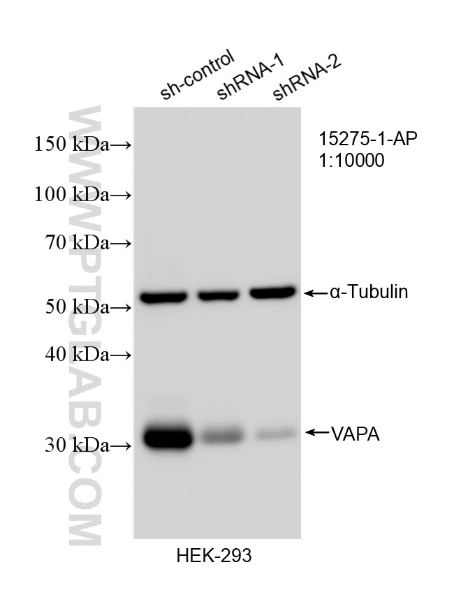
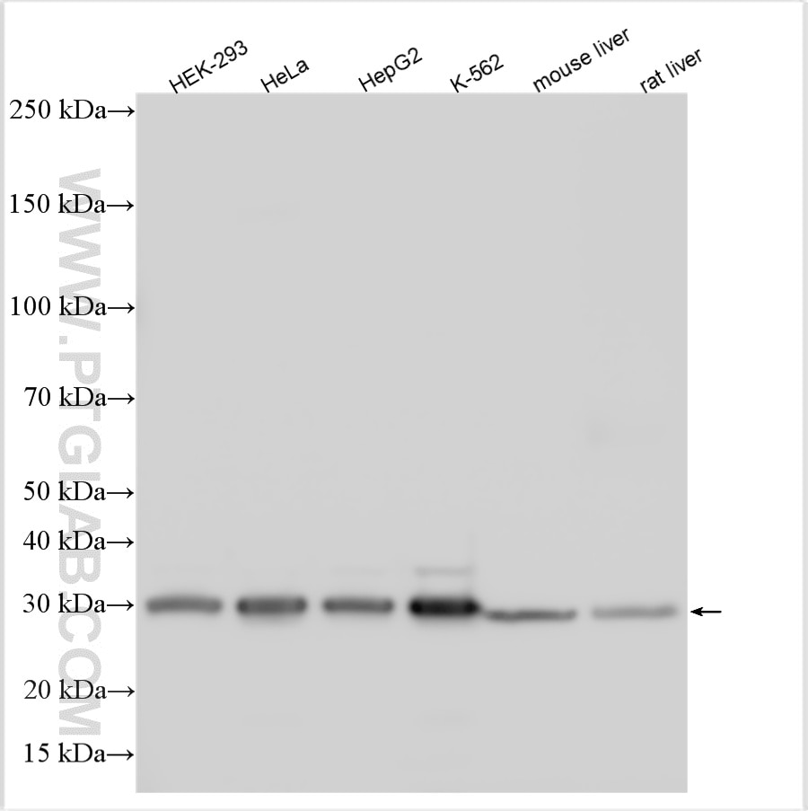
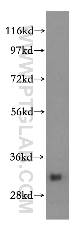
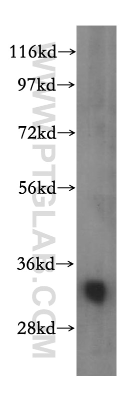
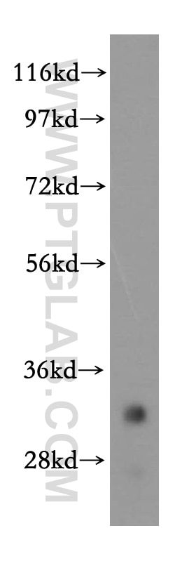
| Positive WB detected in | HEK-293 cells, mouse skeletal muscle tissue, human kidney tissue, human brain tissue, HepG2 cells, mouse liver tissue, rat liver tissue, HeLa cells, K-562 cells |
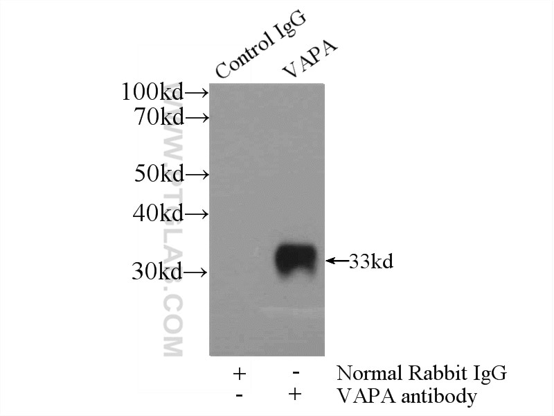
| Positive IP detected in | HEK-293 cells |
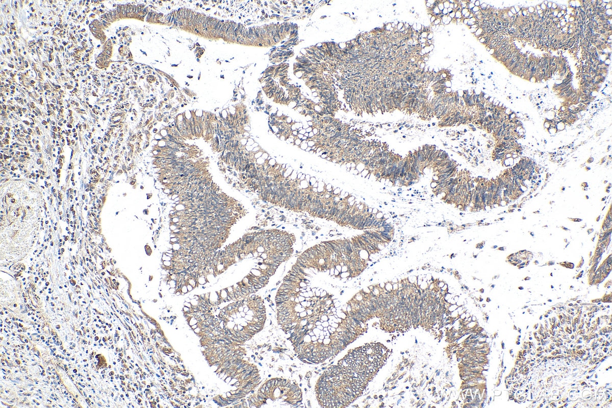
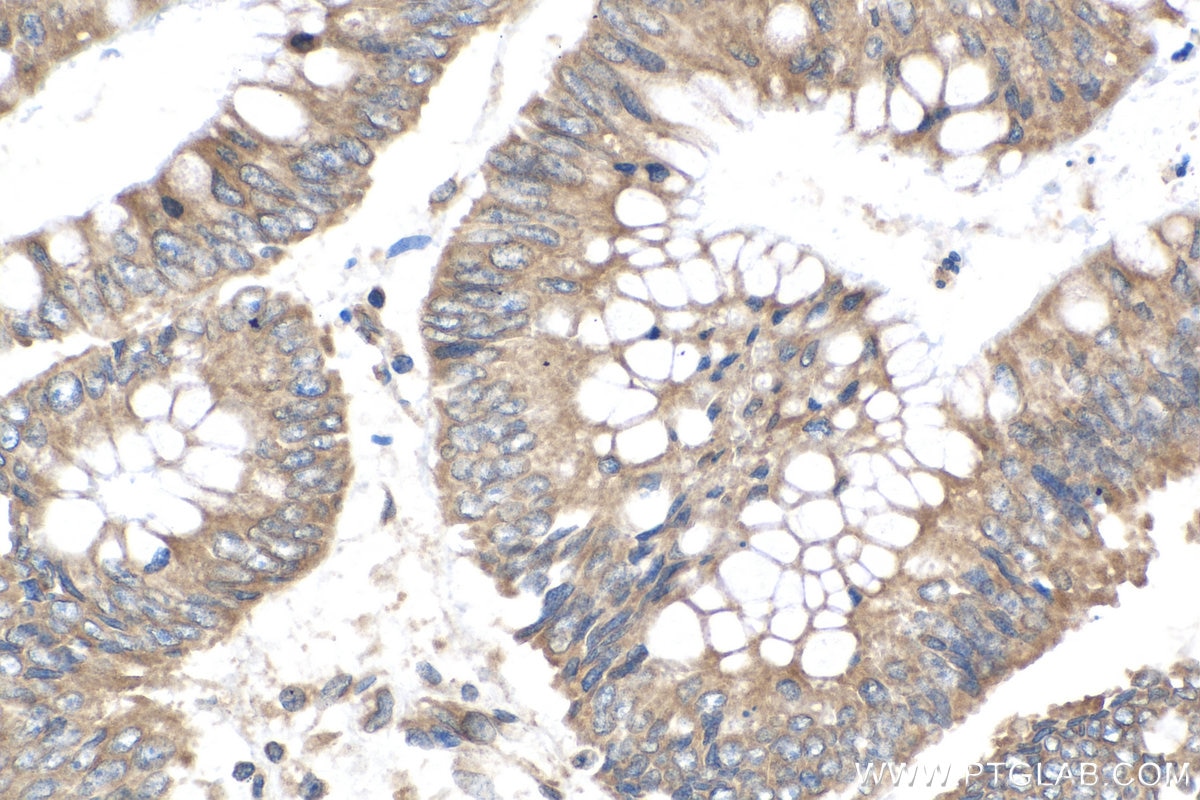
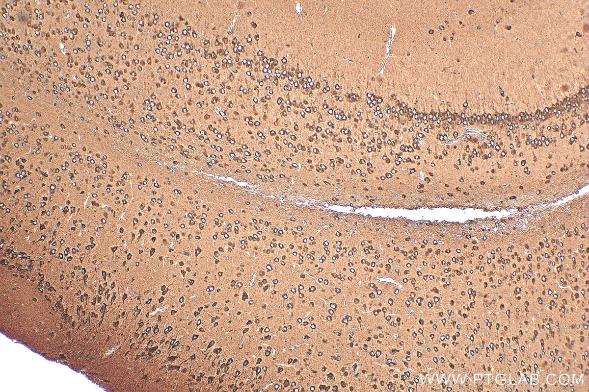
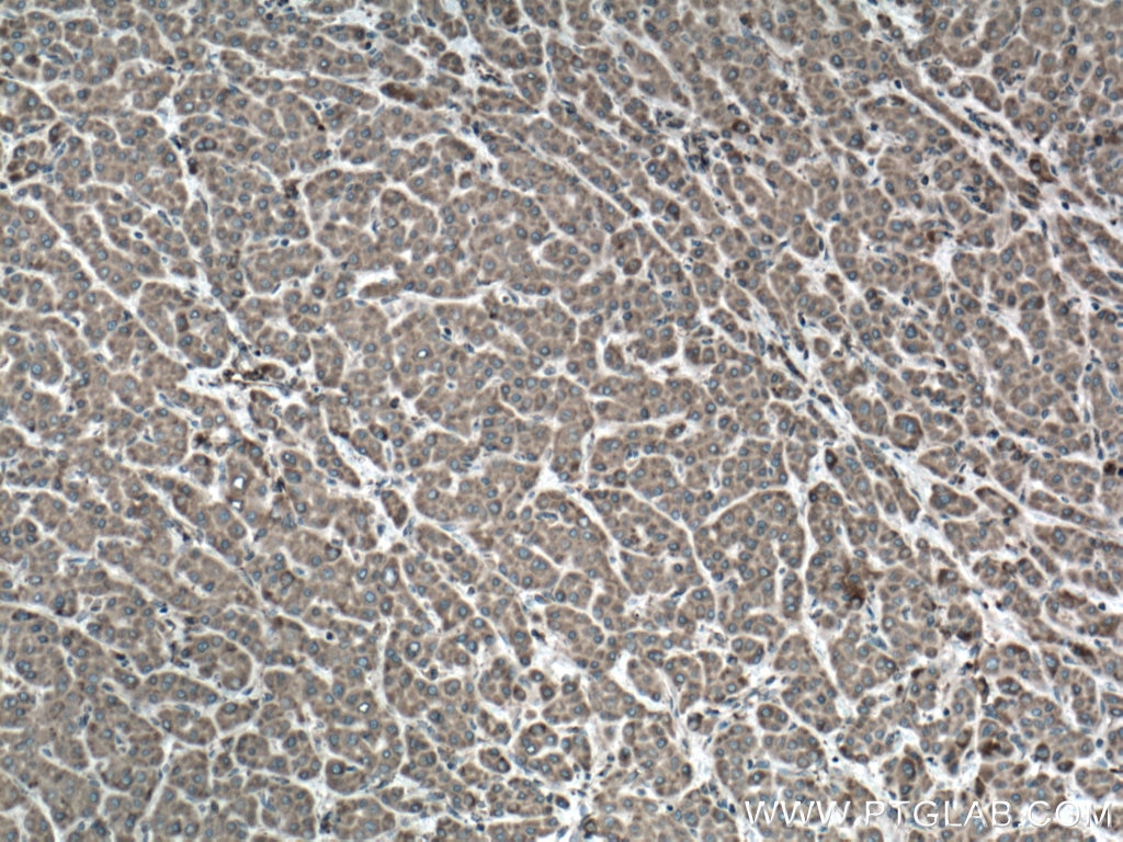
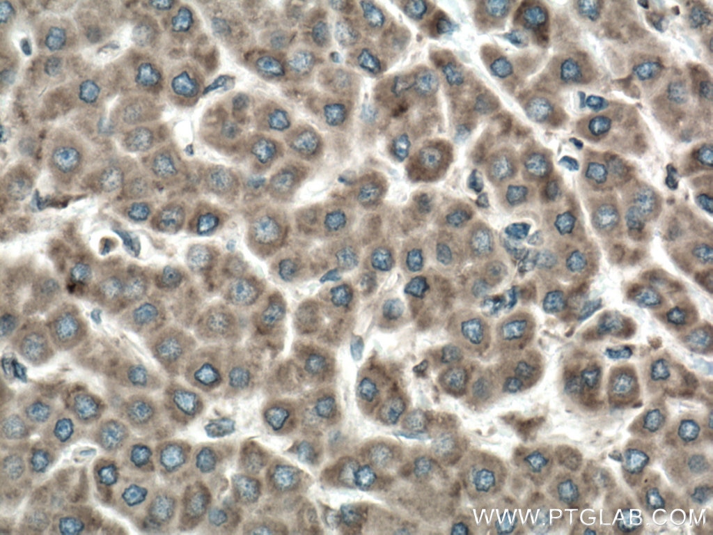
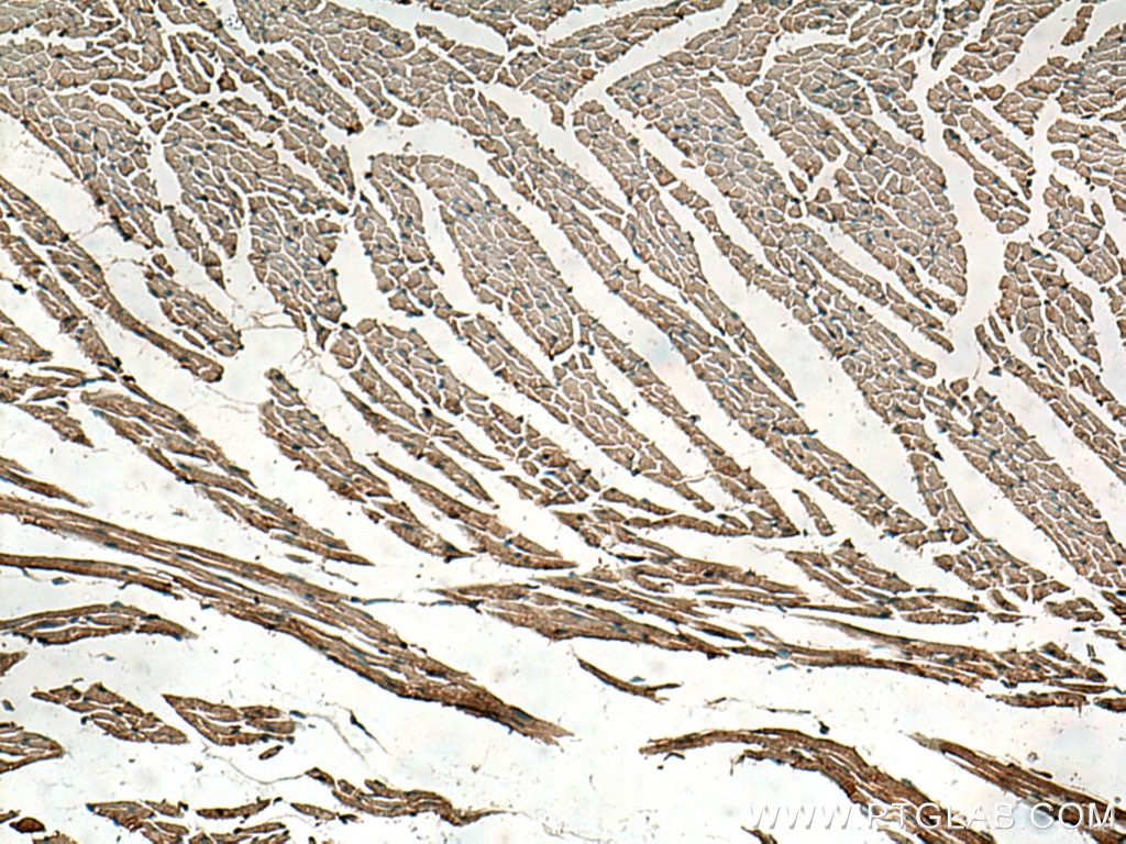
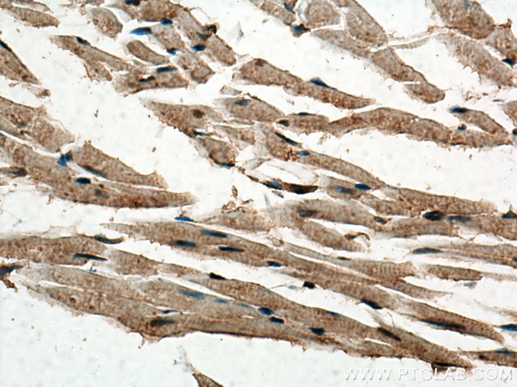
| Positive IHC detected in | human colon cancer tissue, human liver cancer tissue, mouse brain tissue, mouse heart tissue Note: suggested antigen retrieval with TE buffer pH 9.0; (*) Alternatively, antigen retrieval may be performed with citrate buffer pH 6.0 |
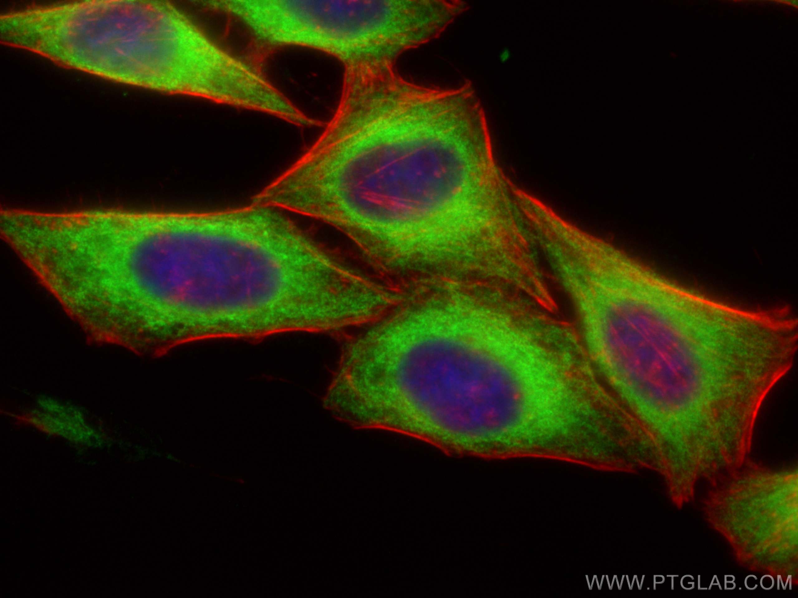
| Positive IF/ICC detected in | HepG2 cells |
Recommended dilution
| Application | Dilution |
|---|---|
| Western Blot (WB) | WB : 1:5000-1:50000 |
| Immunoprecipitation (IP) | IP : 0.5-4.0 ug for 1.0-3.0 mg of total protein lysate |
| Immunohistochemistry (IHC) | IHC : 1:200-1:1600 |
| Immunofluorescence (IF)/ICC | IF/ICC : 1:200-1:800 |
| It is recommended that this reagent should be titrated in each testing system to obtain optimal results. | |
| Sample-dependent, Check data in validation data gallery. | |
Product Information
15275-1-AP targets VAPA in WB, IHC, IF/ICC, IP, COIP, ELISA applications and shows reactivity with human, mouse, rat samples.
| Tested Reactivity | human, mouse, rat |
| Cited Reactivity | human, mouse, rat, monkey |
| Host / Isotype | Rabbit / IgG |
| Class | Polyclonal |
| Type | Antibody |
| Immunogen |
CatNo: Ag7393 Product name: Recombinant human VAPA protein Source: e coli.-derived, PGEX-4T Tag: GST Domain: 1-221 aa of BC002992 Sequence: MASASGAMAKHEQILVLDPPTDLKFKGPFTDVVTTNLKLRNPSDRKVCFKVKTTAPRRYCVRPNSGIIDPGSTVTVSVMLQPFDYDPNEKSKHKFMVQTIFAPPNTSDMEAVWKEAKPDELMDSKLRCVFEMPNENDKLNDMEPSKAVPLNASKQDGPMPKPHSVSLNDTETRKLMEECKRLQGEMMKLSEENRHLRDEGLRLRKVAHSDKPGSTSTASFR Predict reactive species |
| Full Name | VAMP (vesicle-associated membrane protein)-associated protein A, 33kDa |
| Calculated Molecular Weight | 28 kDa |
| Observed Molecular Weight | 28-30 kDa |
| GenBank Accession Number | BC002992 |
| Gene Symbol | VAPA |
| Gene ID (NCBI) | 9218 |
| RRID | AB_2256991 |
| Conjugate | Unconjugated |
| Form | Liquid |
| Purification Method | Antigen affinity purification |
| UNIPROT ID | Q9P0L0 |
| Storage Buffer | PBS with 0.02% sodium azide and 50% glycerol, pH 7.3. |
| Storage Conditions | Store at -20°C. Stable for one year after shipment. Aliquoting is unnecessary for -20oC storage. 20ul sizes contain 0.1% BSA. |
Background Information
VAPA (Vesicle-associated membrane protein-associated protein A), also known as VAP33, is a ubiquitously expressed integral membrane protein of the VAMP-associated protein (VAP) family. It is present in the plasma membrane and in intracellular vesicles, playing a role in vesicle trafficking.
Protocols
| Product Specific Protocols | |
|---|---|
| IF protocol for VAPA antibody 15275-1-AP | Download protocol |
| IHC protocol for VAPA antibody 15275-1-AP | Download protocol |
| IP protocol for VAPA antibody 15275-1-AP | Download protocol |
| WB protocol for VAPA antibody 15275-1-AP | Download protocol |
| Standard Protocols | |
|---|---|
| Click here to view our Standard Protocols |
Publications
What published studies show
The VAPA Polyclonal antibody (Cat# 15275-1-AP) has 42 citations. These appear in journals including Cell, Nature Communications, Nature Microbiology, Autophagy, Neuron, Molecular Cell, EMBO Journal, Current Biology, Journal of Cell Biology, Cell Reports, Journal of Biological Chemistry, and others. Research fields span ER biology, autophagy, membrane contact sites, lipid droplet homeostasis, synaptic plasticity, ciliopathy, viral infection, cholesterol metabolism, Golgi turnover, and neurodegenerative disease.
| Species | Application | Title |
|---|---|---|
Cell Calcium transients on the ER surface trigger liquid-liquid phase separation of FIP200 to specify autophagosome initiation sites
| ||
Nat Microbiol Toxoplasma effector TgROP1 establishes membrane contact sites with the endoplasmic reticulum during infection. | ||
Autophagy USP20 deubiquitinates and stabilizes the reticulophagy receptor RETREG1/FAM134B to drive reticulophagy | ||
Nat Commun Elevated synaptic PKA activity and abnormal striatal dopamine signaling in Akap11 mutant mice, a genetic model of schizophrenia and bipolar disorder. | ||
Nat Commun Bipolar and schizophrenia risk gene AKAP11 encodes an autophagy receptor coupling the regulation of PKA kinase network homeostasis to synaptic transmission. | ||
Nat Commun VAP spatially stabilizes dendritic mitochondria to locally support synaptic plasticity |
Reviews
What customers say
Users consistently report strong, specific signals in Western blot and immunofluorescence, with reliable performance across multiple experiments. One reviewer noted that while immunoprecipitation works on WB, it lacks sensitivity for Coomassie visualization. Another found the antibody less effective on heart tissue sections. Overall, the product is well-regarded for its specificity and signal strength in WB and IF, with minor limitations in IP sensitivity and certain tissue applications.
The reviews below have been submitted by verified Proteintech customers who received an incentive for providing their feedback.
Ziwei (Verified Customer) (01-04-2026) | works as it should be.
|
Ziwei (Verified Customer) (12-14-2025) | The antibody demonstrates reliable performance across multiple experiments.
|
Janathan (Verified Customer) (12-21-2023) | Really good and strong signal in WB and IF. IP works on WB, but not sensitive enough for visualizing with coomassie
|
Wei (Verified Customer) (09-22-2020) | The antibody have high specific band in western blot, but not so good for heart sections.
|
















