"VDAC1/Porin Antibodies" Comparison
View side-by-side comparison of VDAC1/Porin antibodies from other vendors to find the one that best suits your research needs.
Tested Applications
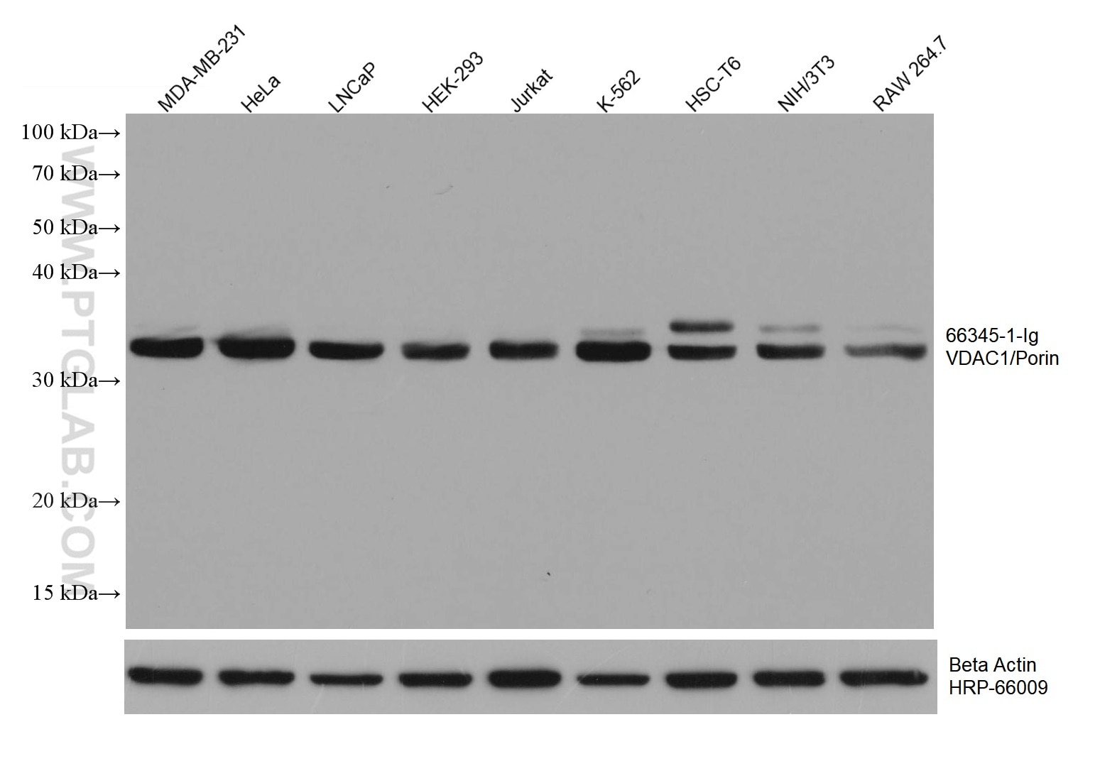
| Positive WB detected in | MDA-MB-231 cells, HeLa cells, LNCaP cells, HEK-293 cells, Jurkat cells, K-562 cells, HSC-T6 cells, NIH/3T3 cells, RAW264.7 cells |
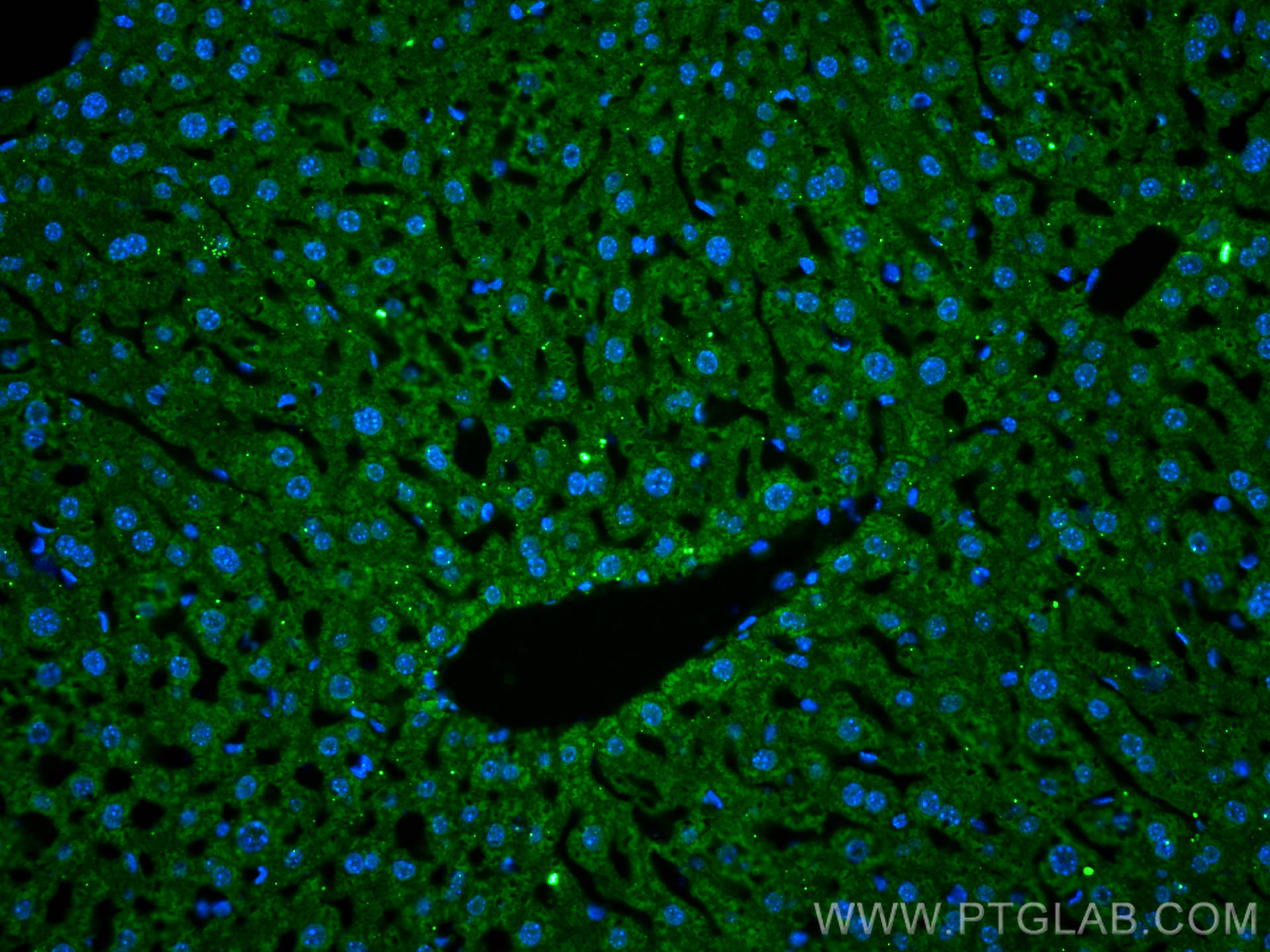
| Positive IF-P detected in | mouse liver tissue |
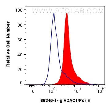
| Positive FC (Intra) detected in | HepG2 cells |
Recommended dilution
| Application | Dilution |
|---|---|
| Western Blot (WB) | WB : 1:5000-1:50000 |
| Immunofluorescence (IF)-P | IF-P : 1:200-1:800 |
| Flow Cytometry (FC) (INTRA) | FC (INTRA) : 0.80 ug per 10^6 cells in a 100 µl suspension |
| It is recommended that this reagent should be titrated in each testing system to obtain optimal results. | |
| Sample-dependent, Check data in validation data gallery. | |
Published Applications
| WB | See 49 publications below |
| IHC | See 3 publications below |
| IF | See 15 publications below |
| IP | See 3 publications below |
| CoIP | See 4 publications below |
Product Information
66345-1-Ig targets VDAC1/Porin in WB, IHC, IF-P, FC (Intra), IP, CoIP, ELISA applications and shows reactivity with human, mouse, rat samples.
| Tested Reactivity | human, mouse, rat |
| Cited Reactivity | human, mouse, rat, pig |
| Host / Isotype | Mouse / IgG3 |
| Class | Monoclonal |
| Type | Antibody |
| Immunogen |
Peptide Predict reactive species |
| Full Name | voltage-dependent anion channel 1 |
| Calculated Molecular Weight | 31 kDa |
| Observed Molecular Weight | 35-37 kDa |
| GenBank Accession Number | NM_003374 |
| Gene Symbol | Porin |
| Gene ID (NCBI) | 7416 |
| RRID | AB_2881725 |
| Conjugate | Unconjugated |
| Form | Liquid |
| Purification Method | Protein A purification |
| UNIPROT ID | P21796 |
| Storage Buffer | PBS with 0.02% sodium azide and 50% glycerol, pH 7.3. |
| Storage Conditions | Store at -20°C. Stable for one year after shipment. Aliquoting is unnecessary for -20oC storage. 20ul sizes contain 0.1% BSA. |
Background Information
VDAC1, also named as VDAC, porin 31HM, porin 31HL and plasmalemmal porin, belongs to the eukaryotic mitochondrial porin family. It adopts an open conformation at low or zero membrane potential and a closed conformation at potentials above 30-40 mV, to form a channel through the mitochondrial outer membrane and also the plasma membrane. Unlike other membrane transport proteins, porins are large enough to allow passive diffusion. Studies have shown that VDAC1 is subject to both phosphorylation and acetylation (PMID: 23233904). The apparent molecular weight of VDAC1 is 30-37 kDa (PMID: 14573604; 23754752; 25681439). Hypoxic conditions were found to trigger cleavage of the VDAC1 C-terminal to yield a 26-kDa truncated but active form (PMID: 22389449; 23233904).
Protocols
| Product Specific Protocols | |
|---|---|
| FC protocol for VDAC1/Porin antibody 66345-1-Ig | Download protocol |
| IF protocol for VDAC1/Porin antibody 66345-1-Ig | Download protocol |
| WB protocol for VDAC1/Porin antibody 66345-1-Ig | Download protocol |
| Standard Protocols | |
|---|---|
| Click here to view our Standard Protocols |
Publications
| Species | Application | Title |
|---|---|---|
Adv Sci (Weinh) SUCLG2 Regulates Mitochondrial Dysfunction through Succinylation in Lung Adenocarcinoma | ||
Free Radic Biol Med The interaction of Atg4B and Bcl-2 plays an important role in Cd-induced crosstalk between apoptosis and autophagy through disassociation of Bcl-2-Beclin1 in A549 cells. | ||
Diabetes PACS-2 Ameliorates Tubular Injury by Facilitating Endoplasmic Reticulum-Mitochondria Contact and Mitophagy in Diabetic Nephropathy. | ||
Oxid Med Cell Longev The lncRNA-AK046375 Upregulates Metallothionein-2 by Sequestering miR-491-5p to Relieve the Brain Oxidative Stress Burden after Traumatic Brain Injury. | ||
Cell Death Discov Idebenone improves motor dysfunction, learning and memory by regulating mitophagy in MPTP-treated mice. |