Tested Applications
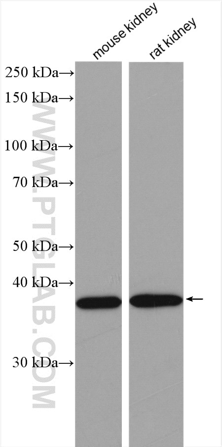
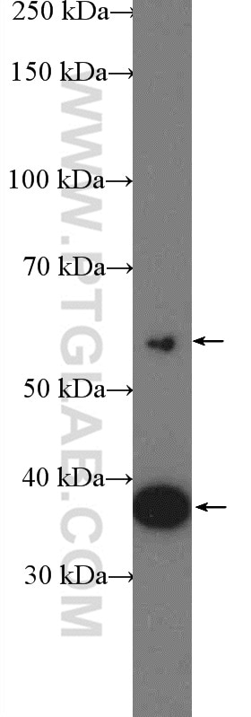
| Positive WB detected in | mouse kidney tissue, HEK-293 cells, rat kidney tissue |
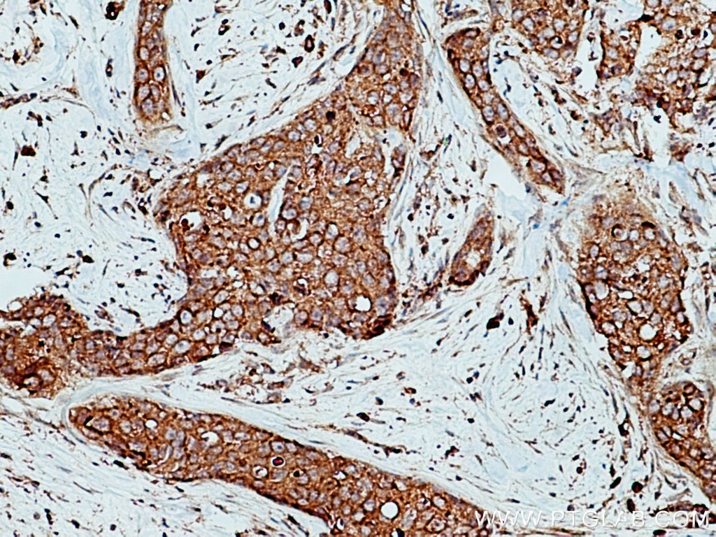
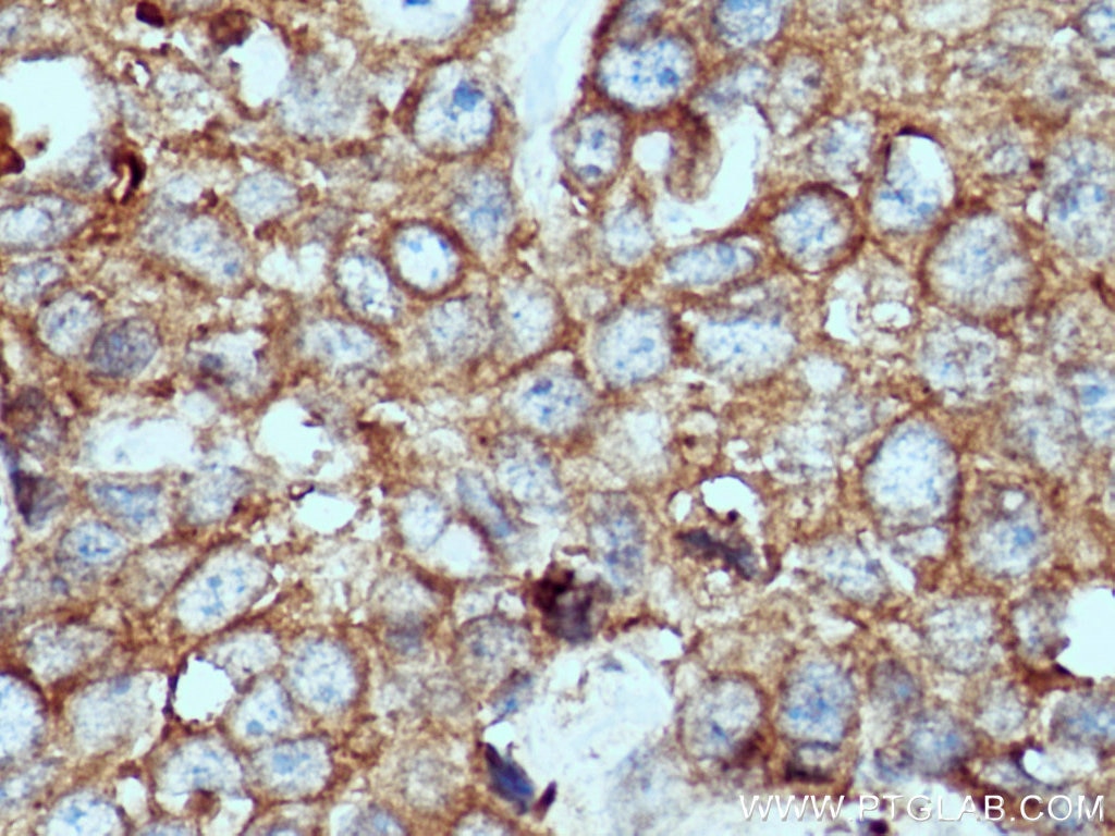
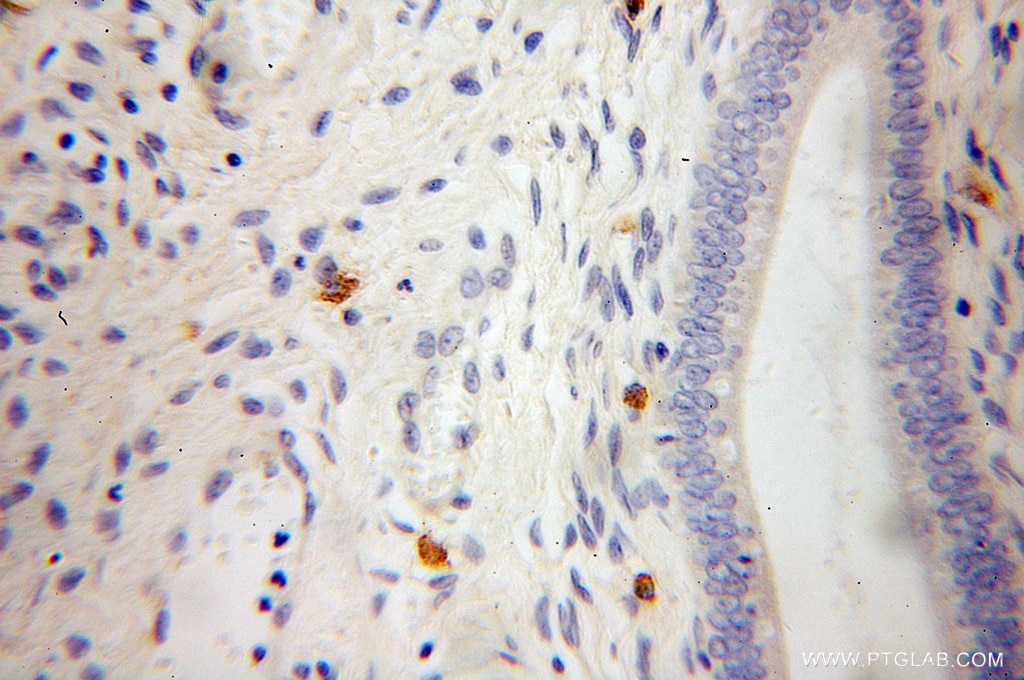
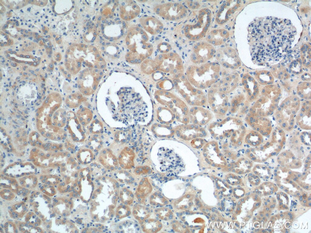
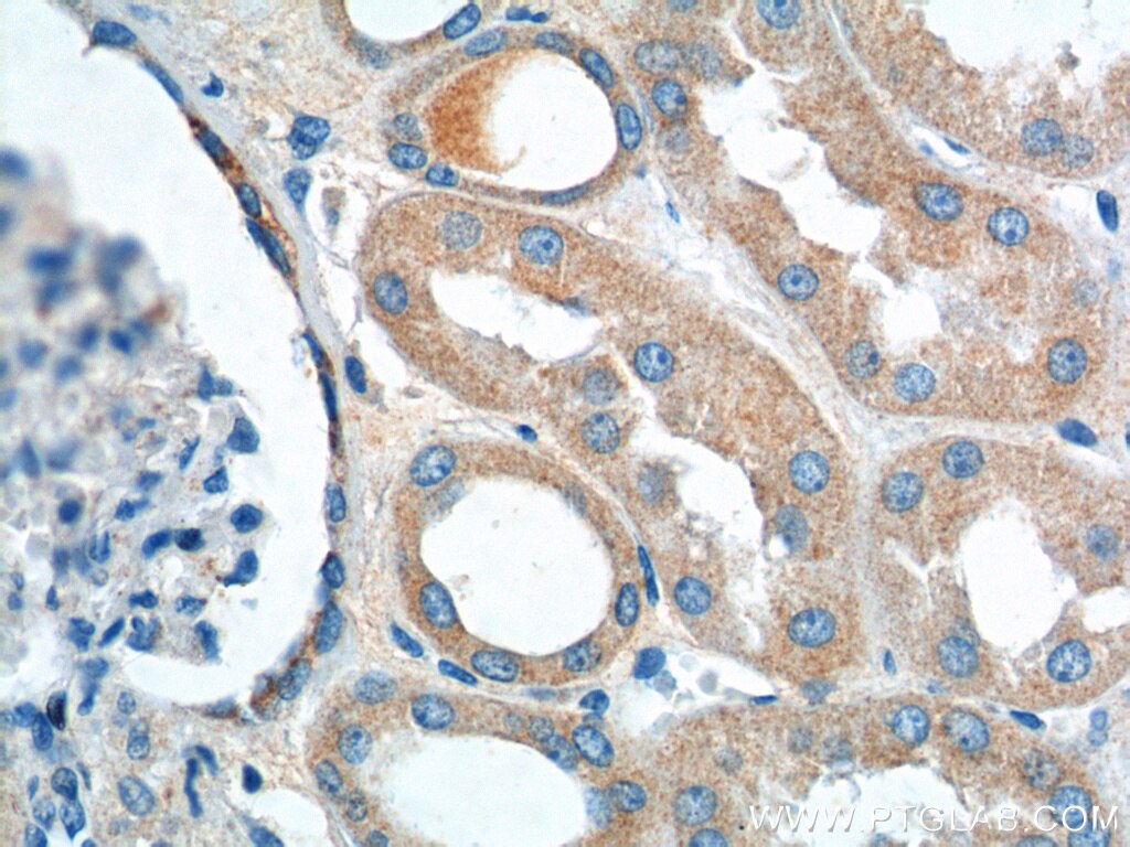
| Positive IHC detected in | human breast cancer tissue, human cervical cancer tissue, human kidney tissue Note: suggested antigen retrieval with TE buffer pH 9.0; (*) Alternatively, antigen retrieval may be performed with citrate buffer pH 6.0 |
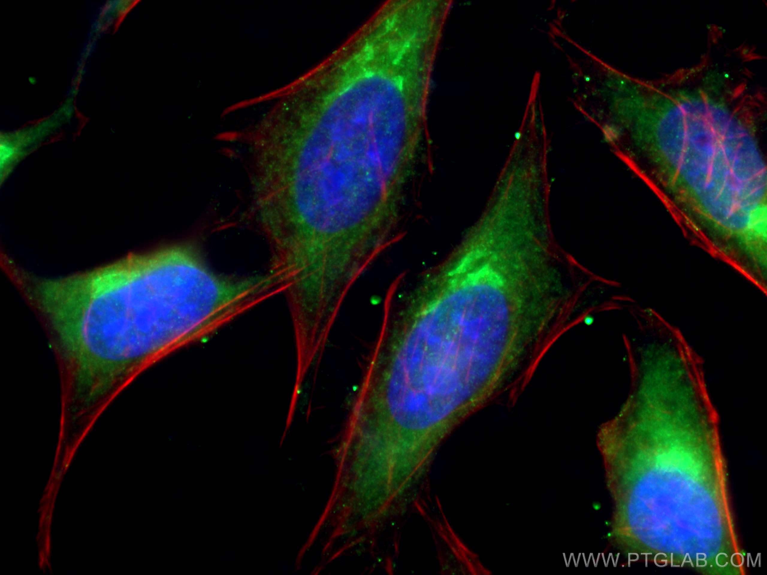
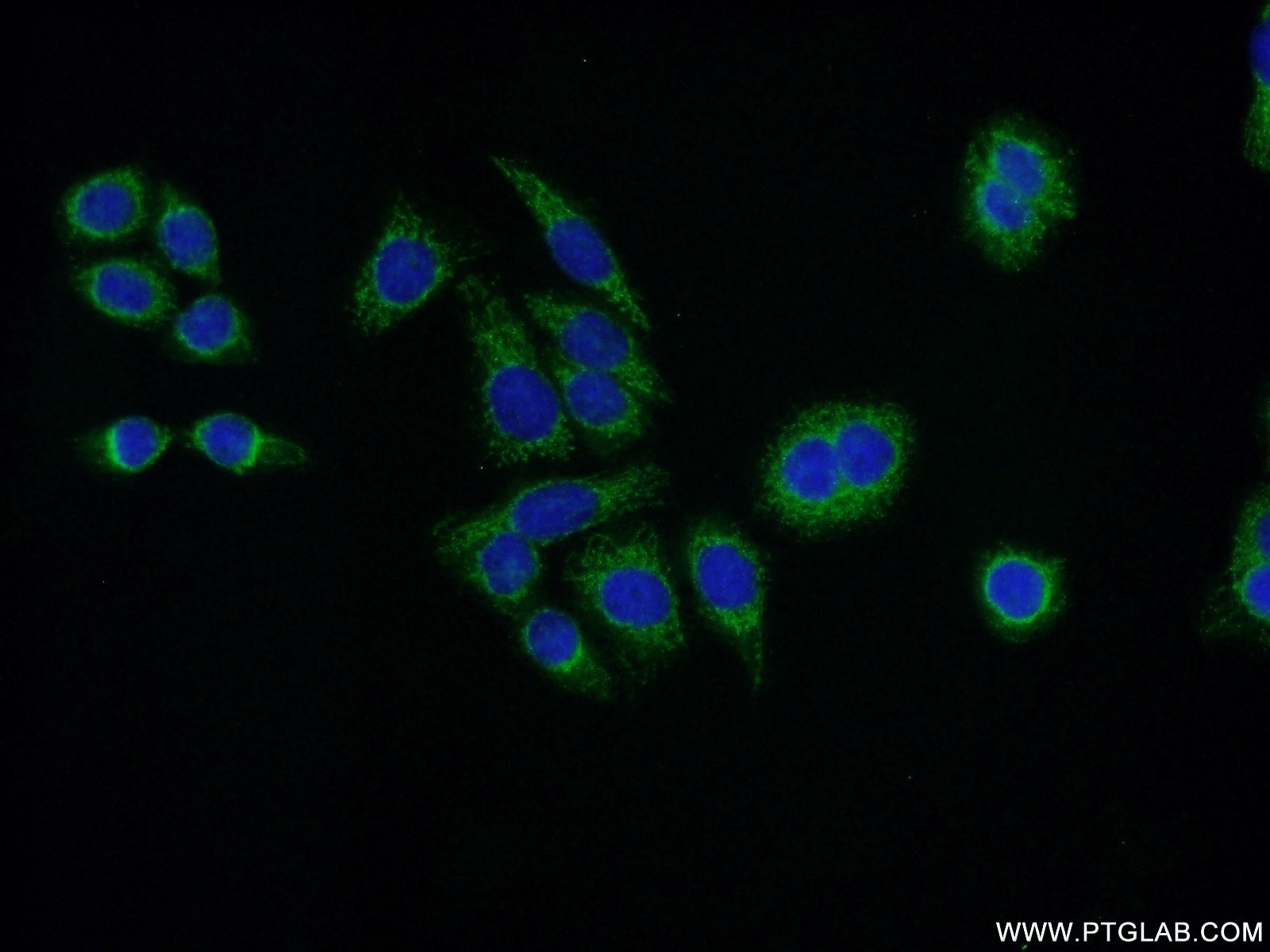
| Positive IF/ICC detected in | HeLa cells |
Recommended dilution
| Application | Dilution |
|---|---|
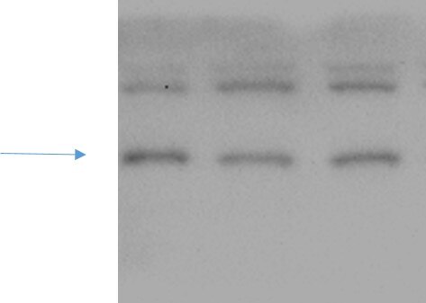
| Western Blot (WB) | WB : 1:500-1:1000 |
| Immunohistochemistry (IHC) | IHC : 1:50-1:500 |
| Immunofluorescence (IF)/ICC | IF/ICC : 1:50-1:500 |
| It is recommended that this reagent should be titrated in each testing system to obtain optimal results. | |
| Sample-dependent, Check data in validation data gallery. | |
Published Applications
| KD/KO | See 1 publications below |
| WB | See 9 publications below |
| IHC | See 1 publications below |
| IF | See 2 publications below |
Product Information
12804-1-AP targets VPS26A in WB, IHC, IF/ICC, ELISA applications and shows reactivity with human, mouse, rat samples.
| Tested Reactivity | human, mouse, rat |
| Cited Reactivity | human, mouse |
| Host / Isotype | Rabbit / IgG |
| Class | Polyclonal |
| Type | Antibody |
| Immunogen |
CatNo: Ag3391 Product name: Recombinant human VPS26A protein Source: e coli.-derived, PGEX-4T Tag: GST Domain: 41-327 aa of BC022505 Sequence: LFYDGESVSGKVNLAFKQPGKRLEHQGIRIEFVGQIELFNDKSNTHEFVNLVKELALPGELTQSRSYDFEFMQVEKPYESYIGANVRLRYFLKVTIVRRLTDLVKEYDLIVHQLATYPDVNNSIKMEVGIEDCLHIEFEYNKSKYHLKDVIVGKIYFLLVRIKIQHMELQLIKKEITGIGPSTTTETETIAKYEIMDGAPVKGESIPIRLFLAGYDPTPTMRDVNKKFSVRYFLNLVLVDEEDRRYFKQQEIILWRKAPEKLRKQRTNFHQRFESPESQASAEQPEM Predict reactive species |
| Full Name | vacuolar protein sorting 26 homolog A (S. pombe) |
| Calculated Molecular Weight | 38 kDa |
| Observed Molecular Weight | 38 kDa |
| GenBank Accession Number | BC022505 |
| Gene Symbol | VPS26A |
| Gene ID (NCBI) | 9559 |
| RRID | AB_2215033 |
| Conjugate | Unconjugated |
| Form | Liquid |
| Purification Method | Antigen affinity purification |
| UNIPROT ID | O75436 |
| Storage Buffer | PBS with 0.02% sodium azide and 50% glycerol, pH 7.3. |
| Storage Conditions | Store at -20°C. Stable for one year after shipment. Aliquoting is unnecessary for -20oC storage. 20ul sizes contain 0.1% BSA. |
Background Information
In mammals, there are two paralogues of yeast Vps26, VPS26A and VPS26B (PMID: 16190980). VPS26 is a component of the retromer complex composed of VPS26 (VPS26A or VPS26B), VPS29, VPS35, SNX1, and SNX2. VPS26A and VPS26B subunits define distinct retromer complexes (PMID: 21920005). The retromer complex is important in recycling transmembrane receptors from endosomes to the trans-Golgi network (TGN).
Protocols
| Product Specific Protocols | |
|---|---|
| IF protocol for VPS26A antibody 12804-1-AP | Download protocol |
| IHC protocol for VPS26A antibody 12804-1-AP | Download protocol |
| WB protocol for VPS26A antibody 12804-1-AP | Download protocol |
| Standard Protocols | |
|---|---|
| Click here to view our Standard Protocols |
Publications
| Species | Application | Title |
|---|---|---|
Cell Metab Phospholipase PLA2G6, a Parkinsonism-Associated Gene, Affects Vps26 and Vps35, Retromer Function, and Ceramide Levels, Similar to α-Synuclein Gain. | ||
bioRxiv Noncanonical roles of ATG5 and membrane atg8ylation in retromer assembly and function | ||
Adv Sci (Weinh) Loss of VMP1 Impairs Tight Junction Recycling and Aggravates Intestinal Barrier Dysfunction in Inflammatory Bowel Disease. | ||
Ann Neurol Retromer proteins reduced in Down syndrome and the Dp16 model: impact of APP dose and preclinical studies of a γ-secretase modulator | ||
Cell Death Dis Targeting sorting nexin 3 to treat pulmonary fibrosis by dual modulating Wnt/β-catenin signaling. |
Reviews
The reviews below have been submitted by verified Proteintech customers who received an incentive for providing their feedback.
FH Xin (Verified Customer) (10-10-2022) | It is Ok to detect endogenous VPS26A although there are some non-specific bands.
 |
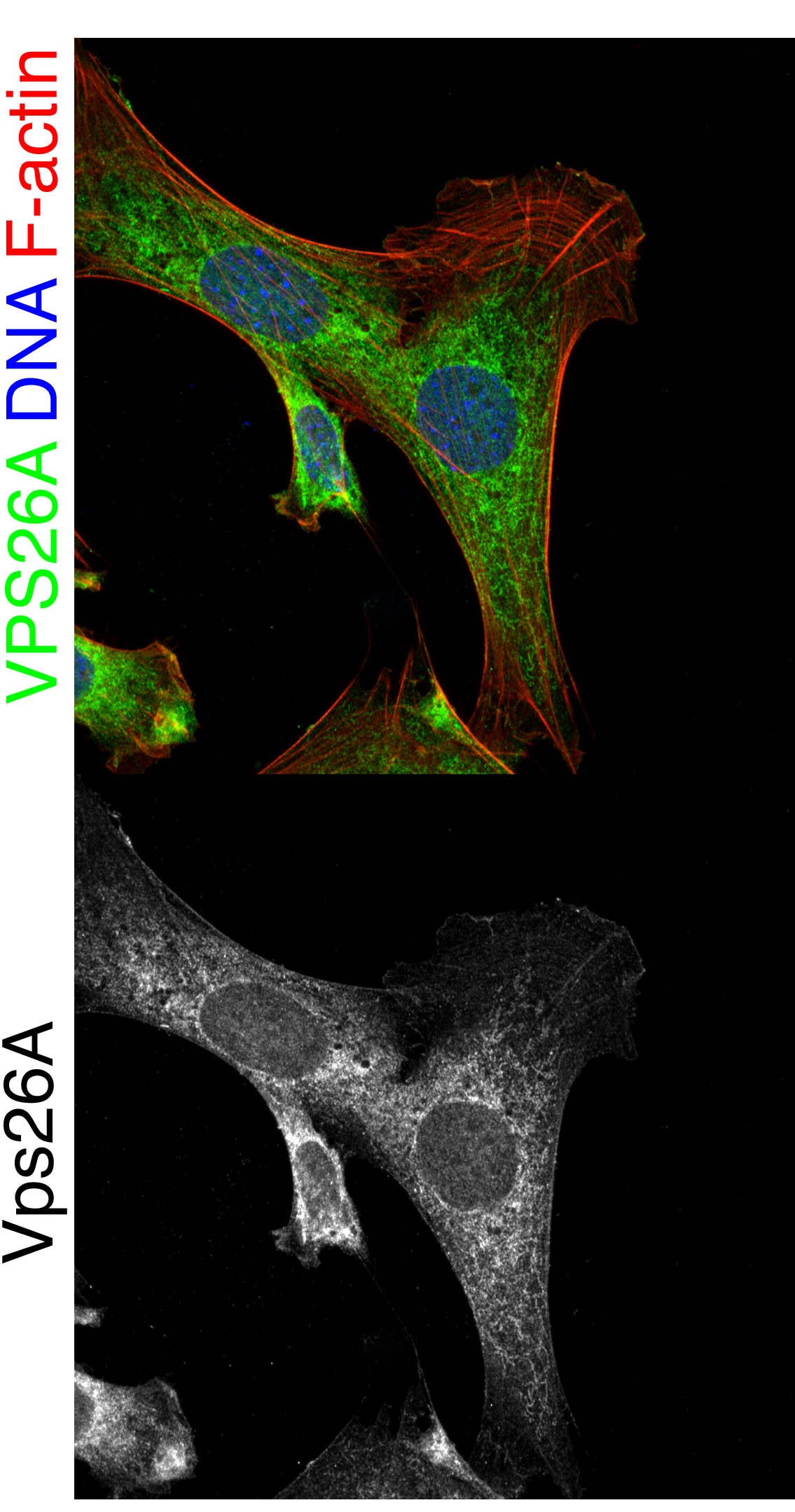
FH Stephen (Verified Customer) (09-07-2019) | Oc-2 cells fixed with 4% PFA Perm. by 0.3% tx100 for 5 min blocked with 1% BSA in 1XPBS for 2 hours vps26a antibody incubated 1:300 overnight at 4 degrees in 1%BSA in 1x PBS. Co-stained with DAPI (blue) and Ph647 (red) to visualize DNA and F-actin respectively.
 |