Tested Applications
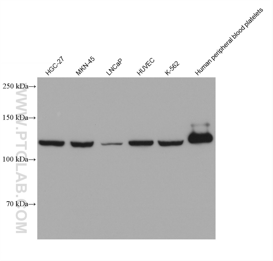
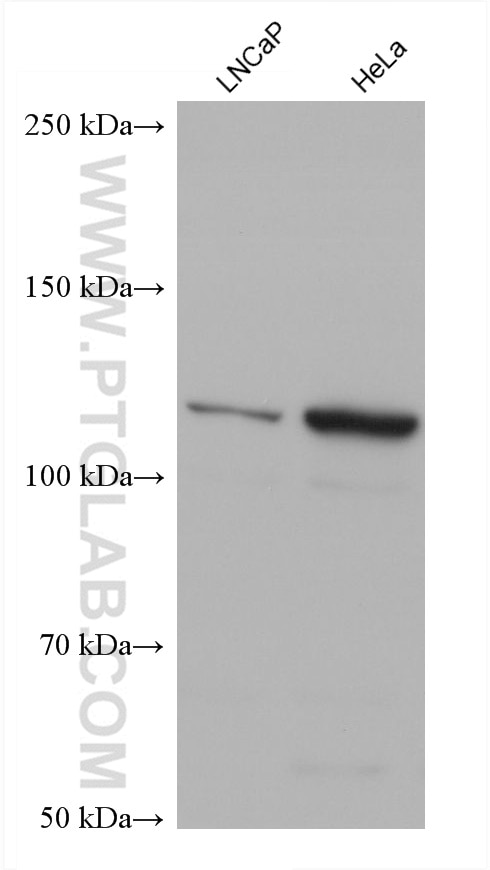
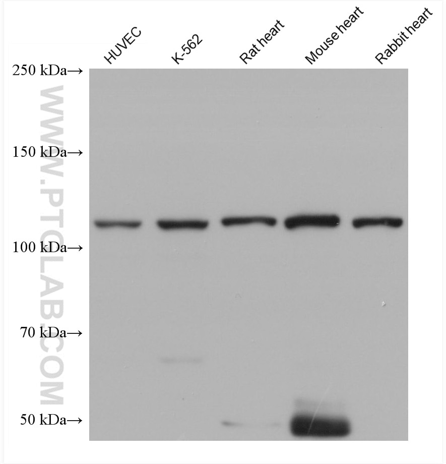
| Positive WB detected in | HGC-27 cells, HUVEC cells, LNCaP cells, MKN-45 cells, K-562 cells, human peripheral blood platelets, rat heart tissue, mouse heart tissue, rabbit heart tissue, HeLa cells |
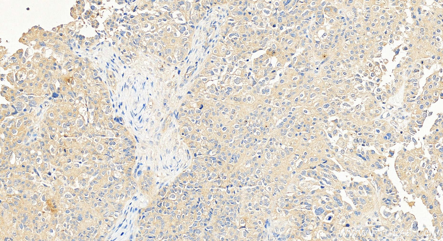
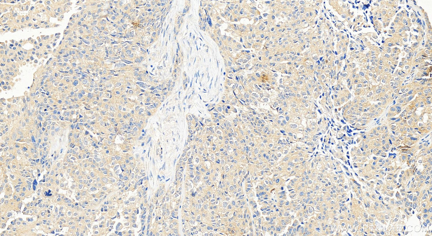
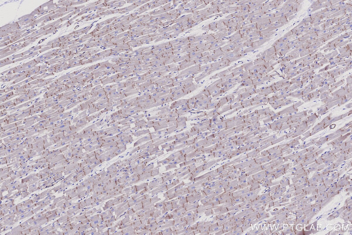
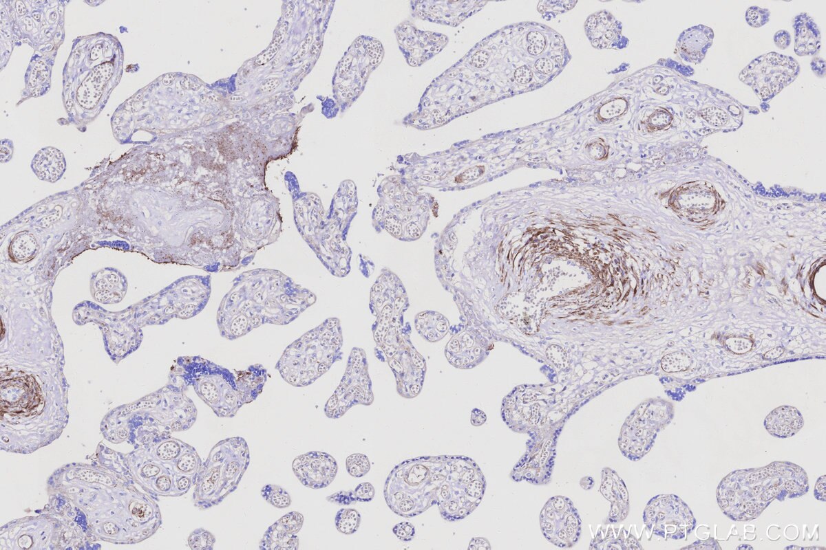
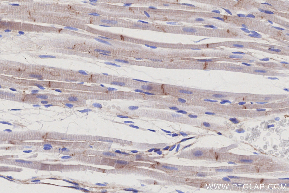
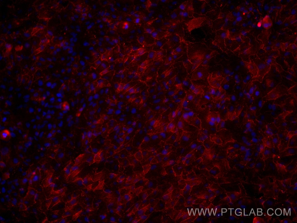
| Positive IHC detected in | human ovary cancer tissue Note: suggested antigen retrieval with TE buffer pH 9.0; (*) Alternatively, antigen retrieval may be performed with citrate buffer pH 6.0 |
| Positive IF/ICC detected in | hTERT-RPE1 cells |
Recommended dilution
| Application | Dilution |
|---|---|
| Western Blot (WB) | WB : 1:5000-1:50000 |
| Immunohistochemistry (IHC) | IHC : 1:1000-1:4000 |
| Immunofluorescence (IF)/ICC | IF/ICC : 1:1000-1:4000 |
| It is recommended that this reagent should be titrated in each testing system to obtain optimal results. | |
| Sample-dependent, Check data in validation data gallery. | |
Published Applications
| WB | See 12 publications below |
Product Information
66305-2-Ig targets Vinculin in WB, IHC, IF/ICC, ELISA applications and shows reactivity with human, mouse, rat, rabbit samples.
| Tested Reactivity | human, mouse, rat, rabbit |
| Cited Reactivity | human, mouse, rat |
| Host / Isotype | Mouse / IgG1 |
| Class | Monoclonal |
| Type | Antibody |
| Immunogen |
CatNo: Ag24946 Product name: Recombinant human Vinculin protein Source: e coli.-derived, PET30a Tag: 6*His Domain: 777-1066 aa of BC039174 Sequence: PKFREAVKAASDELSKTISPMVMDAKAVAGNISDPGLQKSFLDSGYRILGAVAKVREAFQPQEPDFPPPPPDLEQLRLTDELAPPKPPLPEGEVPPPRPPPPEEKDEEFPEQKAGEVINQPMMMAARQLHDEARKWSSKGNDIIAAAKRMALLMAEMSRLVRGGSGTKRALIQCAKDIAKASDEVTRLAKEVAKQCTDKRIRTNLLQVCERIPTISTQLKILSTVKATMLGRTNISDEESEQATEMLVHNAQNLMQSVKETVREAEAASIKIRTDAGFTLRWVRKTPWYQ Predict reactive species |
| Full Name | vinculin |
| Calculated Molecular Weight | 1133 aa, 124 kDa |
| Observed Molecular Weight | 124 kDa |
| GenBank Accession Number | BC039174 |
| Gene Symbol | Vinculin |
| Gene ID (NCBI) | 7414 |
| RRID | AB_3670375 |
| Conjugate | Unconjugated |
| Form | Liquid |
| Purification Method | Protein G purification |
| UNIPROT ID | P18206 |
| Storage Buffer | PBS with 0.02% sodium azide and 50% glycerol, pH 7.3. |
| Storage Conditions | Store at -20°C. Stable for one year after shipment. Aliquoting is unnecessary for -20oC storage. 20ul sizes contain 0.1% BSA. |
Background Information
Vinculin belongs to the vinculin/alpha-catenin family. It is an actin filament (F-actin)-binding protein which involved in cell-matrix adhesion and cell-cell adhesion. Vinculin regulates cell-surface E-cadherin expression and potentiates mechanosensing by the E-cadherin complex. It may also play important roles in cell morphology and locomotion. Vinculin is a 117-kDa, 1,066-amino-acid protein that is ubiquitously expressed. Its splice variant, metavinculin (124 kDa), is muscle-specific.
Protocols
| Product Specific Protocols | |
|---|---|
| IF protocol for Vinculin antibody 66305-2-Ig | Download protocol |
| IHC protocol for Vinculin antibody 66305-2-Ig | Download protocol |
| WB protocol for Vinculin antibody 66305-2-Ig | Download protocol |
| Standard Protocols | |
|---|---|
| Click here to view our Standard Protocols |
Publications
| Species | Application | Title |
|---|---|---|
Adv Sci (Weinh) RFWD3 Reprograms Nucleotide Metabolism Through PHGDH to Induce Chemoresistance In Osteosarcoma | ||
Front Med (Lausanne) ACADL and ADH1B signify ketone body metabolic reprogramming in osteoarthritic synovium: insights from bioinformatics and animal model studies. | ||
J Clin Immunol Novel Compound Heterozygous Mutations in HOIP Result in Autoinflammation and Immunodeficiency. | ||
Phytomedicine Unraveling ancient wisdom: The molecular mechanism of Gujin Luyan Xuming decoction in combating cerebral ischemia-reperfusion injury | ||
Transl Psychiatry Knockdown of Ddx3x in mPFC induces autistic-like phenotype in mice via altered synaptic plasticity. | ||
Mech Ageing Dev Diminished CEACAM1 level plays a critical role in age-related hepatic fibrosis. |
Reviews
The reviews below have been submitted by verified Proteintech customers who received an incentive for providing their feedback.
FH Matthieu (Verified Customer) (09-24-2025) | Band is very clear at the expected size
|
FH Manon (Verified Customer) (09-24-2025) | Good housekeeping antibody
|
FH Emilie (Verified Customer) (07-23-2025) | Used for Western blot on human fibroblasts and gives a clear, well-defined band. I recommend this product.
|