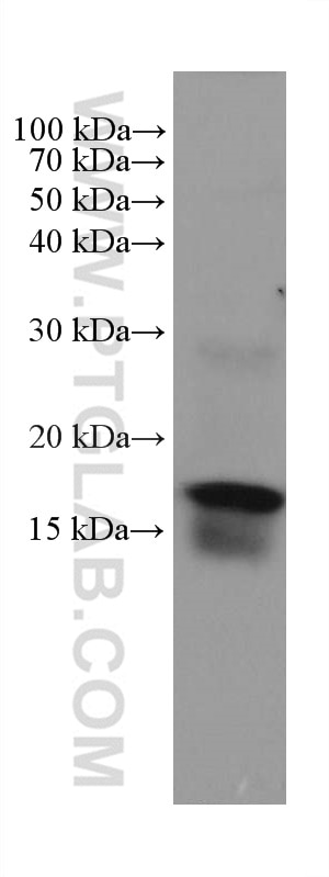
mouse colon tissue were subjected to SDS PAGE followed by western blot with 67389-1-Ig (ZG16 antibody) at dilution of 1:5000 incubated at room temperature for 1.5 hours.
mouse colon tissue were subjected to SDS PAGE followed by western blot with 67389-1-Ig (ZG16 antibody) at dilution of 1:5000 incubated at room temperature for 1.5 hours.
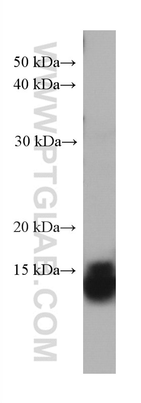
WB analysis of human colon using 67389-1-Ig
human colon tissue were subjected to SDS PAGE followed by western blot with 67389-1-Ig (ZG16 antibody) at dilution of 1:3000 incubated at room temperature for 1.5 hours.
human colon tissue were subjected to SDS PAGE followed by western blot with 67389-1-Ig (ZG16 antibody) at dilution of 1:3000 incubated at room temperature for 1.5 hours.
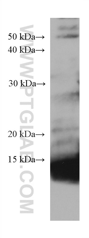
WB analysis of pig colon using 67389-1-Ig
pig colon tissue were subjected to SDS PAGE followed by western blot with 67389-1-Ig (ZG16 antibody) at dilution of 1:3000 incubated at room temperature for 1.5 hours.
pig colon tissue were subjected to SDS PAGE followed by western blot with 67389-1-Ig (ZG16 antibody) at dilution of 1:3000 incubated at room temperature for 1.5 hours.
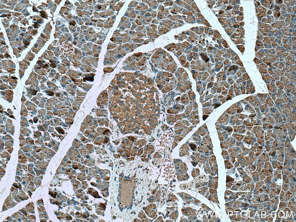
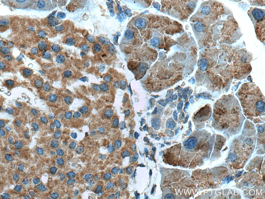
IHC staining of mouse pancreas using 67389-1-Ig
Immunohistochemical analysis of paraffin-embedded mouse pancreas tissue slide using 67389-1-Ig (ZG16 antibody) at dilution of 1:500 (under 10x lens). Heat mediated antigen retrieval with Tris-EDTA buffer (pH 9.0).
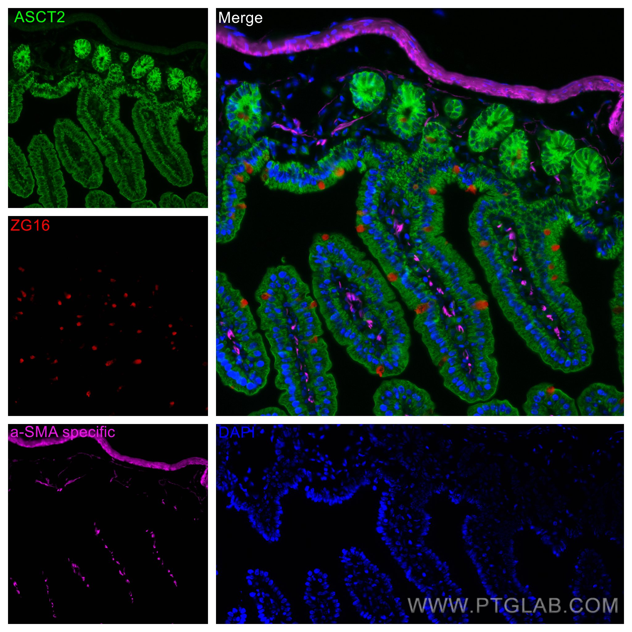
Immunofluorescent analysis of (4% PFA) fixed mouse small intestine tissue using ZG16 antibody (67389-1-Ig, Clone: 1A7B9 ) at dilution of 1:400 and CoraLite®594-Conjugated AffiniPure Goat Anti-Mouse IgG(H+L) (SA00013-3), ASCT2 antibody (20350-1-AP, green), CoraLite® Plus 647 smooth muscle actin specific antibody (CL647-80008, Clone: 5H7, Magenta).
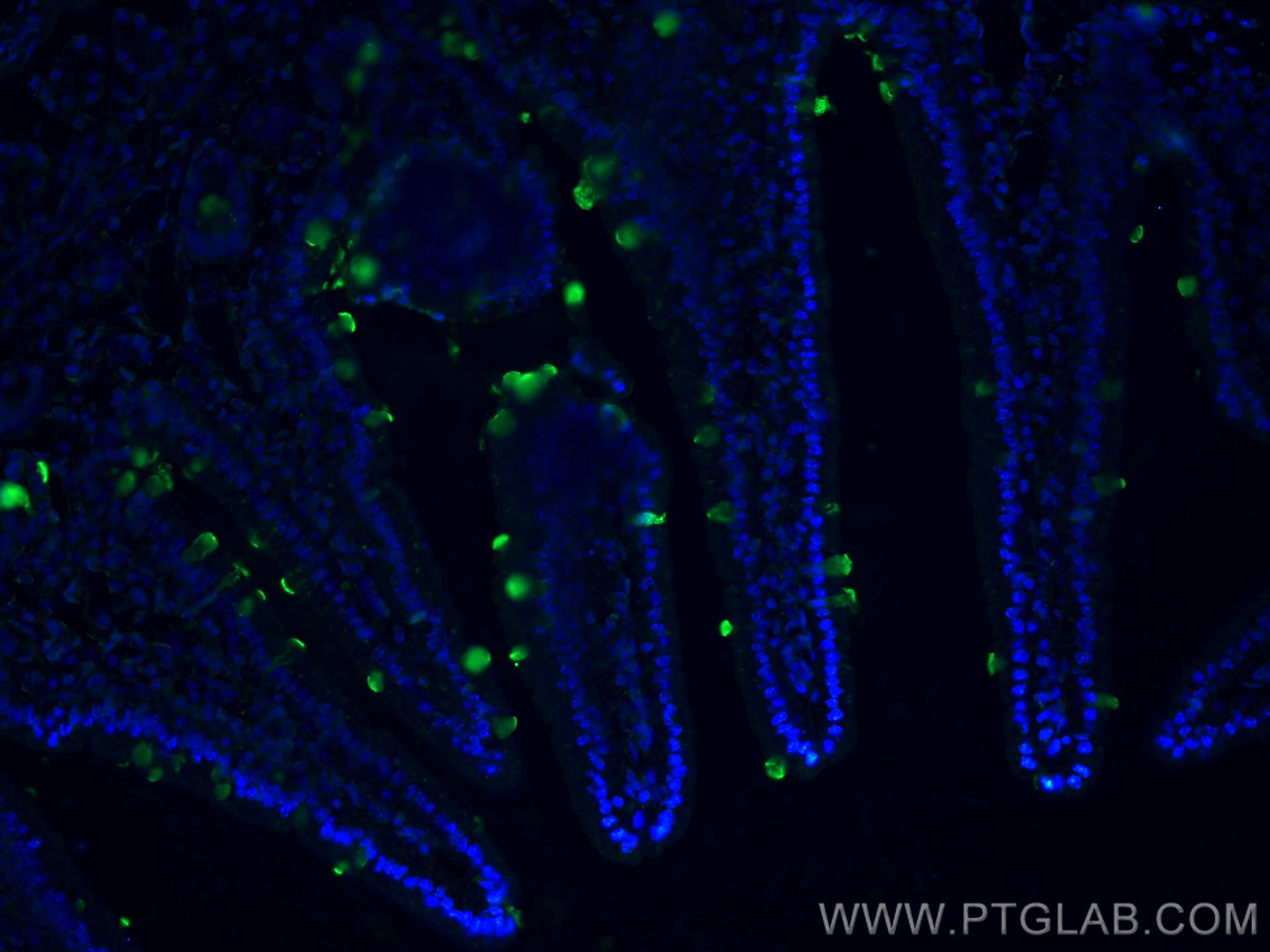
IF Staining of mouse small intestine using 67389-1-Ig
Immunofluorescent analysis of (4% PFA) fixed mouse small intestine tissue using ZG16 antibody (67389-1-Ig, Clone: 1A7B9 ) at dilution of 1:400 and CoraLite®488-Conjugated AffiniPure Goat Anti-Mouse IgG(H+L).
Immunofluorescent analysis of (4% PFA) fixed mouse small intestine tissue using ZG16 antibody (67389-1-Ig, Clone: 1A7B9 ) at dilution of 1:400 and CoraLite®488-Conjugated AffiniPure Goat Anti-Mouse IgG(H+L).
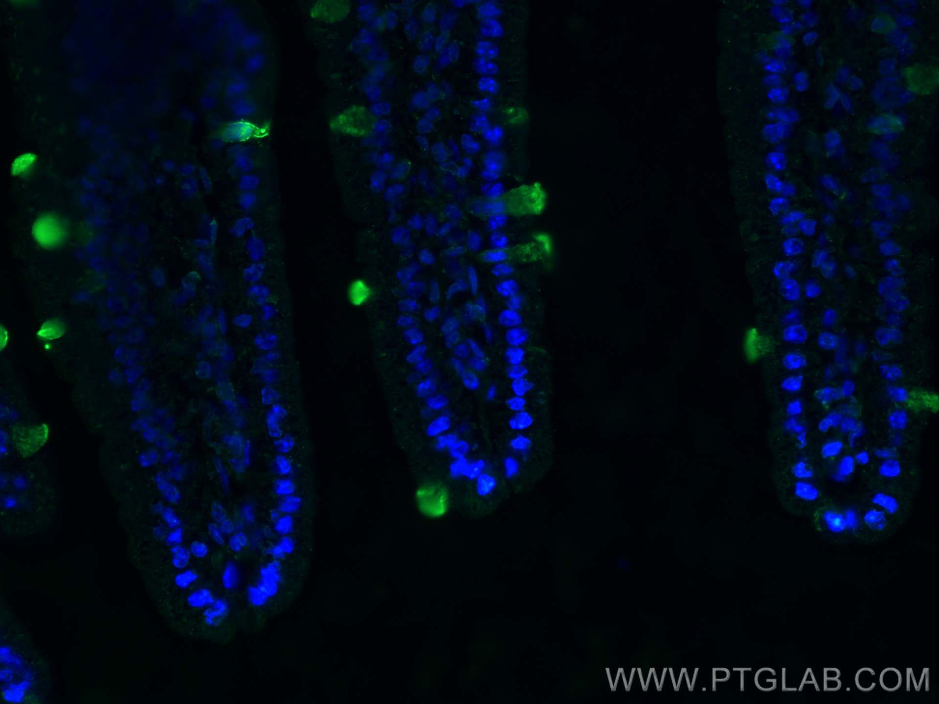
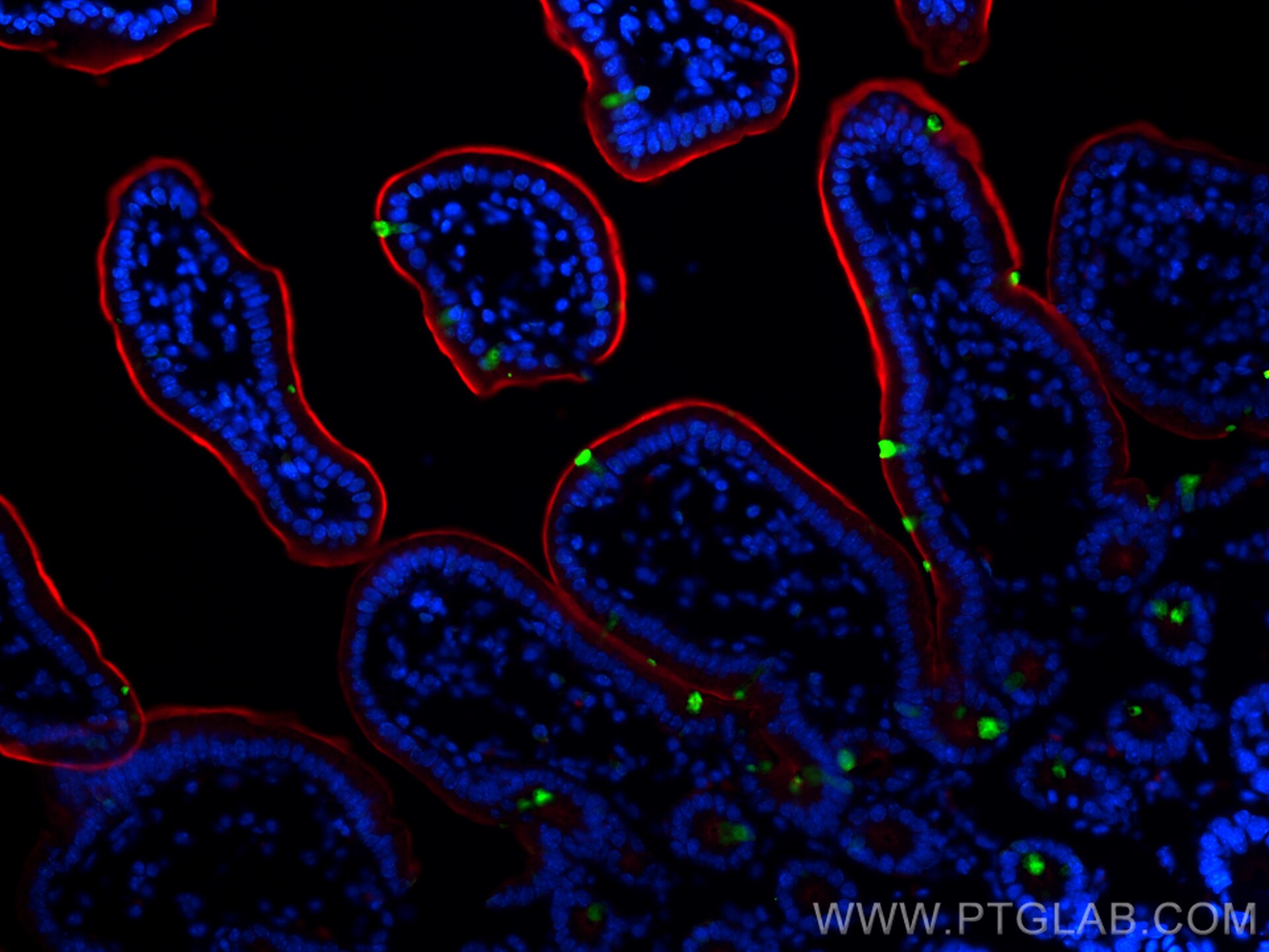
IF Staining of mouse small intestine using 67389-1-Ig
Immunofluorescent analysis of (4% PFA) fixed frozen OCT-embedded mouse small intestine tissue using ZG16 antibody (67389-1-Ig, Clone: 1A7B9 ) at dilution of 1:400 and CoraLite®488-Conjugated Goat Anti-Mouse IgG(H+L) (SA00013-1), CD163 antibody (83285-4-RR, Clone: 240233A3, red).
The Proteintech guarantee covers Proteintech antibodies in any species and any application, including those not listed on the datasheet. If the antibody doesn’t perform, you can receive a hassle-free refund or credit note.
mouse colon tissue, human colon tissue, pig colon tissue
Positive IHC detected in
mouse pancreas tissue Note: suggested antigen retrieval with TE buffer pH 9.0; (*) Alternatively, antigen retrieval may be performed with citrate buffer pH 6.0
Positive IF-P detected in
mouse small intestine tissue
Positive IF-Fro detected in
mouse small intestine tissue
Recommended dilution
Application
Dilution
Western Blot (WB)
WB : 1:2000-1:10000
Immunohistochemistry (IHC)
IHC : 1:250-1:1000
Immunofluorescence (IF)-P
IF-P : 1:200-1:800
Immunofluorescence (IF)-FRO
IF-FRO : 1:200-1:800
It is recommended that this reagent should be titrated in each testing system to obtain optimal results.
Sample-dependent, Check data in validation data gallery.
PBS with 0.02% sodium azide and 50% glycerol, pH 7.3.
Storage Conditions
Store at -20°C. Stable for one year after shipment. Aliquoting is unnecessary for -20oC storage. 20ul sizes contain 0.1% BSA.
Background Information
Zymogen granule protein 16 (ZG16) has a Jacalin-like lectin domain, which is mainly expressed by mucus-secreting cells, including goblet cells in the intestine. It's reported that ZG16 expression was significantly decreased in colorectal cancer compared to normal tissue. ZG16 gene expression and copy number variations (CNV) were associated with multiple molecular and clinicopathological features of CRC including MSI, MLH1 silencing and so on. It's found that ZG16 is negatively correlated with lymphatic invasive and distant metastasis. Besides, overexpression of ZG16 blocks PD-L1 expression in colorectal cancer, and promotes NK cells survival and proliferation. Very importantly, ZG16 suppresses colorectal tumor growth via the immune system. (PMID: 33360840, 21893569)
mouse colon tissue were subjected to SDS PAGE followed by western blot with 67389-1-Ig (ZG16 antibody) at dilution of 1:5000 incubated at room temperature for 1.5 hours.
WB analysis of human colon using 67389-1-Ig
human colon tissue were subjected to SDS PAGE followed by western blot with 67389-1-Ig (ZG16 antibody) at dilution of 1:3000 incubated at room temperature for 1.5 hours.
WB analysis of pig colon using 67389-1-Ig
pig colon tissue were subjected to SDS PAGE followed by western blot with 67389-1-Ig (ZG16 antibody) at dilution of 1:3000 incubated at room temperature for 1.5 hours.
IHC Figures
IHC staining of mouse pancreas using 67389-1-Ig
Immunohistochemical analysis of paraffin-embedded mouse pancreas tissue slide using 67389-1-Ig (ZG16 antibody) at dilution of 1:500 (under 10x lens). Heat mediated antigen retrieval with Tris-EDTA buffer (pH 9.0).
IHC staining of mouse pancreas using 67389-1-Ig
Immunohistochemical analysis of paraffin-embedded mouse pancreas tissue slide using 67389-1-Ig (ZG16 antibody) at dilution of 1:500 (under 40x lens). Heat mediated antigen retrieval with Tris-EDTA buffer (pH 9.0).
IF-P Figures
IF Staining of mouse small intestine using 67389-1-Ig
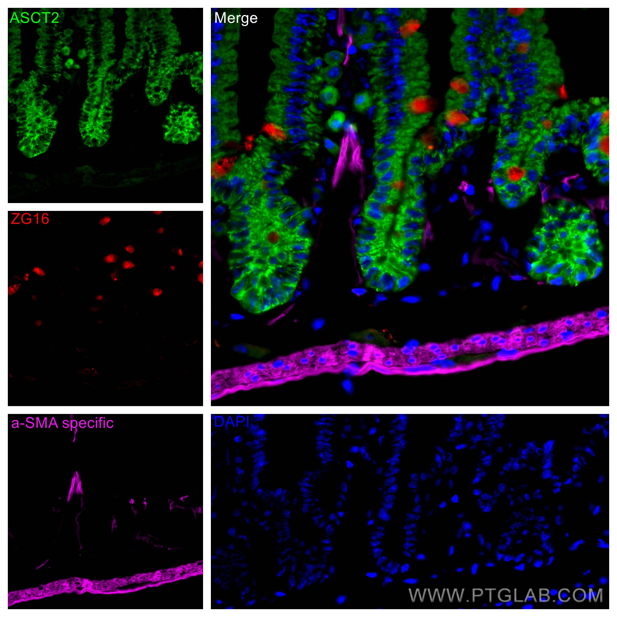
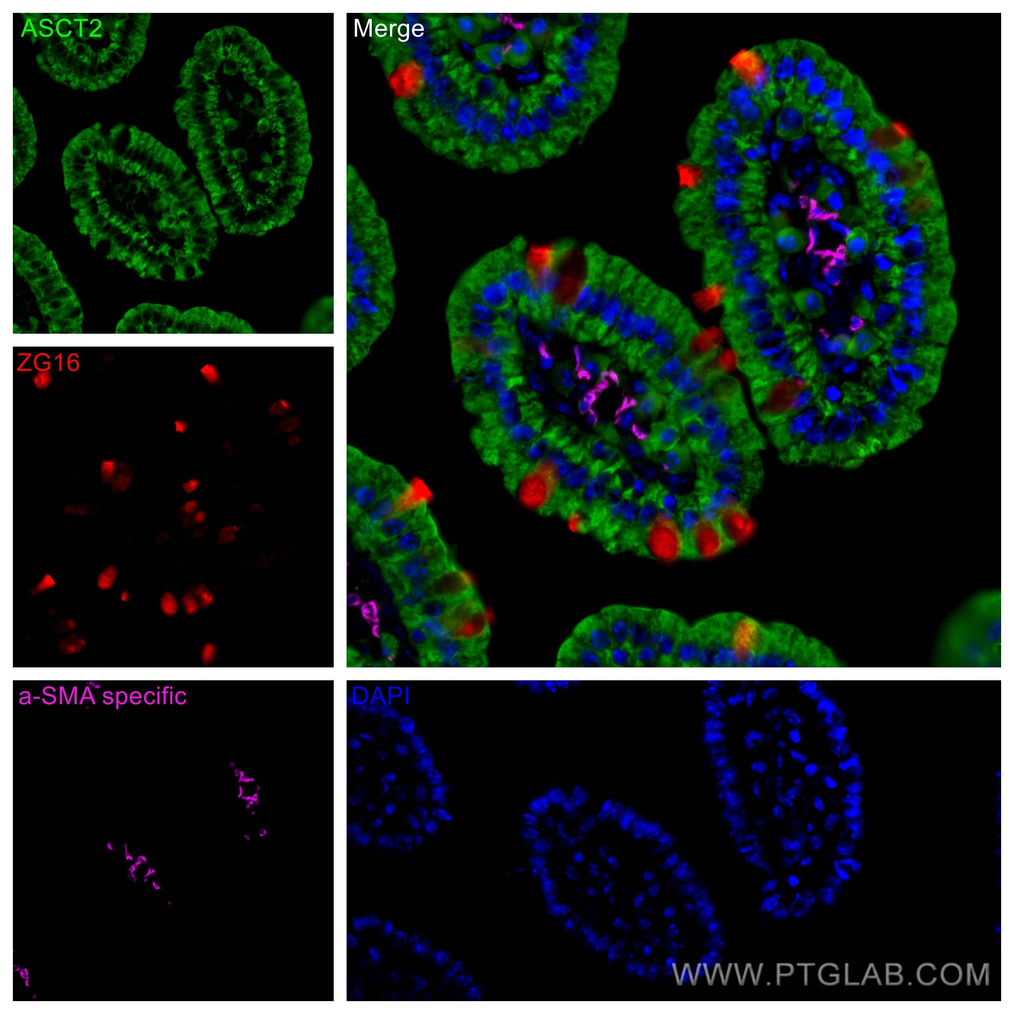
Immunofluorescent analysis of (4% PFA) fixed mouse small intestine tissue using ZG16 antibody (67389-1-Ig, Clone: 1A7B9 ) at dilution of 1:400 and CoraLite®594-Conjugated AffiniPure Goat Anti-Mouse IgG(H+L) (SA00013-3), ASCT2 antibody (20350-1-AP, green), CoraLite® Plus 647 smooth muscle actin specific antibody (CL647-80008, Clone: 5H7, Magenta).
IF Staining of mouse small intestine using 67389-1-Ig
Immunofluorescent analysis of (4% PFA) fixed mouse small intestine tissue using ZG16 antibody (67389-1-Ig, Clone: 1A7B9 ) at dilution of 1:400 and CoraLite®488-Conjugated AffiniPure Goat Anti-Mouse IgG(H+L).
IF Staining of mouse small intestine using 67389-1-Ig
Immunofluorescent analysis of (4% PFA) fixed mouse small intestine tissue using ZG16 antibody (67389-1-Ig, Clone: 1A7B9 ) at dilution of 1:400 and CoraLite®488-Conjugated AffiniPure Goat Anti-Mouse IgG(H+L).
IF-FRO Figures
IF Staining of mouse small intestine using 67389-1-Ig
Immunofluorescent analysis of (4% PFA) fixed frozen OCT-embedded mouse small intestine tissue using ZG16 antibody (67389-1-Ig, Clone: 1A7B9 ) at dilution of 1:400 and CoraLite®488-Conjugated Goat Anti-Mouse IgG(H+L) (SA00013-1), CD163 antibody (83285-4-RR, Clone: 240233A3, red).
The species listed in Tested Reactivity are in-house verified and applicable species. For unlisted species, please refer to the homology analysis of the immunogen sequence and related species. For rabbit polyclonal antibodies, homology >70% is recommended. For mouse monoclonal antibodies and rabbit recombinant antibodies, homology >90% is recommended. Generally, the higher the homology, the greater the applicability. However, there will be certain differences in protein expression in different species, tissues or cells. Therefore, the homology analysis results are for reference only and do not serve as a guarantee.
At Proteintech, we pride ourselves on our antibody quality, customer service and transparency. As such, we are comparing our antibodies with other vendors, enabling easy identification and comparisons of key data to help you choose the suitable antibody for your needs.
We have selected the top cited antibodies from these vendors for you to compare.
 at dilution of 1:5000 incubated at room temperature for 1.5 hours.")
 at dilution of 1:3000 incubated at room temperature for 1.5 hours.")
 at dilution of 1:3000 incubated at room temperature for 1.5 hours.")
 at dilution of 1:500 (under 10x lens). Heat mediated antigen retrieval with Tris-EDTA buffer (pH 9.0).")
 at dilution of 1:500 (under 40x lens). Heat mediated antigen retrieval with Tris-EDTA buffer (pH 9.0).")
 fixed mouse small intestine tissue using ZG16 antibody (67389-1-Ig, Clone: 1A7B9 ) at dilution of 1:400 and CoraLite®594-Conjugated AffiniPure Goat Anti-Mouse IgG(H+L) (SA00013-3), ASCT2 antibody (20350-1-AP, green), CoraLite® Plus 647 smooth muscle actin specific antibody (CL647-80008, Clone: 5H7, Magenta).")
 fixed mouse small intestine tissue using ZG16 antibody (67389-1-Ig, Clone: 1A7B9 ) at dilution of 1:400 and CoraLite®594-Conjugated AffiniPure Goat Anti-Mouse IgG(H+L) (SA00013-3), ASCT2 antibody (20350-1-AP, green), CoraLite® Plus 647 smooth muscle actin specific antibody (CL647-80008, Clone: 5H7, Magenta).")
 fixed mouse small intestine tissue using ZG16 antibody (67389-1-Ig, Clone: 1A7B9 ) at dilution of 1:400 and CoraLite®594-Conjugated AffiniPure Goat Anti-Mouse IgG(H+L) (SA00013-3), ASCT2 antibody (20350-1-AP, green), CoraLite® Plus 647 smooth muscle actin specific antibody (CL647-80008, Clone: 5H7, Magenta).")
 fixed mouse small intestine tissue using ZG16 antibody (67389-1-Ig, Clone: 1A7B9 ) at dilution of 1:400 and CoraLite®488-Conjugated AffiniPure Goat Anti-Mouse IgG(H+L).")
 fixed mouse small intestine tissue using ZG16 antibody (67389-1-Ig, Clone: 1A7B9 ) at dilution of 1:400 and CoraLite®488-Conjugated AffiniPure Goat Anti-Mouse IgG(H+L).")
 fixed frozen OCT-embedded mouse small intestine tissue using ZG16 antibody (67389-1-Ig, Clone: 1A7B9 ) at dilution of 1:400 and CoraLite®488-Conjugated Goat Anti-Mouse IgG(H+L) (SA00013-1), CD163 antibody (83285-4-RR, Clone: 240233A3, red).")