Tested Applications
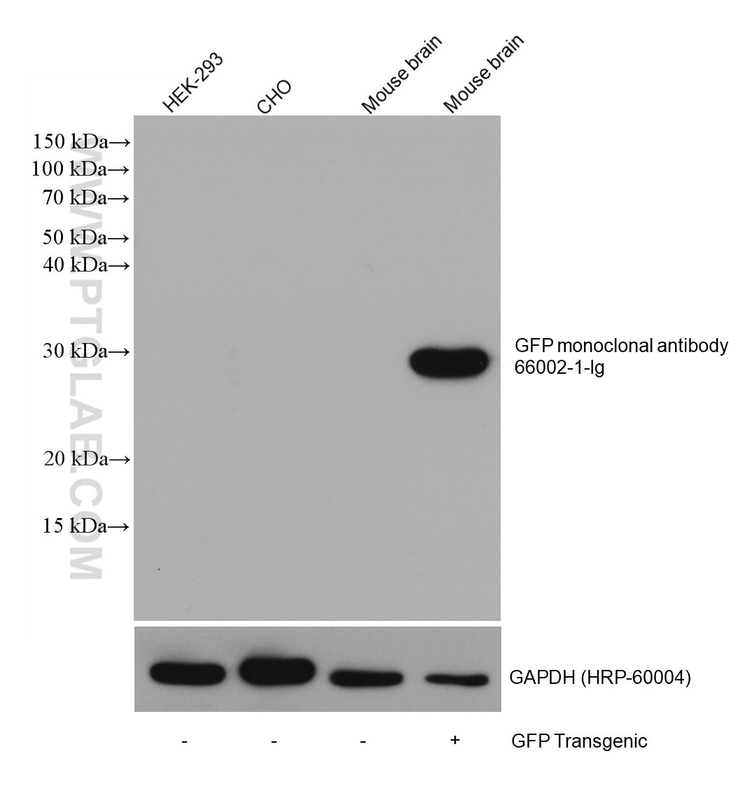
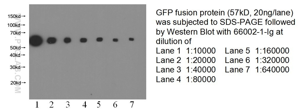
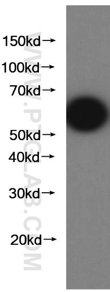
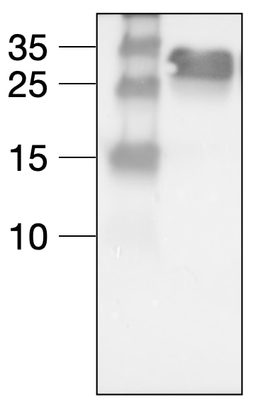
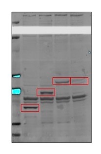
| Positive WB detected in | GFP transgenic mouse brain tissue, Recombinant protein |
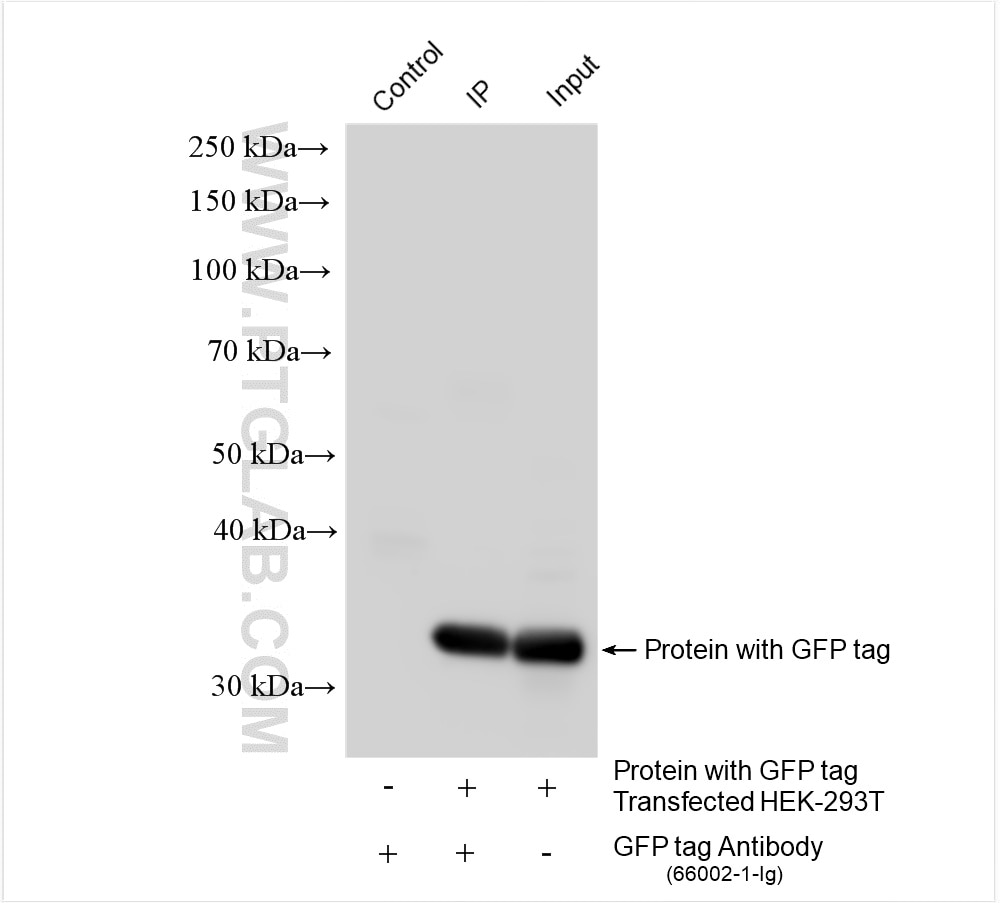
| Positive IP detected in | Transfected HEK-293T cells |
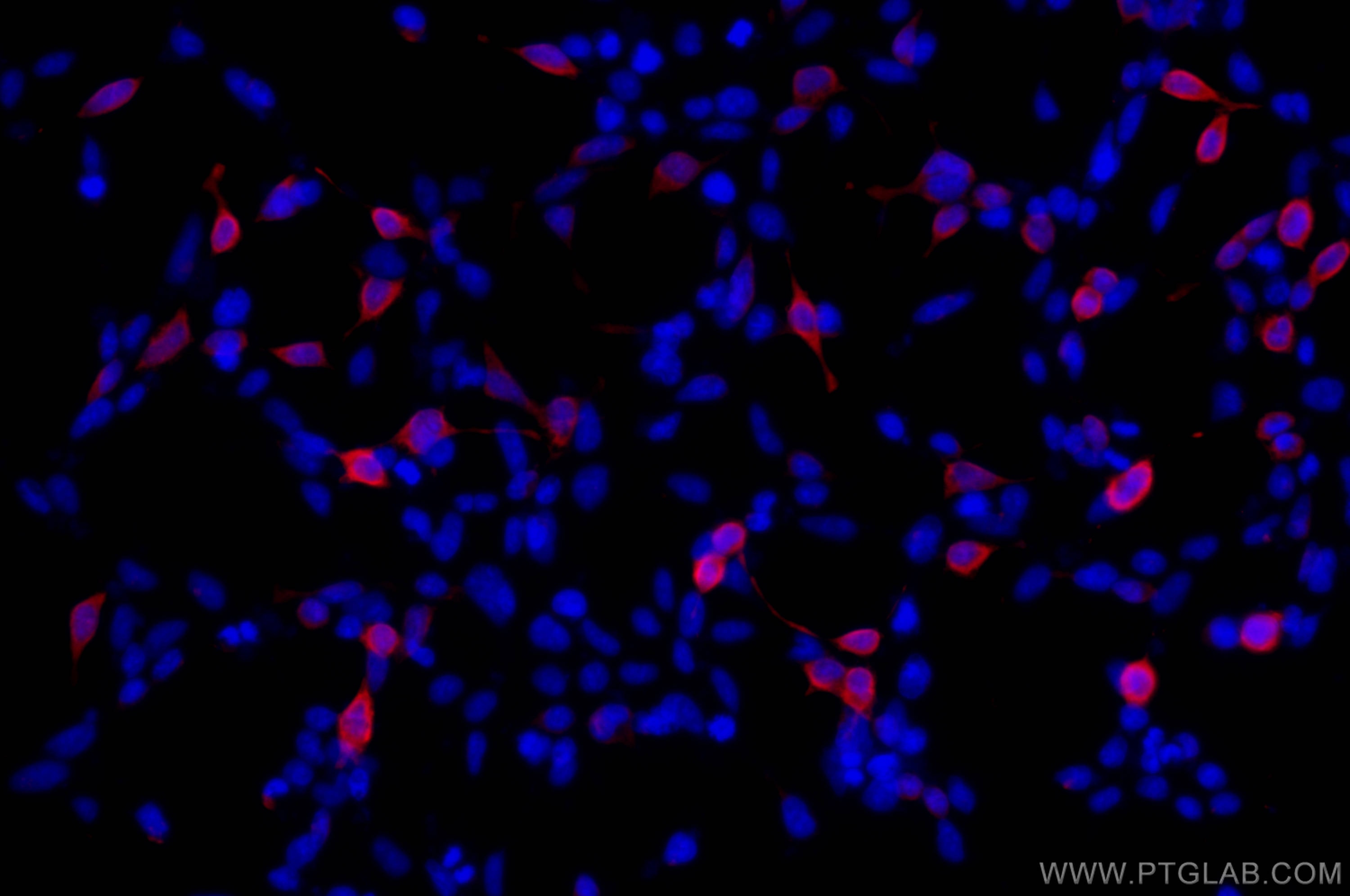
| Positive IF/ICC detected in | Transfected HEK-293 cells |
Recommended dilution
| Application | Dilution |
|---|---|
| Western Blot (WB) | WB : 1:20000-1:100000 |
| Immunoprecipitation (IP) | IP : 0.5-4.0 ug for 1.0-3.0 mg of total protein lysate |
| Immunofluorescence (IF)/ICC | IF/ICC : 1:400-1:1600 |
| It is recommended that this reagent should be titrated in each testing system to obtain optimal results. | |
| Sample-dependent, Check data in validation data gallery. | |
Product Information
66002-1-Ig targets GFP tag in WB, IHC, IF/ICC, IP, CoIP, ChIP, RIP, ELISA applications and shows reactivity with recombinant protein samples.
| Tested Reactivity | recombinant protein |
| Cited Reactivity | human, mouse, rat, pig, silkworm |
| Host / Isotype | Mouse / IgG2a |
| Class | Monoclonal |
| Type | Antibody |
| Immunogen |
CatNo: Ag2128 Product name: Recombinant aequorea victoria GFP tag protein Source: e coli.-derived, PGEX-4T Tag: GST Domain: 1-238 aa of M62653 Sequence: MSKGEELFTGVVPILVELDGDVNGHKFSVSGEGEGDATYGKLTLKFICTTGKLPVPWPTLVTTFSYGVQCFSRYPDHMKQHDFFKSAMPEGYVQERTIFFKDDGNYKTRAEVKFEGDTLVNRIELKGIDFKEDGNILGHKLEYNYNSHNVYIMADKQKNGIKVNFKIRHNIEDGSVQLADHYQQNTPIGDGPVLLPDNHYLSTQSALSKDPNEKRDHMVLLEFVTAAGITHGMDELYK Predict reactive species |
| Full Name | GFP tag |
| Calculated Molecular Weight | 26 kDa |
| GenBank Accession Number | M62653 |
| Gene Symbol | |
| Gene ID (NCBI) | |
| RRID | AB_11182611 |
| Conjugate | Unconjugated |
| Form | Liquid |
| Purification Method | Protein A purification |
| UNIPROT ID | P42212 |
| Storage Buffer | PBS with 0.02% sodium azide and 50% glycerol, pH 7.3. |
| Storage Conditions | Store at -20°C. Stable for one year after shipment. Aliquoting is unnecessary for -20oC storage. 20ul sizes contain 0.1% BSA. |
Background Information
Green Fluorescent Proteins (GFPs) encompass a diverse range of proteins carrying a green chromophore, originating from various species and forming different protein lineages.
Wildtype GFP consists of 238 amino acid residues (26.9 kDa). GFP was first identified in the jellyfish Aequorea victoria. It emits green light with a peak wavelength of 509 nm upon excitation by blue light at 395 nm.
When fused with other proteins, GFP serves as a versatile reporter protein e.g. for quantifying expression levels or facilitates visualization of subcellular localization through fluorescence microscopy.
This antibody is a mouse (IgG2a) monoclonal antibody against GFP reactive to GFP, eGFP, eYFP, YFP and CFP.
Protocols
| Product Specific Protocols | |
|---|---|
| IF protocol for GFP tag antibody 66002-1-Ig | Download protocol |
| IP protocol for GFP tag antibody 66002-1-Ig | Download protocol |
| WB protocol for GFP tag antibody 66002-1-Ig | Download protocol |
| Standard Protocols | |
|---|---|
| Click here to view our Standard Protocols |
Publications
| Species | Application | Title |
|---|---|---|
Cell Discov Glc7/PP1 dephosphorylates histone H3T11 to regulate autophagy and telomere silencing in response to nutrient availability | ||
Cell Host Microbe Microbiota-derived urocanic acid triggered bytyrosine kinase inhibitors potentiates cancer immunotherapy efficacy | ||
Cell Metab Protein O-GlcNAcylation and hexokinase mitochondrial dissociation drive heart failure with preserved ejection fraction | ||
Nat Genet Pathogenic SPTBN1 variants cause an autosomal dominant neurodevelopmental syndrome. |
Reviews
The reviews below have been submitted by verified Proteintech customers who received an incentive for providing their feedback.
FH Danyan (Verified Customer) (04-08-2026) | It's specific and sensitive, in both wester blot and immunofluorescence applications. Having relied on this particular antibody in our lab for many years, we’ve found its performance to be exceptionally stable and reproducible across countless experiments.
|
FH Danyan (Verified Customer) (03-19-2026) | The GFP tag mouse monoclonal antibody performs exceptionally well in our hands. It produces a clear, sharp hand at the expected molecular weight in western blot with very low nonspecific binding.
|
FH Danyan (Verified Customer) (02-27-2026) | we are very satisfied with this GFP tag mouse monoclonal antibody. The signal is strong and specific. It works reliable in western blot and immunofluorescence applications. Highly recommended!
 |
FH Danyan (Verified Customer) (02-09-2026) | the GFP tag monoclonal antibody performed well in our experiments with clear and consistent signal. the product quality is reliable and shipping was timely.
|
FH Prakash (Verified Customer) (10-28-2025) | It works well in western blot.
|
FH Javier (Verified Customer) (09-09-2025) | Perfect antibody for Wb and IP assays.
|
FH Ioana (Verified Customer) (08-27-2025) | This antibody works well in Western Blotting and amplifying signal after PFA fixation during IF.
|
FH Anastasia (Verified Customer) (08-07-2024) | Zebrafish embryos expressing cytosolic GFP
 |
FH Amy (Verified Customer) (02-11-2023) | Detected EGFP-tagged constructs expressed in HEK293T cells at similar strength to other antibodies however with slightly more background.
 |
FH Tom (Verified Customer) (02-03-2023) | Works great. No problem. Love proteintech.
|
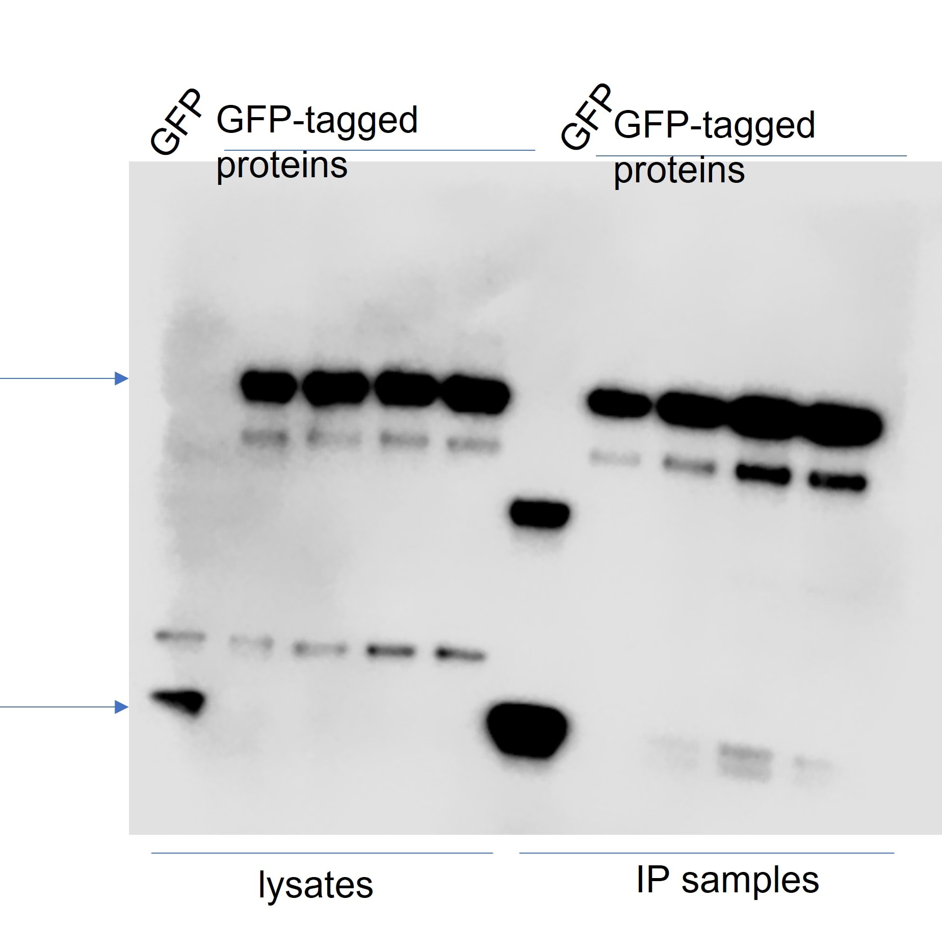
FH Tatyana (Verified Customer) (01-09-2023) | Good signal, antibody can be reused. Overexpressed GFP-tagged proteins (and GFP alone) were pulled down using GFP-Trap beads and WB was done on lysates and IP samples. Antibody was diluted in 4% milk (and could be reused several times).
 |
FH Sheila (Verified Customer) (07-22-2022) | It works very well
|
FH Juliana (Verified Customer) (01-28-2022) | works great for Western blot!
|
FH Prasanna (Verified Customer) (01-04-2021) | Of the 3 antibodies tested for GFP this one gave the cleanest and strongest signal by western blot. When I saw it on sale, I bought 8!
|
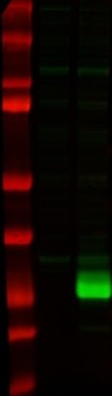
FH Lana (Verified Customer) (12-22-2020) | SDS-PAGE: 15 ug/ul RIPA protein lysate, 4-12% Bis-Tris gradient gel.Transfer: Immobilon-FL transfer membranes (Millipore) for 2h at 80V, 4C.Blocking: SEA Block Blocking Buffer 1h, room T.Primary Ab: O/N incubation at 4C, 1:5000.Secondary Ab: IRDye 800CW Goat anti-Mouse, 1:15000.Lines of WB image: 1 – protein ladder, 2 – HEK293 whole cell lysate, negative transfection, 3 – whole cell lysate of cells transfected with eGFP.
 |
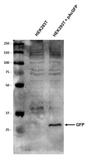
FH Thomas (Verified Customer) (11-19-2020) | HEK293T and HEK293T stably transfected with pAcGFP plasmid. 10ug total protein loaded per well. Membrane blocked 1 hour in 5% BSA prior to anti-GFP (1:2000) o/n at 4 degrees. Goat anti-mouse HRP secondary (1:10,000) used.
 |
FH Jane (Verified Customer) (03-02-2020) | Adenovirus-GFP infected cardiomyocytes stained with GFP antibody, signal is strong and effective
|
FH LUNFENG (Verified Customer) (01-27-2020) | GOOD
|
FH Jie (Verified Customer) (01-27-2020) | Worked for western blot with GFP-LC3 transfected cardiomyocytes
|
FH Paul (Verified Customer) (01-15-2020) | Works well for Westerns.
|
FH Laura (Verified Customer) (01-15-2020) | Good antibody for Western Blot.
|
FH Aamir (Verified Customer) (01-08-2020) | Worked well for WB
|
FH Benjamin (Verified Customer) (01-07-2020) | Easily detects recombinant GFP protein via western blot with very little background.
|
FH Jing (Verified Customer) (01-03-2020) | Used this to detect tranduce efficiency of the AAV-GFP virus. with 5% non-fat milk, there are two bands around 20-30kd, not sure which one is correct. And the antibody is relative weak, has to use 1:500 dilution.
|
FH Shan (Verified Customer) (12-25-2019) | The GFP antibody showed great sensitivity for WB and it was easily detecable. But it was insteresting that when the SDS PAGE gel separating gel concentration reeached to 12%, you can see two bands at ~30kDa and ~20kDa.
 |
FH Jason (Verified Customer) (11-04-2019) | This is a good antibody for detecting GFP tag on Western blot, using 1:1000 dilution. The price is unbeatable, worth each penny.
|
FH Hend (Verified Customer) (10-14-2019) | antibody used in western blot against egfp labelled protien and clear band was detected.
|