
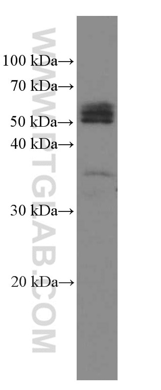

Tested Applications
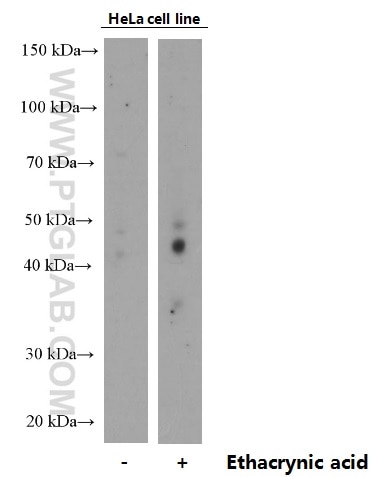
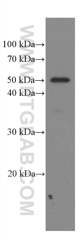
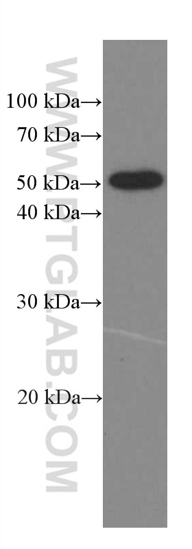
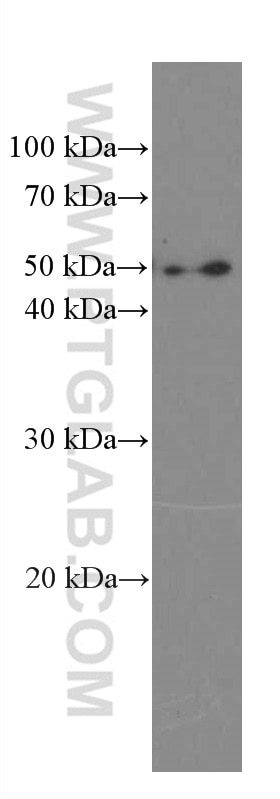
| Positive WB detected in | EA treated HeLa cells, HEK-293 cells, human placenta tissue, Neuro-2a cells, C6 cells |
Recommended dilution
| Application | Dilution |
|---|---|
| Western Blot (WB) | WB : 1:500-1:2000 |
| It is recommended that this reagent should be titrated in each testing system to obtain optimal results. | |
| Sample-dependent, Check data in validation data gallery. | |
Product Information
66318-1-Ig targets Phospho-TDP43 (Ser409/410) in WB, IHC, IF, ELISA applications and shows reactivity with human, mouse, rat samples.
| Tested Reactivity | human, mouse, rat |
| Cited Reactivity | human, mouse, rat, canine |
| Host / Isotype | Mouse / IgG1 |
| Class | Monoclonal |
| Type | Antibody |
| Immunogen |
Peptide Predict reactive species |
| Full Name | TAR DNA binding protein |
| Calculated Molecular Weight | 43 kDa |
| Observed Molecular Weight | 45-50 kDa, 35 kDa |
| GenBank Accession Number | NM_007375 |
| Gene Symbol | TDP-43 |
| Gene ID (NCBI) | 23435 |
| RRID | AB_2881699 |
| Conjugate | Unconjugated |
| Form | Liquid |
| Purification Method | Protein G purification |
| UNIPROT ID | Q13148 |
| Storage Buffer | PBS with 0.02% sodium azide and 50% glycerol, pH 7.3. |
| Storage Conditions | Store at -20°C. Stable for one year after shipment. Aliquoting is unnecessary for -20oC storage. 20ul sizes contain 0.1% BSA. |
Background Information
The TARDBP gene encodes the TDP-43 protein, initially found to repress HIV-1 transcription by binding TAR DNA. TDP-43 has since been shown to bind RNA as well as DNA, and have multiple functions in transcriptional repression, translational regulation and pre-mRNA splicing. For instance, it is reported to regulate alternate splicing of the CTFR gene. In 2006 Neumann et al. found that hyperphosphorylated, ubiquitinated and/or cleaved forms of TDP-43, collectively known as pathological TDP-43, play a major role in the disease mechanisms of ubiquitin-positive, tau- and alpha-synuclein-negative frontotemporal dementia (FTLD-U) and in amyotrophic lateral sclerosis (ALS). Various forms of TDP-43 exist, including 18-35 kDa of cleaved C-terminal fragments, 45-50 kDa phospho-protein, 55 kDa glycosylated form, 75 kDa hyperphosphorylated form, and 90-300 kDa cross-linked form. (PMID: 17023659,19823856, 21666678, 22193176). 66318-1-Ig is a phospho-dependent monoclonal antibody specifically recognizing phospho-TDP43 (409/410). It does not react with native TDP-43.
Protocols
| Product Specific Protocols | |
|---|---|
| WB protocol for Phospho-TDP43 (Ser409/410) antibody 66318-1-Ig | Download protocol |
| Standard Protocols | |
|---|---|
| Click here to view our Standard Protocols |
Publications
What published studies show
The Phospho-TDP43 (Ser409/410) Monoclonal antibody (Cat# 66318-1-Ig) has 25 citations. These appear in journals including Nature Cell Biology, Nature Communications, Molecular Cell, PNAS, Acta Neuropathologica, iScience, Brain Communications, and Neuropathology. The research spans neurodegenerative diseases—particularly amyotrophic lateral sclerosis (ALS), frontotemporal dementia, Alzheimer's disease, and spinal muscular atrophy—focusing on TDP-43 phosphorylation, aggregation, phase separation, and biomarker development.
| Species | Application | Title |
|---|---|---|
Nat Cell Biol Caspase-2 is a condensate-mediated deubiquitinase in protein quality control | ||
Nat Commun Halogen doped graphene quantum dots modulate TDP-43 phase separation and aggregation in the nucleus | ||
Mol Cell Stress Induces Dynamic, Cytotoxicity-Antagonizing TDP-43 Nuclear Bodies via Paraspeckle LncRNA NEAT1-Mediated Liquid-Liquid Phase Separation. | ||
Adv Sci (Weinh) Quantitative Profiling of Nanoscopic Protein Aggregates Reveals Specific Fingerprint of TDP-43-Positive Assemblies in Motor Neuron Disease. | ||
Proc Natl Acad Sci U S A De novo design of protein binders to stabilize monomeric TDP-43 and inhibit its pathological aggregation. | ||
Biomed Pharmacother Involvement of CB1 and CB2 receptors in neuroprotective effects of cannabinoids in experimental TDP-43 related frontotemporal dementia using male mice |
Reviews
What customers say
All three reviews rate this antibody 5 stars. Users report reliable performance in Western blot (1:1000) and immunofluorescence (1:200) on iPSC-derived motor neurons, with one noting "very nice staining by IF" and strong WB results. A third reviewer confirmed good performance in immunohistochemistry. Overall, customers find the antibody consistent and effective across multiple applications, particularly for neuronal studies.
The reviews below have been submitted by verified Proteintech customers who received an incentive for providing their feedback.
Emilie (Verified Customer) (09-24-2025) | I tried it in Western blot (1:1000) and immunofluorescence (1:200), and it gave reliable results.
|
Manon (Verified Customer) (09-23-2025) | The antibody shows very nice staining by IF and also works well by WB
|
Alejandro (Verified Customer) (04-08-2020) | Good product
|






