Tested Applications
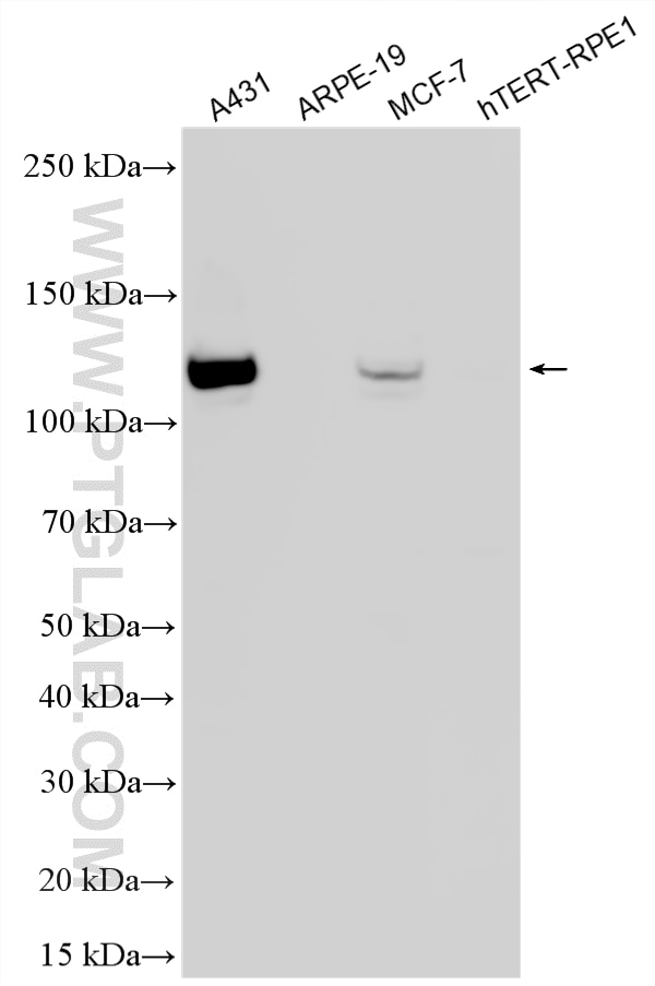
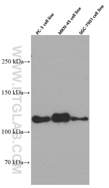
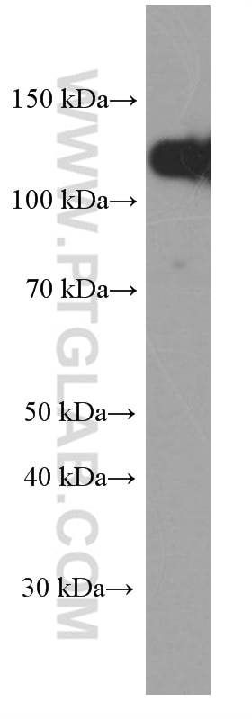
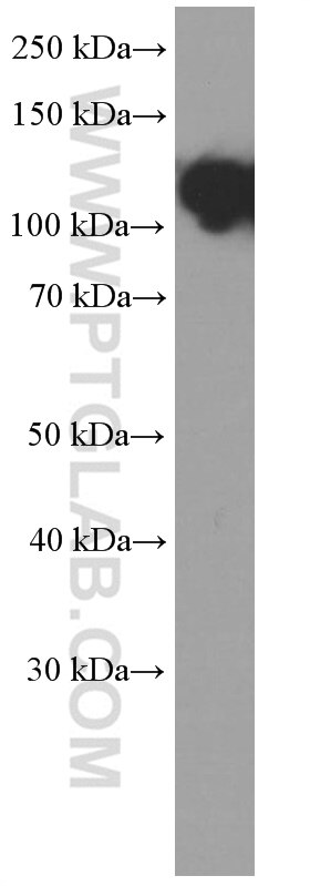
| Positive WB detected in | PC-3 cells, A431 cells, MCF-7 cells, pig brain tissue, MKN-45 cells, SGC-7901 cells |
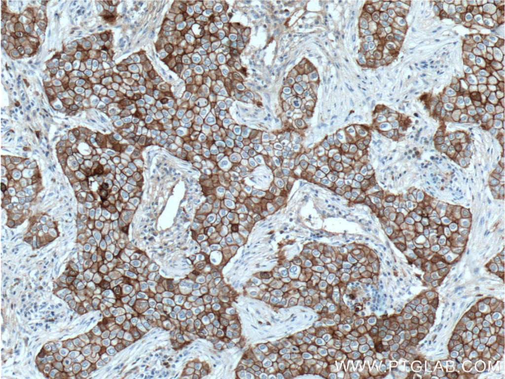
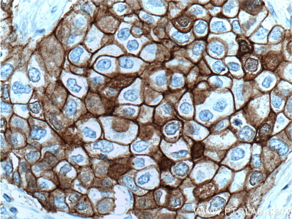
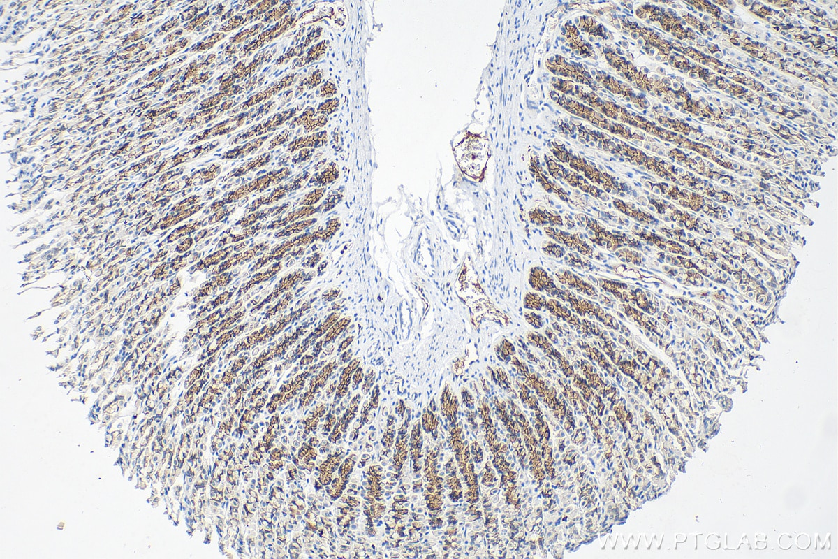
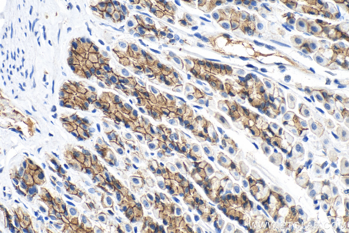
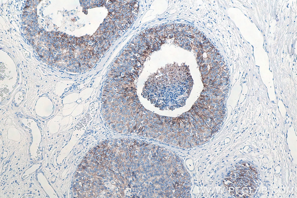
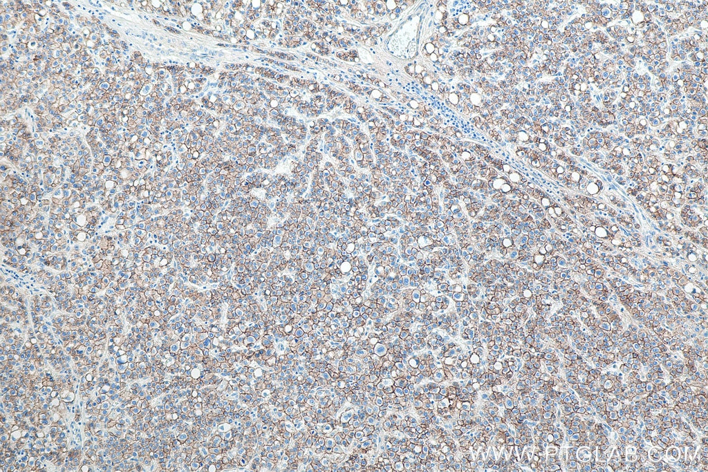
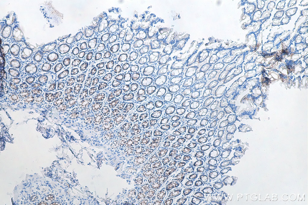
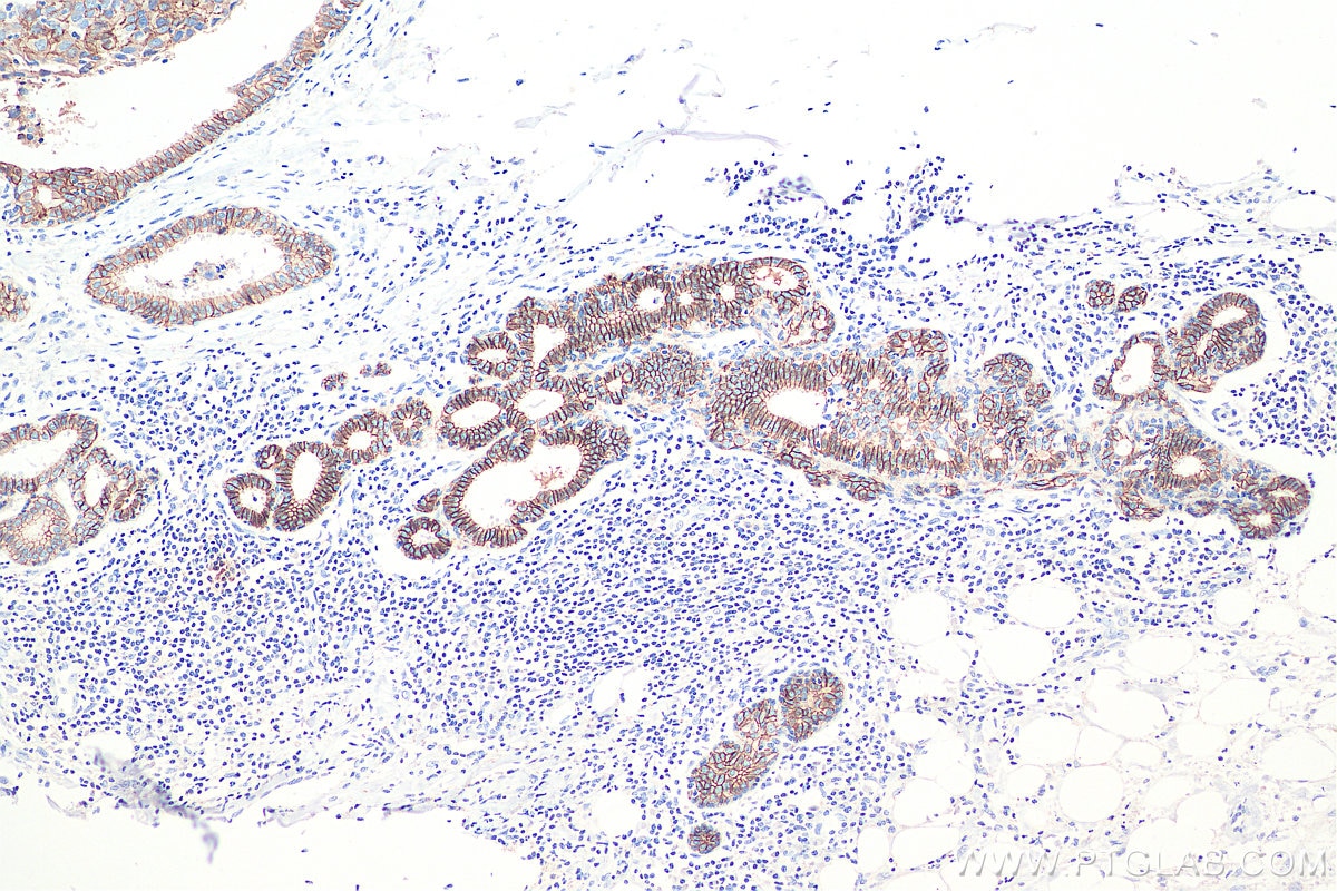
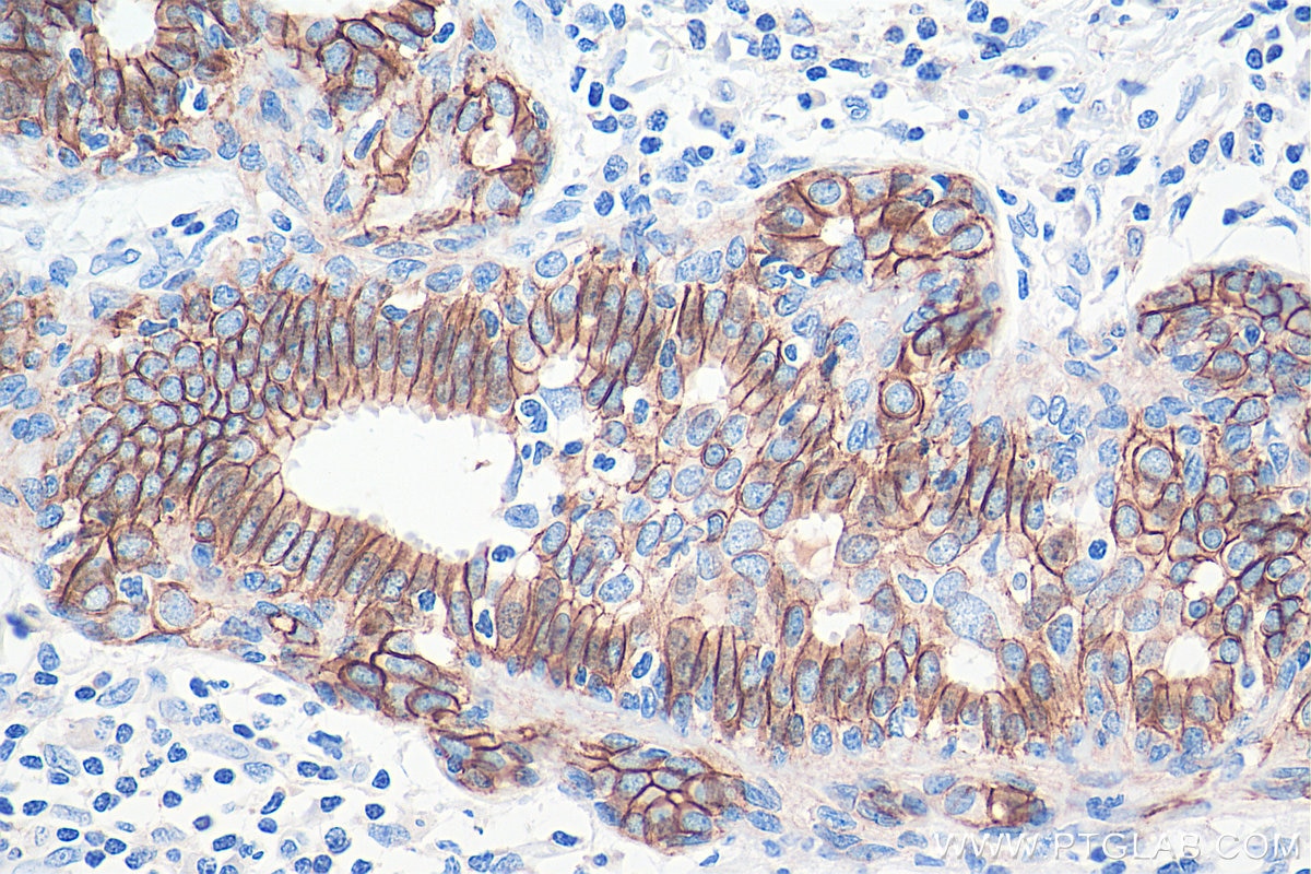
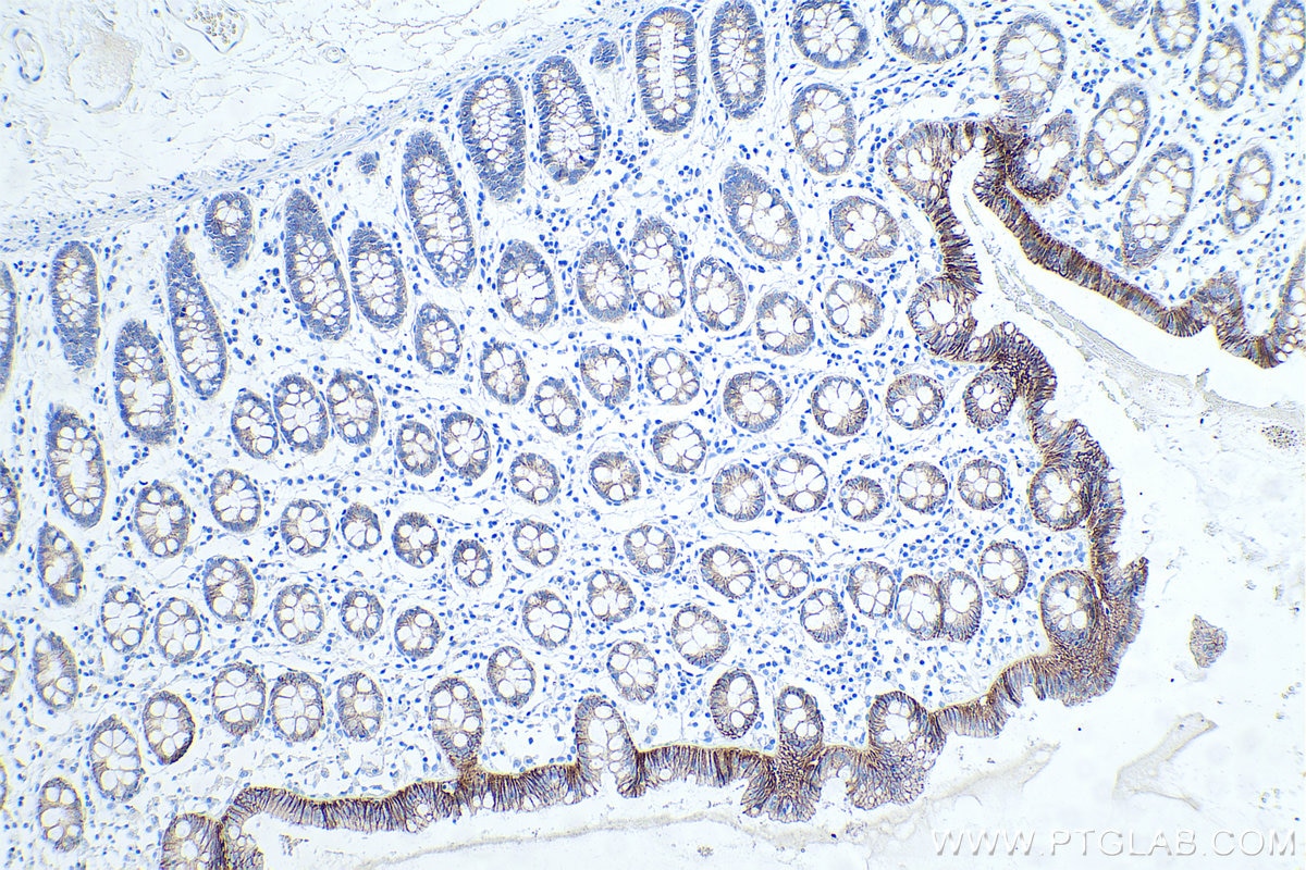
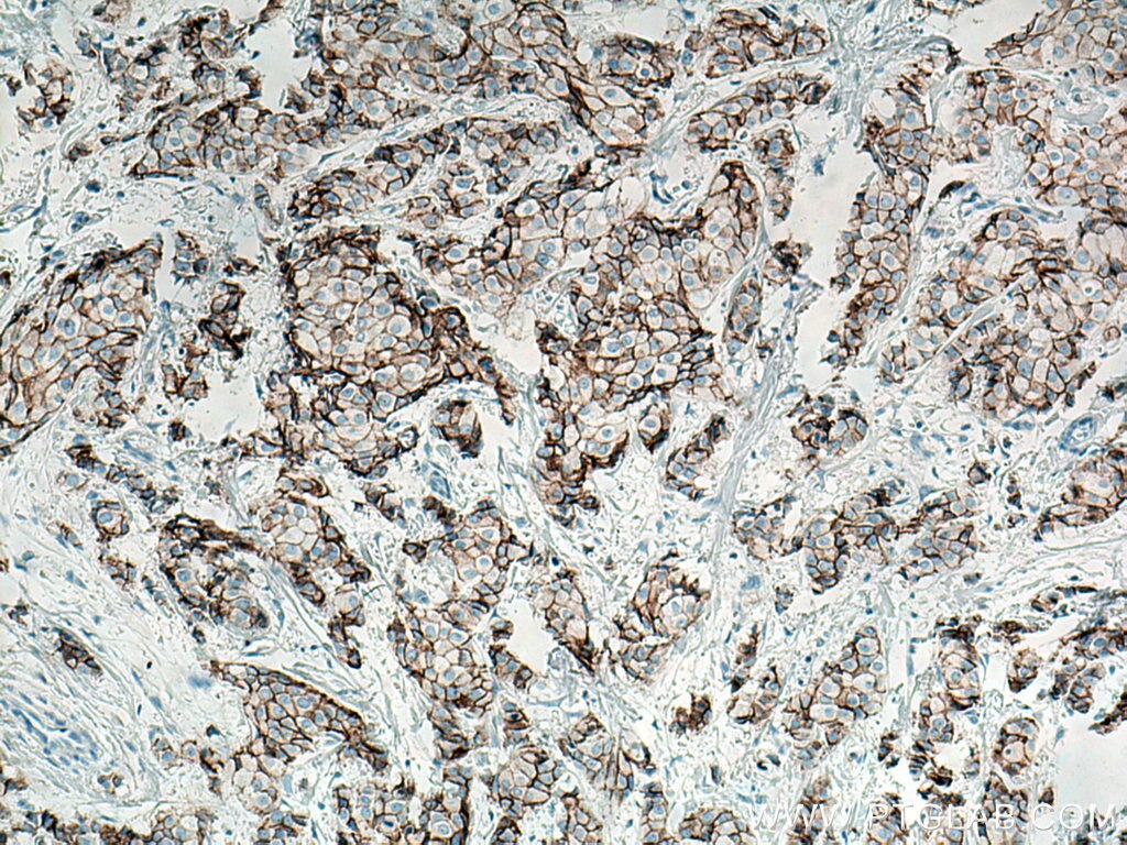
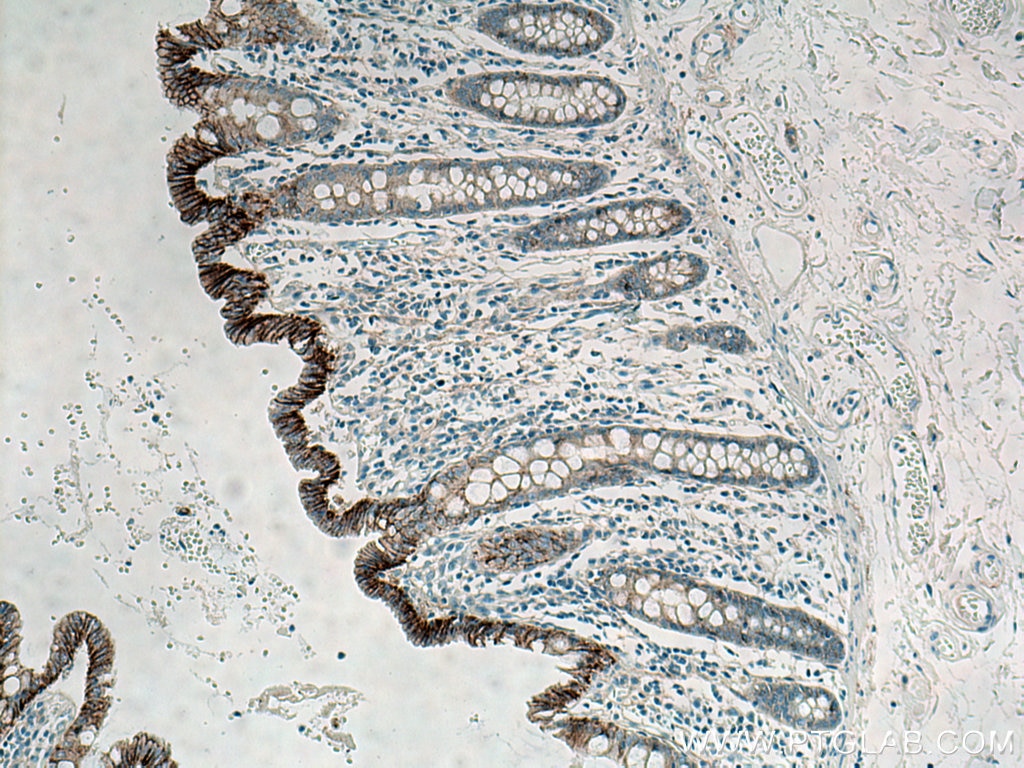
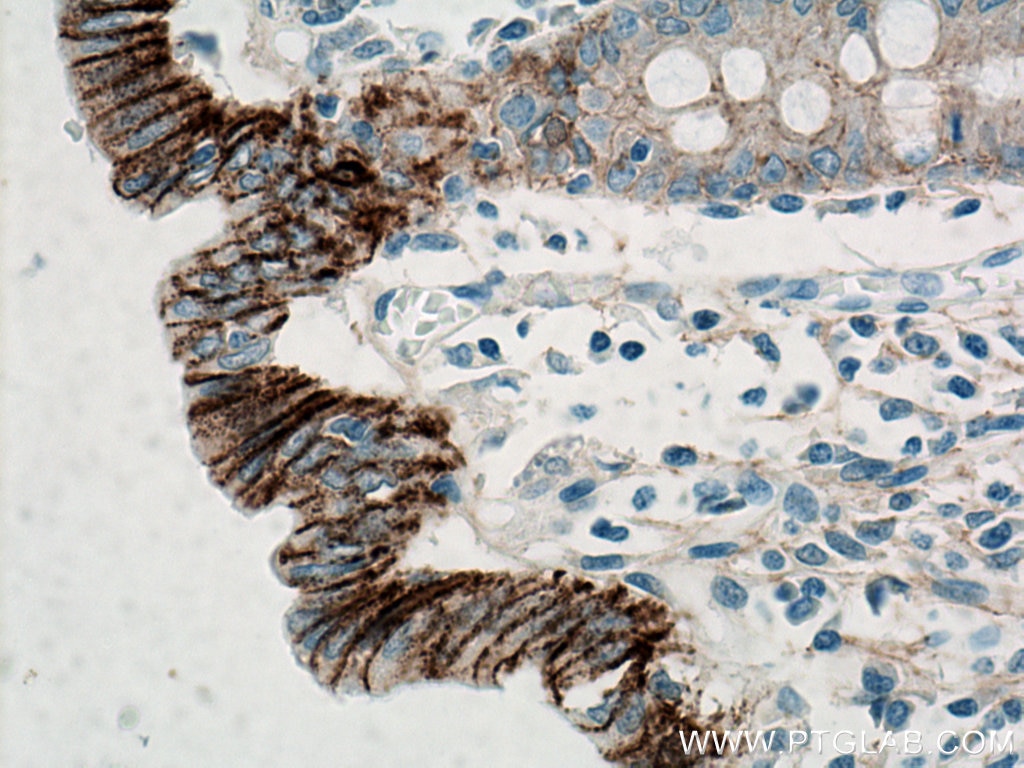
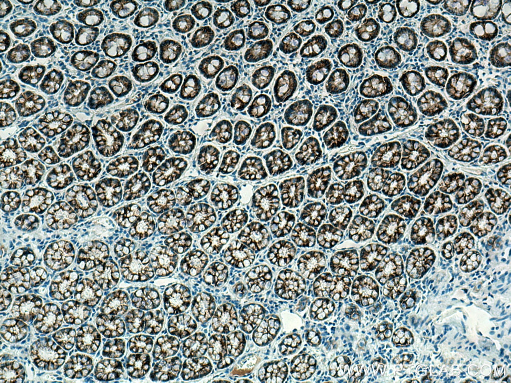
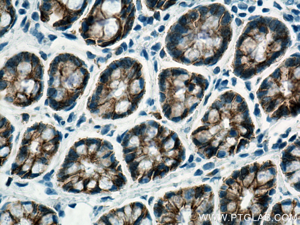
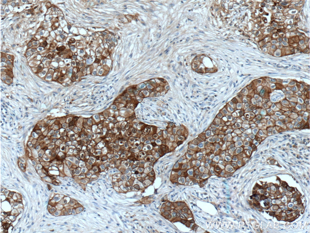
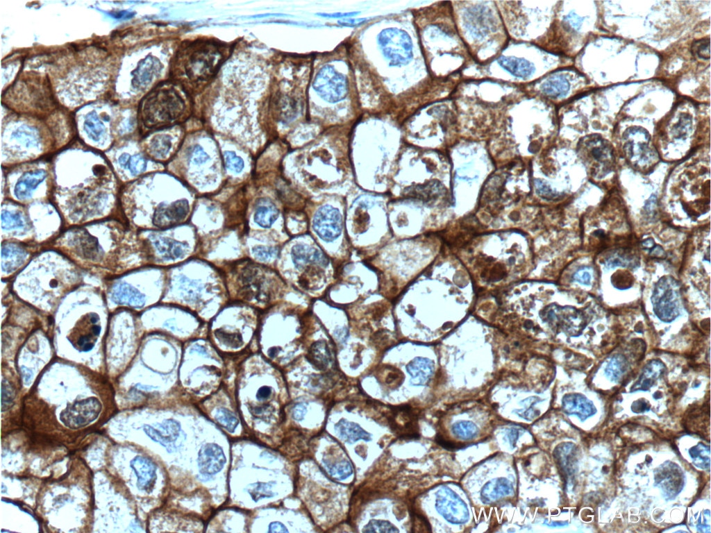
| Positive IHC detected in | human breast cancer tissue, human colon tissue, rat colon tissue, rat stomach tissue Note: suggested antigen retrieval with TE buffer pH 9.0; (*) Alternatively, antigen retrieval may be performed with citrate buffer pH 6.0 |
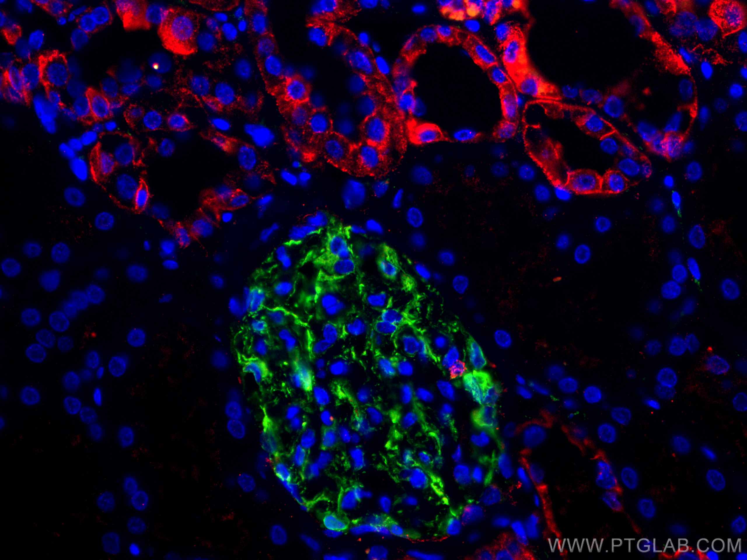
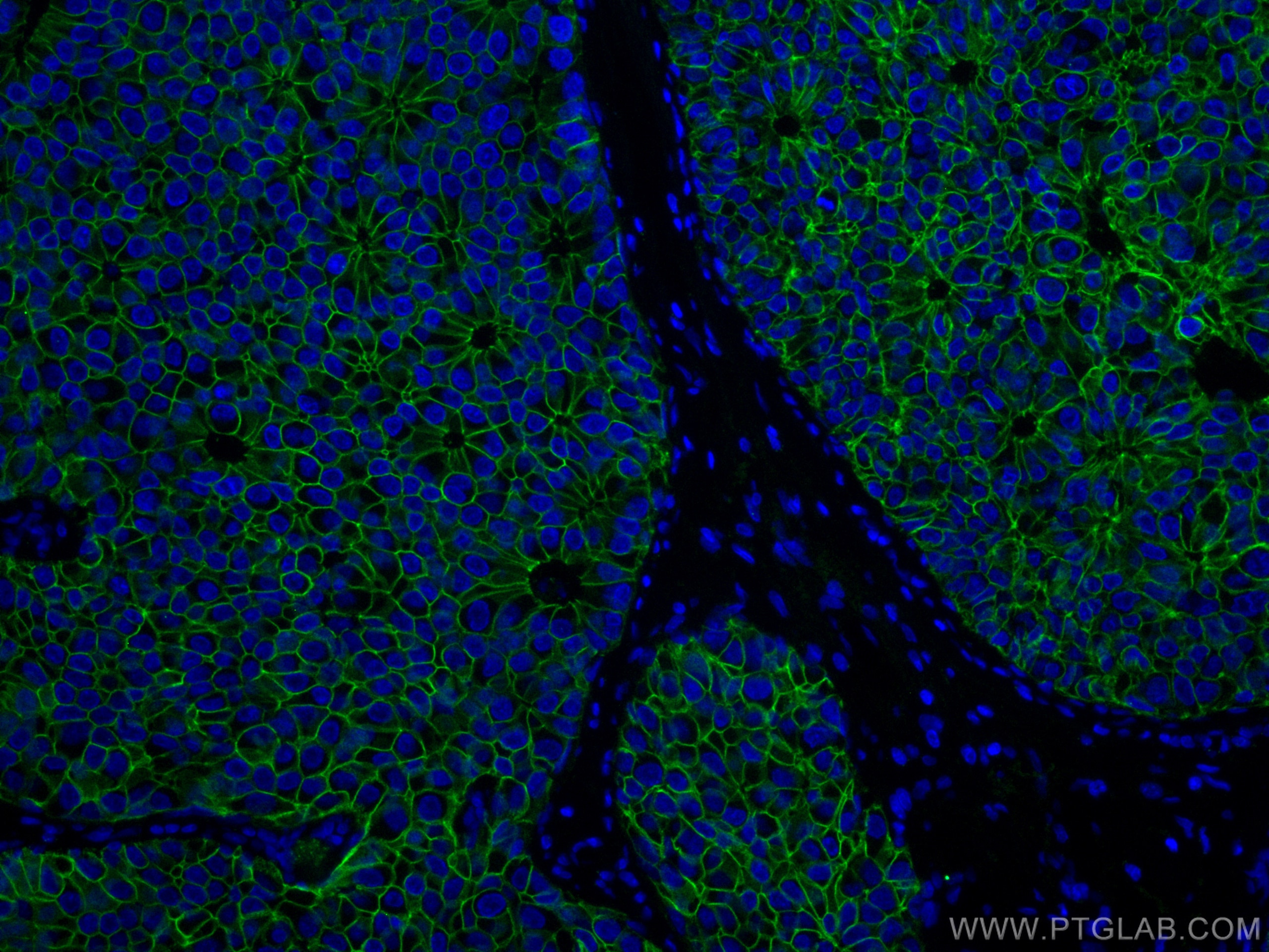
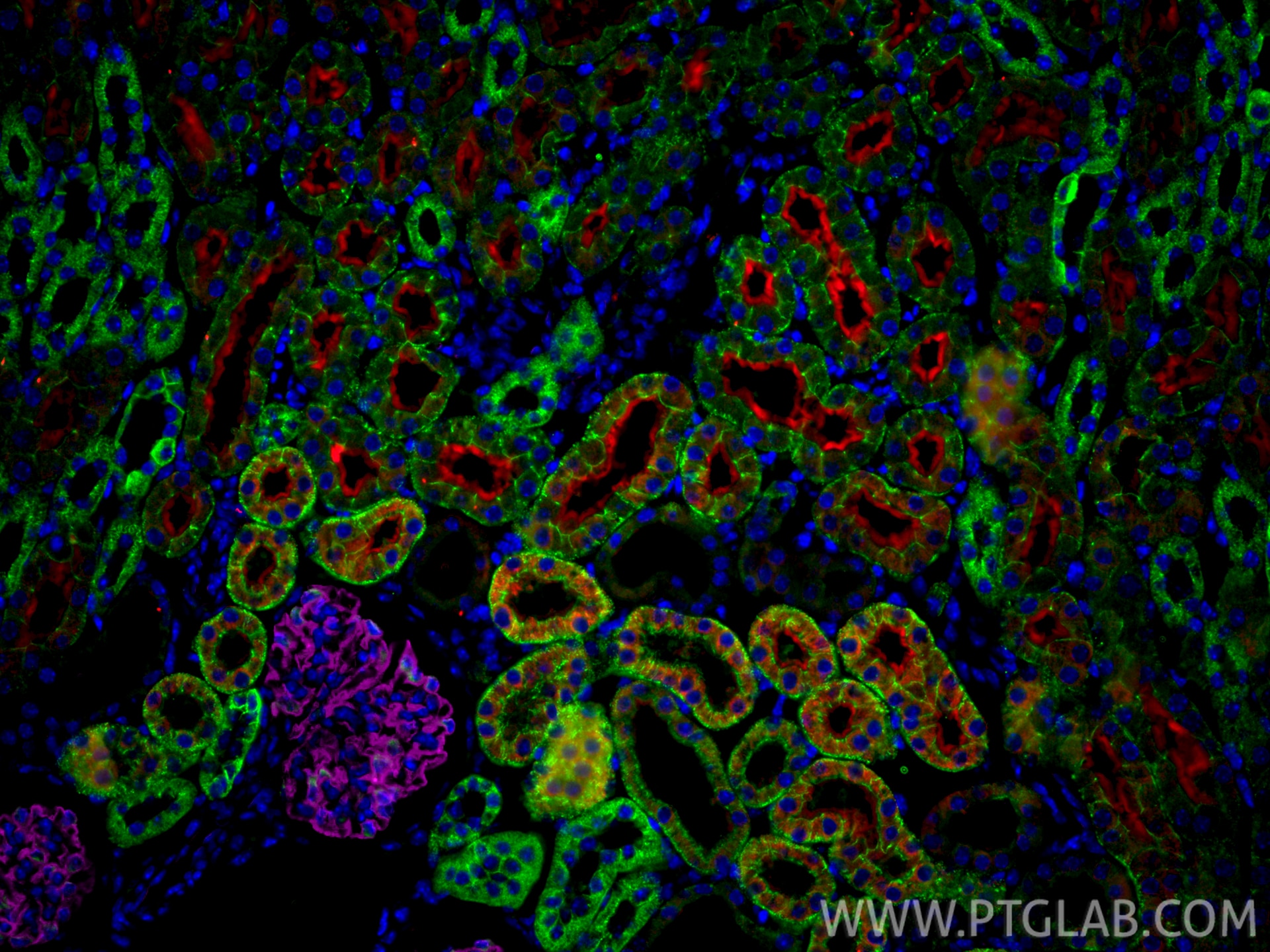
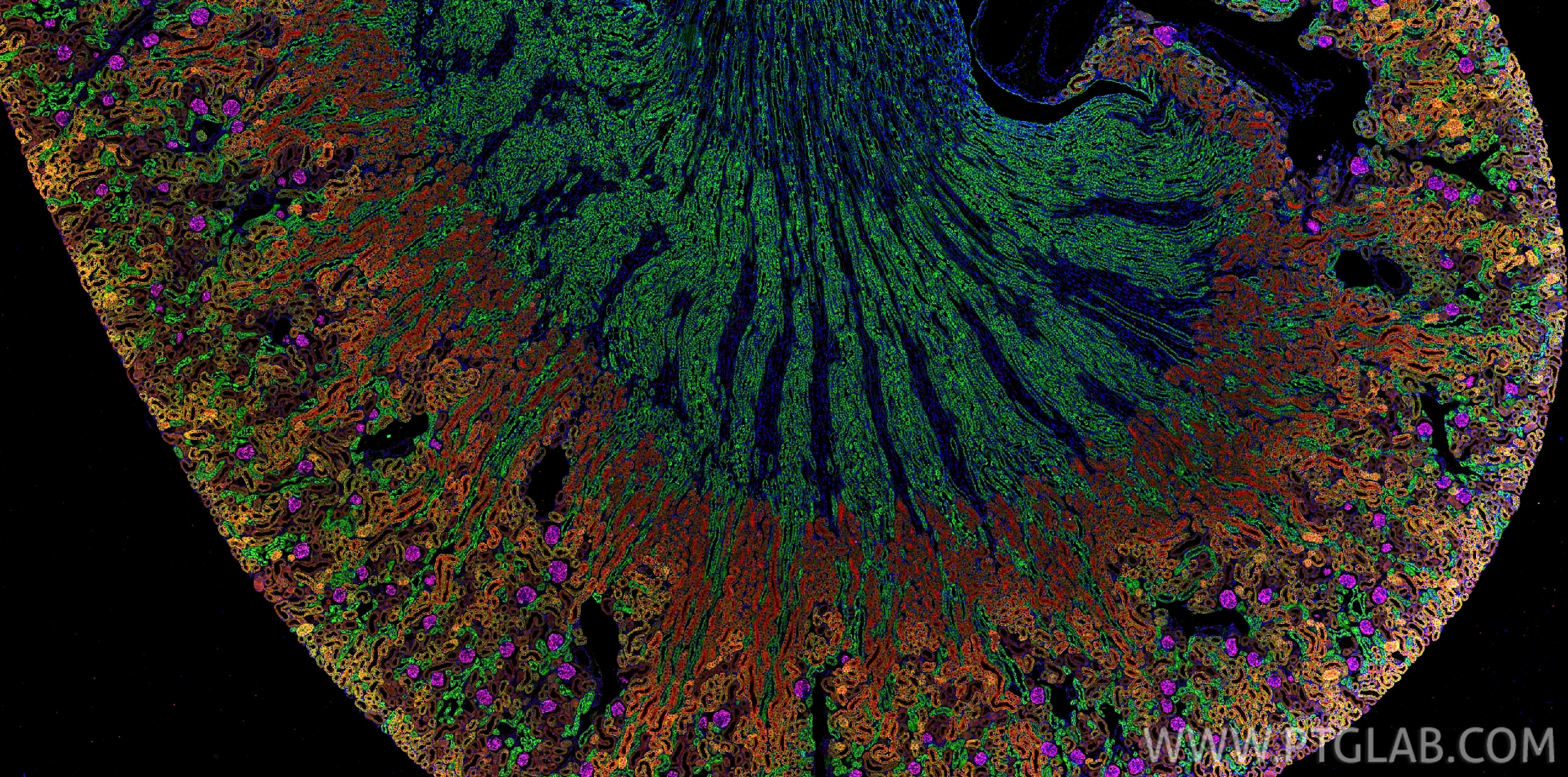
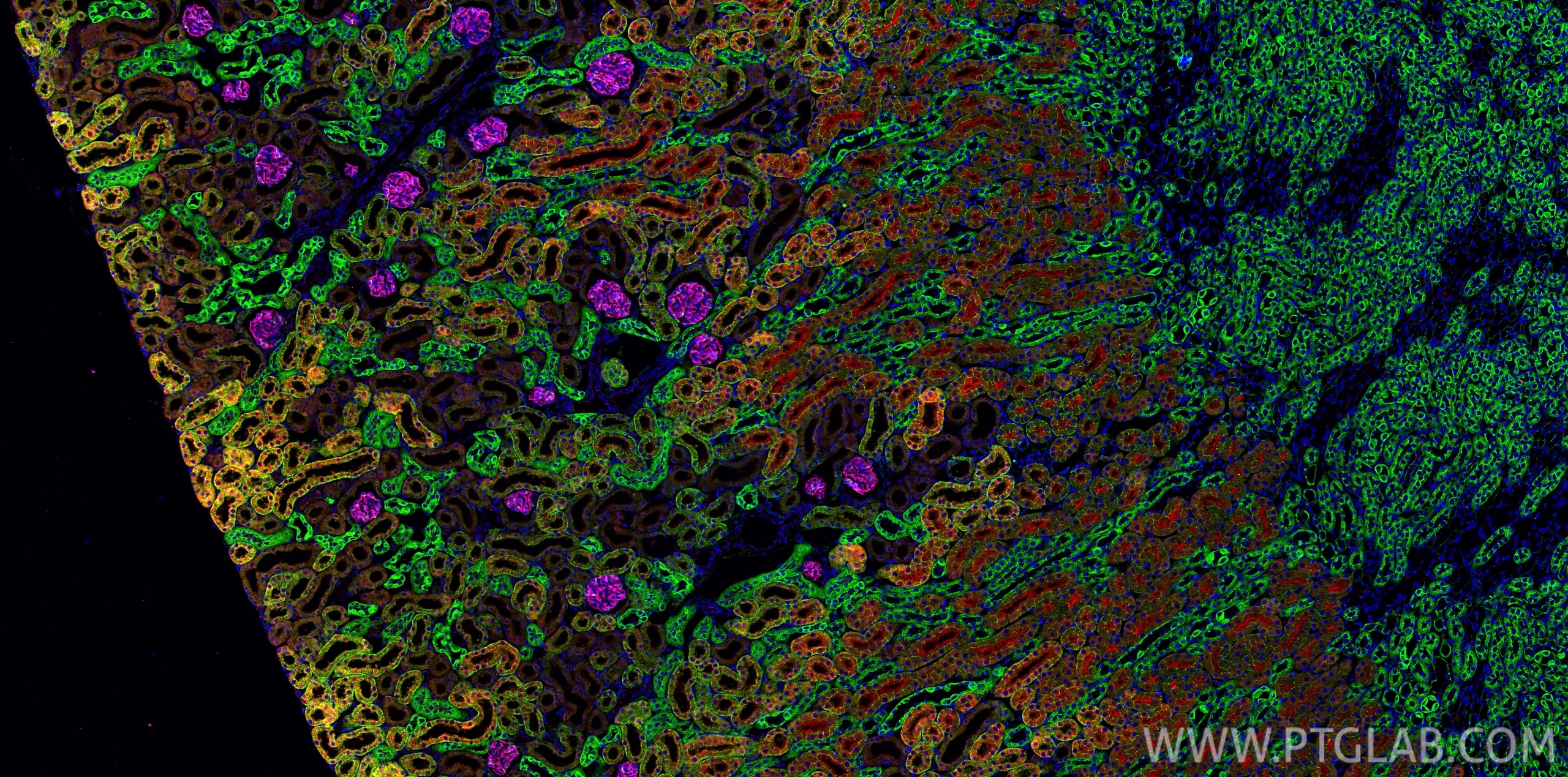
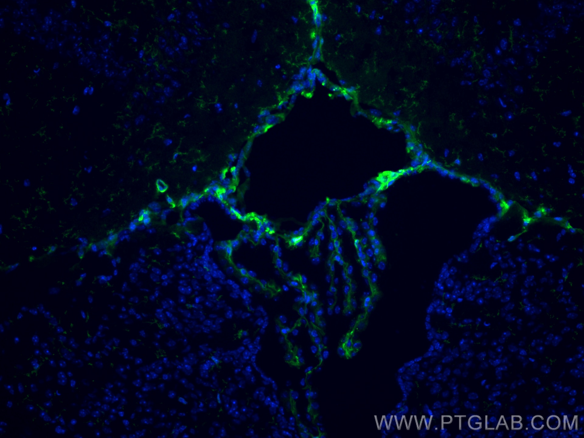
| Positive IF-P detected in | human breast cancer tissue, human kidney tissue, mouse kidney tissue |
| Positive IF-Fro detected in | mouse brain tissue |
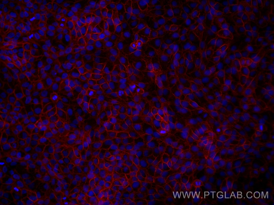
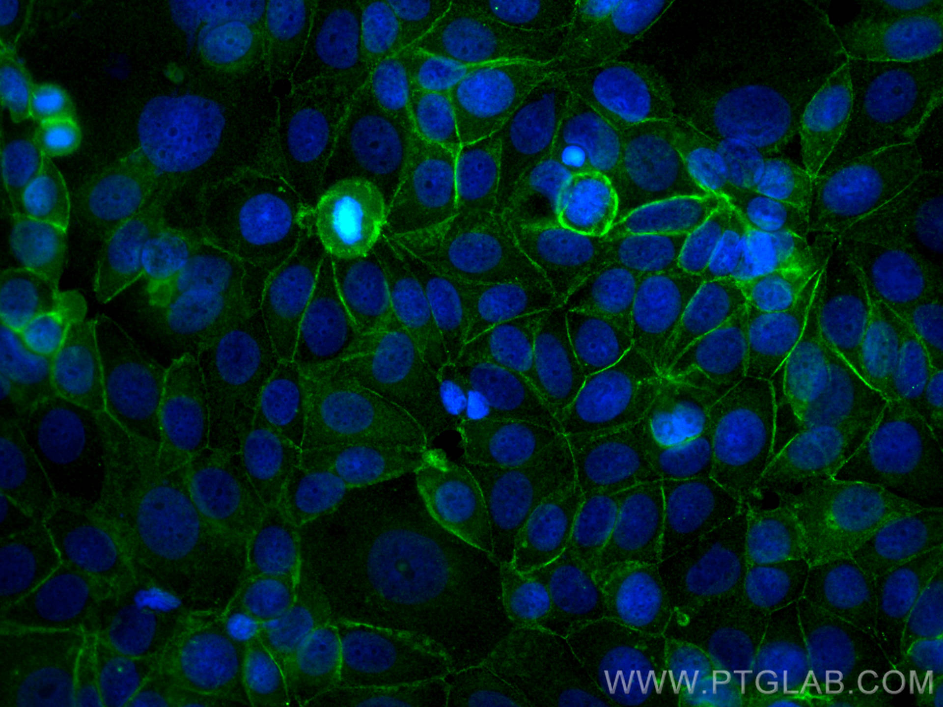
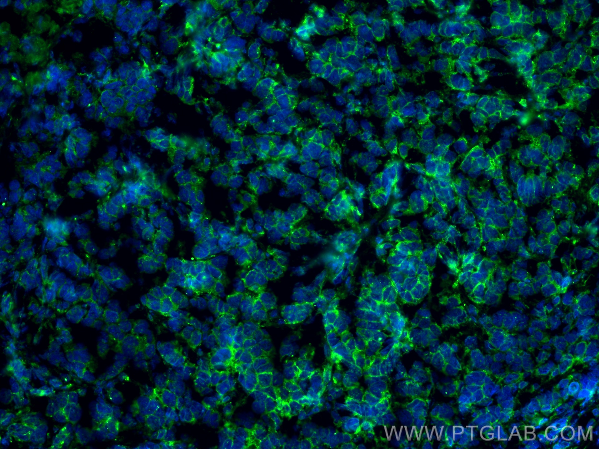
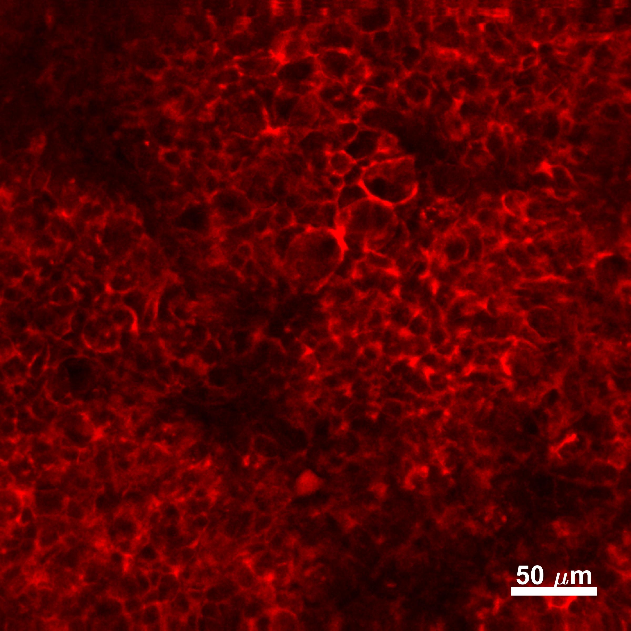
| Positive IF/ICC detected in | MCF-7 cells, mouse breast cancer, HaCaT cells |
Recommended dilution
| Application | Dilution |
|---|---|
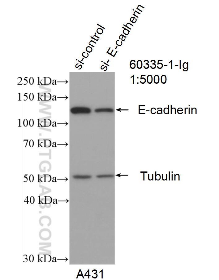
| Western Blot (WB) | WB : 1:2000-1:8000 |
| Immunohistochemistry (IHC) | IHC : 1:1000-1:4000 |
| Immunofluorescence (IF)-P | IF-P : 1:200-1:800 |
| Immunofluorescence (IF)-FRO | IF-FRO : 1:200-1:800 |
| Immunofluorescence (IF)/ICC | IF/ICC : 1:200-1:800 |
| It is recommended that this reagent should be titrated in each testing system to obtain optimal results. | |
| Sample-dependent, Check data in validation data gallery. | |
Published Applications
| WB | See 233 publications below |
| IHC | See 37 publications below |
| IF | See 71 publications below |
| FC | See 1 publications below |
Product Information
60335-1-Ig targets E-cadherin in WB, IHC, IF/ICC, IF-P, IF-Fro, ELISA applications and shows reactivity with human, mouse, rat, pig samples.
| Tested Reactivity | human, mouse, rat, pig |
| Cited Reactivity | human, mouse, rat, pig, canine, monkey |
| Host / Isotype | Mouse / IgG2b |
| Class | Monoclonal |
| Type | Antibody |
| Immunogen |
CatNo: Ag15085 Product name: Recombinant human E-cadherin protein Source: e coli.-derived, PET28a Tag: 6*His Domain: 373-622 aa of BC141838 Sequence: PIFNPTTYKGQVPENEANVVITTLKVTDADAPNTPAWEAVYTILNDDGGQFVVTTNPVNNDGILKTAKGLDFEAKQQYILHVAVTNVVPFEVSLTTSTATVTVDVLDVNEAPIFVPPEKRVEVSEDFGVGQEITSYTAQEPDTFMEQKITYRIWRDTANWLEINPDTGAISTRAELDREDFEHVKNSTYTALIIATDNGSPVATGTGTLLLILSDVNDNAPIPEPRTIFFCERNPKPQVINIIDADLPPI Predict reactive species |
| Full Name | cadherin 1, type 1, E-cadherin (epithelial) |
| Calculated Molecular Weight | 882 aa, 97 kDa |
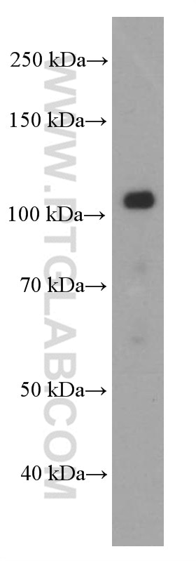
| Observed Molecular Weight | 120 kDa |
| GenBank Accession Number | BC141838 |
| Gene Symbol | E-cadherin |
| Gene ID (NCBI) | 999 |
| RRID | AB_2881444 |
| Conjugate | Unconjugated |
| Form | Liquid |
| Purification Method | Protein A purification |
| UNIPROT ID | P12830 |
| Storage Buffer | PBS with 0.02% sodium azide and 50% glycerol, pH 7.3. |
| Storage Conditions | Store at -20°C. Stable for one year after shipment. Aliquoting is unnecessary for -20oC storage. 20ul sizes contain 0.1% BSA. |
Background Information
Cadherins are a family of transmembrane glycoproteins that mediate calcium-dependent cell-cell adhesion and play an important role in the maintenance of normal tissue architecture. E-cadherin (epithelial cadherin), also known as CDH1 (cadherin 1) or CAM 120/80, is a classical member of the cadherin superfamily which also include N-, P-, R-, and B-cadherins. E-cadherin is expressed on the cell surface in most epithelial tissues. The extracellular region of E-cadherin establishes calcium-dependent homophilic trans binding, providing specific interaction with adjacent cells, while the cytoplasmic domain is connected to the actin cytoskeleton through the interaction with p120-, α-, β-, and γ-catenin (plakoglobin). E-cadherin is important in the maintenance of the epithelial integrity, and is involved in mechanisms regulating proliferation, differentiation, and survival of epithelial cell. E-cadherin may also play a role in tumorigenesis. It is considered to be an invasion suppressor protein and its loss is an indicator of high tumor aggressiveness.
Protocols
| Product Specific Protocols | |
|---|---|
| IHC protocol for E-cadherin antibody 60335-1-Ig | Download protocol |
| WB protocol for E-cadherin antibody 60335-1-Ig | Download protocol |
| IF protocol for E-cadherin antibody 60335-1-Ig | Download protocol |
| Standard Protocols | |
|---|---|
| Click here to view our Standard Protocols |
Publications
| Species | Application | Title |
|---|---|---|
Adv Mater Glycated ECM Derived Carbon Dots Inhibit Tumor Vasculogenic Mimicry by Disrupting RAGE Nuclear Translocation and Its Interaction With HMGB1 | ||
Theranostics FSH induces EMT in ovarian cancer via ALKBH5-regulated Snail m6A demethylation | ||
Theranostics Vitamin D binding protein (VDBP) hijacks twist1 to inhibit vasculogenic mimicry in hepatocellular carcinoma | ||
Biomaterials Urinary exosomes-based Engineered Nanovectors for Homologously Targeted Chemo-Chemodynamic Prostate Cancer Therapy via abrogating IGFR/AKT/NF-kB/IkB signaling. |
Reviews
The reviews below have been submitted by verified Proteintech customers who received an incentive for providing their feedback.
FH Aqib (Verified Customer) (09-02-2024) | It works quite well. recommended
|
FH Saba (Verified Customer) (06-14-2022) | The IF staining was very good and satisfying.
|
FH Silvia (Verified Customer) (02-09-2022) | The antibody worked well on HT-29 cells at 1:800 dilution for IF.
|
FH Joshua (Verified Customer) (12-27-2019) | Caco-2 cells fixed in 4% paraformaldehyde. Stained overnight at 4C. Bright stain, minimal background
 |
FH Louisiane (Verified Customer) (02-06-2019) | Cells were fixed with 4% PFA for 10 min, permeabilized with 0.1% Triton-X100 for 5 min and blocked with 1% FBS/1% BSA in PBS for 3 h. Antibodies were diluted in 1% FBS/1% BSA in PBS. Primary antibody: 2 h. Alexa Fluor anti-mouse secondary antibody (1:250): 1 h.Cells were imaged by confocal microscopy - no labeling was observed.
|